Spin-reorientation-induced band gap in Fe3Sn2: Optical signatures of Weyl nodes
Abstract
Temperature- and frequency-dependent infrared spectroscopy identifies two contributions to the electronic properties of the magnetic kagome metal Fe3Sn2: two-dimensional Dirac fermions and strongly correlated flat bands. The interband transitions within the linearly dispersing Dirac bands appear as a two-step feature along with a very narrow Drude component due to intraband contribution. Low-lying absorption features indicate flat bands with multiple van Hove singularities. Localized charge carriers are seen as a Drude-peak shifted to finite frequencies. The spectral weight is redistributed when the spins are reoriented at low temperatures; a sharp mode appears suggesting the opening of a gap due to the spin reorientation as the sign of additional Weyl nodes in the system.
Magnetic kagome metals are emerging as a new class of materials with special crystal structures, which are supposed to bring together electronic correlations, magnetism, and topological orders Liu et al. (2019). Merging the strong electronic correlations with the topologically nontrivial states makes new types of exotic phenomena possible ranging from fractional quantum Hall effect to axion insulators.
Unfortunately, the realization of the materials in this regard is scarce Nakatsuji et al. (2015); Nayak et al. (2016); Liu et al. (2018); Xu et al. (2018). The FeSn-binary compounds are possible candidates; Fe3Sn2 is one of them, where the linearly dispersing Dirac bands lying below the Fermi energy are confirmed as well as flat bands around . The crystal structure of Fe3Sn2 protects the inversion and three-fold rotational symmetry, while the time reversal symmetry is broken due to the magnetic nature of the system. The unique structural properties of this system give rise the flat-band ferromagnetism Lin et al. (2018), anomalous Hall effect Wang et al. (2016); Li et al. (2019), and topological Dirac states Ye et al. (2018). Due to the strong influence of magnetism, a large tunability of the spin-orbit coupling Yin et al. (2018) and of the massive Dirac fermions Ye et al. (2018); Lin and Chen (2020) was proposed as well as the emergence of further Weyl nodes at the Fermi energy Yao et al. (2018).
Fe3Sn2 consist of bilayer kagome network separated via stanene layers. The bilayer structure gives rise to the interlayer hybridization due to the multiple orbitals of Fe atoms leading to deviations from the ideal single-orbital two-dimensional kagome lattice scenario Mielke (1991a, b, 1992). For instance, the flat bands do not extend over the entire Brillouin zone and show a small dispersion Lin et al. (2018); moreover, correlations among the Dirac bands open a gap at the crossing points that give rise to the correlated massive Dirac fermions Ye et al. (2018). Despite these deviations from the ideal scenario, this system provides a beautiful playground for investigating the interplay between magnetism, strong electronic correlations, and topological orders.
For Fe3Sn2 it is known that even in the absence of an external magnetic field the spins reorient at reduced temperatures; despite the fact that the system orders ferromagnetically at much higher K Kumar et al. (2019). The implications of this reorientation on the electronic structure remain an open question. Considering the large sensitivity of magnetism on temperature, here we employ temperature-dependent broadband infrared spectroscopy for studying this model compound. We look for optical fingerprints of Dirac fermions, localized electrons of the flat bands, and spin reorientation, as they directly probe the interplay between these unique states along with the energy scales. In the absence of an external magnetic field, we investigate the effects of the inherent magnetization on the observed properties. Moreover, the high spectral resolution of our technique even in the low energy range, gives us the opportunity to test theoretical proposals regarding the additional Weyl nodes in the vicinity of .
Single crystals of Fe3Sn2 were synthesized as described elsewhere Wang et al. (2016). Here we performed temperature-dependent optical reflectivity measurements on single crystals of Fe3Sn2 in a broad energy range. Single crystals of Fe3Sn2 were grown using self-flux method as described elsewhere Wang et al. (2016). An as-grown sample with a good surface quality was chosen for the optical spectroscopy study. The (001)-plane lateral dimensions of the sample used in the infrared spectroscopy study is 1000 m800 m 200 m.
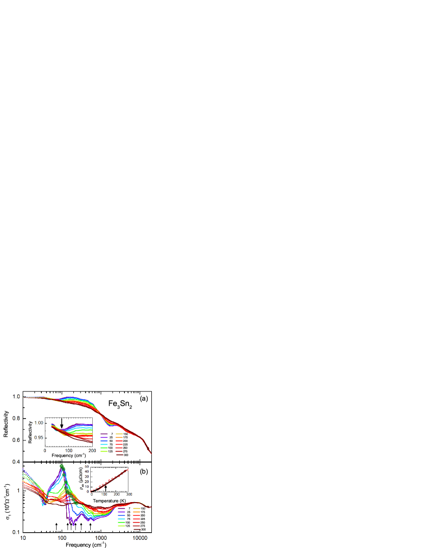
Fig. 1 displays the temperature-dependent reflectivity along with the optical conductivity in the entire measured range. Consistent with the highly metallic nature of the sample, the low-energy reflectivity reaches almost unity and the optical conductivity approaches the dc conductivity values on the order of at low . While the optical properties are basically temperature-independent above approximately 1 eV, a series of interesting features are identified below this energy: (i) A strong suppression of the reflectivity and, concomitantly, the optical conductivity starting from the mid-infrared range. (ii) A peak-like structure (marked with green circles) that shifts to lower energies with decreasing . (iii) A very narrow Drude component that gets even sharper upon cooling. The scattering of this Drude component is very small and barely visible in our measurement window; however, the dc resistivity data [the inset of the Fig. 1(b)] corroborate its existence \bibnoteIt is interesting to examine the scattering rate of this sharp Drude-component. As the momentum-relaxing scattering of the free carriers (in the current case is the Dirac fermions as explained in the text) can be observed in the optical conductivity. Hence, one can directly obtain the corresponding scattering time, , from the real part of the optical conductivity, . The simultaneous fits of the reflectivity and the optical conductivity of our low energy Drude-component reveals a scattering rate [] of 2 cm-1 at 7 K, while it increases to 17 cm-1 at room temperature. These corresponds to the scattering times ps and ps. Considering the reported average Fermi velocity for Fe3Sn2, m/s, the momentum relaxation lengths [] are calculated as m and nm. Considering that the localized carrier response of the flat bands are separated in energy and does not contribute to the low energy dynamics (within the obtained scattering scales), these length scales suggest that Fe3Sn2 readily might be a suitable platform to realize a ballistic transport..
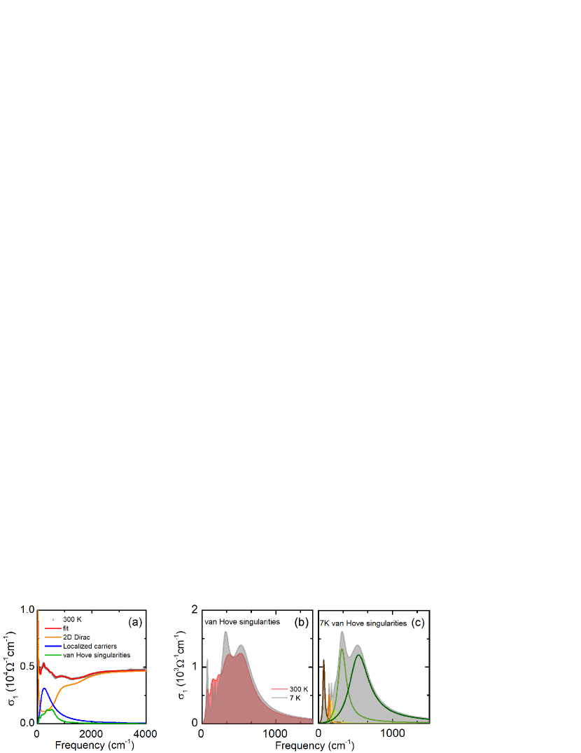
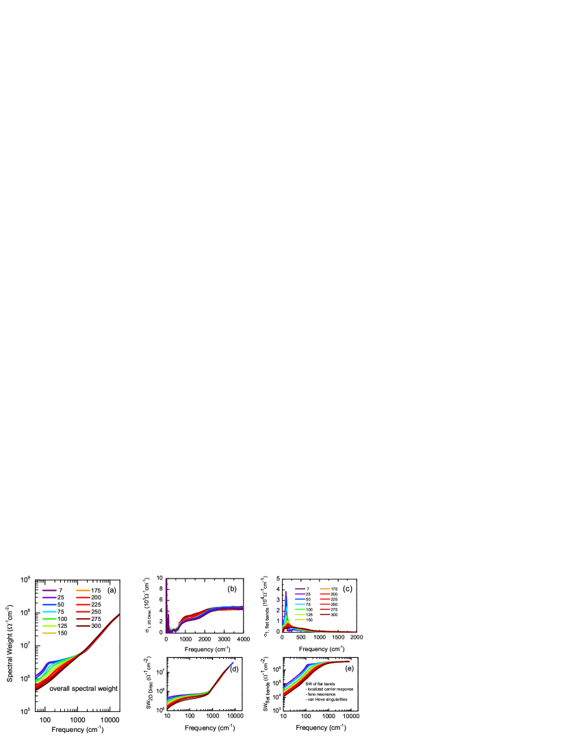
We decomposed the optical conductivity into two main parts shown in Fig. 2(a). While the high-energy absorption and the low-energy Drude-component can be interpreted within the Dirac fermions framework, the strong absorption features in between have to be treated separately. We attribute these features to the charge carriers within the flat bands, as explained later. This decomposition is supported by the analysis of the spectral weight (SW) [inset of Fig. 2(b)]. The transfer of SW takes place within the different contributions; the overall SW, but also the Dirac and the flat-band spectral weights are conserved.
But first let us discuss the Dirac physics and its optical signatures in Fe3Sn2. The results of angle resolved photoemmision spectroscopy (ARPES) Ye et al. (2018); Yao et al. (2018), scanning tunneling microscopy (STM) Lin et al. (2018), and magneto-transport measurements Wang et al. (2016); Liu et al. (2018); Li et al. (2019); Ye et al. (2018); Kumar et al. (2019) evidence the linearly dispersing bands of massless Dirac fermions and flat bands of massive localized electrons. ARPES data indicate that the Dirac bands lie well below (within 0.15 eV); whereas the magneto-transport measurements also verify the existence of a topologically nontrivial state. Moreover, the underlying bilayer structure of the kagome lattice gives rise to correlations among the Dirac fermions causing a correlation gap to open at the Dirac points.
The optical signatures of these Dirac points are clearly visible in our spectra. The mid-infrared absorption and the accompanying low-energy Drude component can be well reproduced by taking into account the intraband and interband responses of two-dimensional Dirac fermions as shown in graphene, for instance Li et al. (2008); Scharf et al. (2013). For a two-dimensional Dirac system with the Dirac point at the Fermi energy and in the absence of other contributions, the optical conductivity is expected to exhibit a frequency-independent behavior. On the other hand, the shift of the Dirac point with respect to results in absorption feature, where the SW is transferred to the intraband response of the Dirac bands. This situation forbids low energy transitions (up to , where defines the energy shift between and the Dirac point) and the optical conductivity starts to increase above a certain energy that is defined as the Pauli edge. In the two-dimensional case, one expects a step-like increase starting from and leveling off to the -independent behavior Gusynin et al. (2006).

In Fig. 2(b), the 7 K spectrum is plotted with the overall fit to our data, and after subtracting the low-lying absorption features. This remaining part represents the inter- and intraband contributions of the Dirac fermions; the resulting blue curve reproduces the high-energy interband transitions very well. In our case, these high-energy intraband transitions involve not one step-like feature, but actually two. Hence, a somewhat more complicated picture is present in this system and the response of the Dirac fermions cannot be explained within a single Dirac cone picture. This conclusion is in line with ARPES results Ye et al. (2018), as depicted by the two-cone picture in Fig. 2(a). In turn, this gives rise to the two-step absorption feature of the optical conductivity. We also like to point out that even the high-temperature spectra reveal signatures of these two step absorption feature. In Fig. 2(b), the and 150 K spectra are given for comparison; the steps due to the Dirac bands are marked by colored dots.
From our spectrum at K we estimate the positions of the Dirac points at = 346 cm-1 (42 meV) and = 983 cm-1 (122 meV); they do not change significantly with temperature. These values are well in line with ARPES measurements Ye et al. (2018), while they do not consider the correlation gap observed in ARPES. Such an energy gap due to correlated Dirac fermions resembles the optical response known from density-wave systems Uykur et al. (2019). However, in the present case we should not see such an effect, as the gap is buried well below .
Let us turn to the low-lying absorption band of the spectra. We associate these features with the response of the flat bands, since they are located in the vicinity of the Fermi energy. We can identify several absorption features in the spectra at 72, 141, 172, 223, 311, 542 cm-1; however, a close look reveals that most of them do not shift with decreasing temperature, but simply get sharper [marked by the arrows in Fig. 1(b)]. We attribute these peaks to van Hove singularities of the flat bands. As shown by STM measurements Lin et al. (2018), flat bands do not extend over the entire Brillouin zone. They possess a small dispersion giving rise to several peaks in the density of states around the Fermi energy. Due to the band dispersion, the transitions across the Fermi energy between these flat bands occur at slightly different energies, as observed.
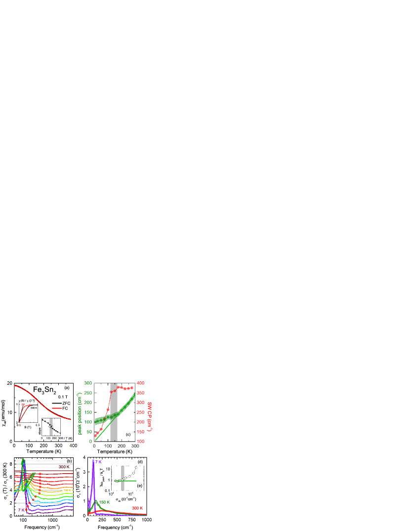
The absorption feature around 100 cm-1 is by far the most prominent one with a distinct dynamics: it changes its shape and strongly shifts in energy upon cooling. Our detailed analysis reveals that this feature is strongly linked to the underlying magnetic structure. Fe3Sn2 possesses flat-band ferromagnetism related to the underlying kagome lattice with a ferromagnetic phase transition at K. Previous magnetization, neutron diffraction, and Mössbauer spectroscopy studies all conclude that the spins are canted towards out of plane at high temperatures up to . With decreasing temperature they tend to reorient towards the kagome plane Trumpy et al. (1970); Caer et al. (1978); Malaman et al. (1978); Caer et al. (1979). However, no agreement has been reached on the temperature range, where this reorientation occurs, and whether the phase transition is of first or a second order. A recent magneto-transport study showed that the spin reorientation takes place in a temperature range between 70-150 K with a transition peak at K Kumar et al. (2019). The nature of this spin reorientation and its implications on the electronic structure remain to be clarified. Characterizing our sample in this regard, in Fig. 3(a) we plot the magnetic susceptibility as a function of temperature. Our findings are consistent with the literature and yield a crossover temperature slightly below 150 K.
Let us turn back to the strong optical absorption around 100 cm-1. To better analyse the evolution of the peak, we plotted the relative optical conductivity [] in Fig. 3(b), where one can trace the energy position of the peak as well as the transfer of spectral-weight to the low-lying absorption features. Although we cannot rule out that appear accidentally, at elevated temperatures a clear isosbestic point (indicated by the red symbols, crossing of the spectra at 300 K spectrum, see Supplementary Materials for the details) of the spectra can be defined along with the SW transfer that is lead by the temperature change Greger et al. (2013). Below the spin reorientation temperature the isosbestic behavior does not hold anymore and the spectra crossing point rapidly shifts to the smaller energies, as the SW accumulates to a very sharp peak structure. From panel (c) we can see that its maximum (green circles) gradually moves to lower energies: while for K it decreases linearly in , the shift tends to saturate at lower temperatures. The drastic change of the isosbestic signature suggests the influence of another mechanism other than the temperature on the SW redistribution.
Since the flat bands possess strongly correlated, localized charge carriers, the so-called localization peak is a plausible assumption for the observed peak. The generalization of the Drude response is commonly discussed in the framework of the strongly correlated electron systems Smith (2001); Fratini et al. (2014); Delacrétaz et al. (2017). The partial localization of the charge carriers leads to a displaced Drude-peak, where the low-energy part of the optical conductivity is strongly suppressed and the peak-like structure appears at finite energies. Literature is rich in this regard, with examples from numerous transition metal oxides Kostic et al. (1998); Lee et al. (2002); Bernhard et al. (2004); Wang et al. (2004); Puchkov et al. (1995); *Hwang2007; Tsvetkov et al. (1997); Osafune et al. (1999); *Uykur2011; Rozenberg et al. (1995); Jönsson et al. (2007); Takenaka et al. (1999); *Takenaka2002; Jaramillo et al. (2014) and organic conductors Dong et al. (1999); Takenaka et al. (2005). Please note that the phenomenological descriptions of the optical conductivity does not rely on any specific nature of the localization, but generally discussed in terms of disorder effects. Here, we employed the model proposed by Fratini et al., where the low-energy Drude-response is modified with the electron backscattering Fratini et al. (2014); *Fratini2016; *Fratini2020, while the high-energy tail of the modified Drude response is still defined with the elastic scattering rate of the free carriers. These optical fingerprints commonly go hand in hand with a linear-in- resistivity (so-called bad metallic behavior), where the shift of the peak scales with the dc-conductivity Delacrétaz et al. (2017).
To better demonstrate the phenomenological description of the peak in Fig. 3(d), we subtract the response of the Dirac bands, as well as the van-Hove singularities from the measured optical conductivity. The solid lines are the best fits to the model suggested by Fratini et al. Fratini et al. (2014). The high-temperature data are well reproduced by taking into account the backscattering correction, where we restrict ourselves with the static limit. We estimate the backscattering time of the electrons, , to be around 13 fs at room temperature and increases to 76 fs at K. Below the transition at 150 K, the description breaks down: the observed feature corresponds to a very sharp Fano-like peak rather than a modified Drude-peak. This is in accord with the breakdown of the scaling relation between the dc conductivity and the linear shift of the peak energy below 150 K, displayed in Fig. 3(e). Indeed, the presence of spin reorientation calls for a different approach at low temperatures. A better accuracy can be satisfied by taking into account a coupling of a Fano resonance to the electronic background. The Fano resonance starts to appear below 150 K and gets more pronounced with decreasing temperature. Along with a stronger coupling to the electronic background, the behavior of the Fano-resonance follows the magnetization of the sample and the spin reorientation.
The appearance of such a sharp peak resembles excitations between electronic bands at ; i.e. it suggests the development of a partial gap in the density of states. This can be explained by additional Weyl nodes recently predicted for Fe3Sn2 Yao et al. (2018). Their existence is linked to the spin directions of the iron atoms within the kagome plane. When the spins reorient within the plane, the Weyl nodes become gapped, for the certain direction of magnetization, while for the other in-plane direction, there should be no gap. Hence, we conclude that the spin reorientation to the in-plane direction opens up the gap at the Weyl points, that we detect by optical means. The gap energy estimated from our measurements is 98 cm-1 (12 meV), which is in accord with the gap energy expected from calculations Yao et al. (2018).
It is not surprising that these Weyl nodes are not identified by ARPES, because the current optical measurements possess a much higher energy resolution. Moreover, since the magnetic properties and the electronic structure are very sensitive to small fields, signatures of the gap might be missed in the magneto-transport measurements. The current optical study was conducted in zero field, taking into account only the inherent magnetization of the compound. This allows us to discover experimentally the proposed gap opening.
In summary, temperature and frequency-dependent infrared studies on the magnetic kagome metal Fe3Sn2 reveal optical fingerprints of strongly correlated flat bands and topologically nontrivial Dirac fermions. The two-step absorption feature that evolves in the frequency-independent optical conductivity indicates the existence of two-dimensional Dirac cones shifted in energy with respect to each other. The flat bands are seen as multiple absorption peaks in the low-energy optical conductivity. One of the peaks exhibits a strong shift in energy. At high temperatures, this peak reveals striking similarities with the displaced Drude-peak as an indication of localization effects. Below the spin reorientation temperature around 120 K, a gap opens around the Fermi energy as signature for theoretically proposed gapped Weyl nodes. On the other hand, spin reorientation seems not to effect the Dirac nodes buried well into the Fermi energy.
Acknowledgements.
Authors acknowledge the fruitful discussions with L. Z. Maulana and A.V. Pronin and the technical support by G. Untereiner. H.C.L. acknowledges support from the National Key R&D Program of China (Grants No. 2016YFA0300504, 2018YFE0202600), and the National Natural Science Foundation of China (No. 11574394, 11774423, 11822412). The work has been supported by the Deutsche Forschungsgemeinschaft (DFG) via DR228/48-1 and DR228/51-1. E.U. acknowledges the European Social Fund and the Baden-Württemberg Stiftung for the financial support of this research project by the Eliteprogramme.References
- Liu et al. (2019) D. F. Liu, A. J. Liang, E. K. Liu, Q. N. Xu, Y. W. Li, C. Chen, D. Pei, W. J. Shi, S. K. Mo, P. Dudin, T. Kim, C. Cacho, G. Li, Y. Sun, L. X. Yang, Z. K. Liu, S. S. P. Parkin, C. Felser, and Y. L. Chen, “Magnetic Weyl semimetal phase in a Kagomé crystal,” Science 365, 1282–1285 (2019).
- Nakatsuji et al. (2015) Satoru Nakatsuji, Naoki Kiyohara, and Tomoya Higo, “Large anomalous Hall effect in a non-collinear antiferromagnet at room temperature,” Nature 527, 212–215 (2015).
- Nayak et al. (2016) Ajaya K. Nayak, Julia Erika Fischer, Yan Sun, Binghai Yan, Julie Karel, Alexander C. Komarek, Chandra Shekhar, Nitesh Kumar, Walter Schnelle, Jürgen Kübler, Claudia Felser, and Stuart S. P. Parkin, “Large anomalous Hall effect driven by a nonvanishing Berry curvature in the noncolinear antiferromagnet Mn3Ge,” Sci. Adv. 2 (2016), 10.1126/sciadv.1501870.
- Liu et al. (2018) Enke Liu, Yan Sun, Nitesh Kumar, Lukas Muechler, Aili Sun, Lin Jiao, Shuo-Ying Yang, Defa Liu, Aiji Liang, Qiunan Xu, Johannes Kroder, Vicky Süß, Horst Borrmann, Chandra Shekhar, Zhaosheng Wang, Chuanying Xi, Wenhong Wang, Walter Schnelle, Steffen Wirth, Yulin Chen, Sebastian T. B. Goennenwein, and Claudia Felser, “Giant anomalous Hall effect in a ferromagnetic kagome-lattice semimetal,” Nat. Phys. 14, 1125–1131 (2018).
- Xu et al. (2018) Qiunan Xu, Enke Liu, Wujun Shi, Lukas Muechler, Jacob Gayles, Claudia Felser, and Yan Sun, “Topological surface Fermi arcs in the magnetic Weyl semimetal ,” Phys. Rev. B 97, 235416 (2018).
- Lin et al. (2018) Zhiyong Lin, Jin-Ho Choi, Qiang Zhang, Wei Qin, Seho Yi, Pengdong Wang, Lin Li, Yifan Wang, Hui Zhang, Zhe Sun, Laiming Wei, Shengbai Zhang, Tengfei Guo, Qingyou Lu, Jun-Hyung Cho, Changgan Zeng, and Zhenyu Zhang, “Flatbands and Emergent Ferromagnetic Ordering in Kagome Lattices,” Phys. Rev. Lett. 121, 096401 (2018).
- Wang et al. (2016) Qi Wang, Shanshan Sun, Xiao Zhang, Fei Pang, and Hechang Lei, “Anomalous Hall effect in a ferromagnetic single crystal with a geometrically frustrated Fe bilayer kagome lattice,” Phys. Rev. B 94, 075135 (2016).
- Li et al. (2019) Hang Li, Bei Ding, Jie Chen, Zefang Li, Zhipeng Hou, Enke Liu, Hongwei Zhang, Xuekui Xi, Guangheng Wu, and Wenhong Wang, “Large topological Hall effect in a geometrically frustrated kagome magnet Fe3Sn2,” Appl. Phys. Lett. 114, 192408 (2019).
- Ye et al. (2018) Linda Ye, Mingu Kang, Junwei Liu, Felix von Cube, Christina R. Wicker, Takehito Suzuki, Chris Jozwiak, Aaron Bostwick, Eli Rotenberg, David C. Bell, Liang Fu, Riccardo Comin, and Joseph G. Checkelsky, “Massive Dirac fermions in a ferromagnetic kagome metal,” Nature 555, 638–642 (2018).
- Yin et al. (2018) Jia-Xin Yin, Songtian S. Zhang, Hang Li, Kun Jiang, Guoqing Chang, Bingjing Zhang, Biao Lian, Cheng Xiang, Ilya Belopolski, Hao Zheng, Tyler A. Cochran, Su-Yang Xu, Guang Bian, Kai Liu, Tay-Rong Chang, Hsin Lin, Zhong-Yi Lu, Ziqiang Wang, Shuang Jia, Wenhong Wang, and M. Zahid Hasan, “Giant and anisotropic many-body spin-orbit tunability in a strongly correlated kagome magnet,” Nature 562, 91–95 (2018).
- Lin and Chen (2020) Zheng-Zhe Lin and Xi Chen, “Tunable massive dirac fermions in ferromagnetic fe3sn2 kagome lattice,” phys. stat. sol. (RRL) , 1900705 (2020).
- Yao et al. (2018) M. Yao, H. Lee, N. Xu, Y. Wang, J. Ma, O. V. Yazyev, Y. Xiong, M. Shi, G. Aeppli, and Y. Soh, “Switchable Weyl nodes in topological Kagome ferromagnet Fe3Sn2,” (2018), arXiv:1810.01514 [cond-mat.str-el] .
- Mielke (1991a) A Mielke, “Ferromagnetic ground states for the Hubbard model on line graphs,” J. Phys. A 24, L73–L77 (1991a).
- Mielke (1991b) A Mielke, “Ferromagnetism in the Hubbard model on line graphs and further considerations,” J. Phys. A 24, 3311–3321 (1991b).
- Mielke (1992) A Mielke, “Exact ground states for the Hubbard model on the Kagome lattice,” J. Phys. A. 25, 4335–4345 (1992).
- Kumar et al. (2019) Neeraj Kumar, Y. Soh, Yihao Wang, and Y. Xiong, “Magnetotransport as a diagnostic of spin reorientation: Kagome ferromagnet as a case study,” Phys. Rev. B 100, 214420 (2019).
- (17) It is interesting to examine the scattering rate of this sharp Drude-component. As the momentum-relaxing scattering of the free carriers (in the current case is the Dirac fermions as explained in the text) can be observed in the optical conductivity. Hence, one can directly obtain the corresponding scattering time, , from the real part of the optical conductivity, . The simultaneous fits of the reflectivity and the optical conductivity of our low energy Drude-component reveals a scattering rate [] of 2 cm-1 at 7 K, while it increases to 17 cm-1 at room temperature. These corresponds to the scattering times ps and ps. Considering the reported average Fermi velocity for Fe3Sn2, m/s, the momentum relaxation lengths [] are calculated as m and nm. Considering that the localized carrier response of the flat bands are separated in energy and does not contribute to the low energy dynamics (within the obtained scattering scales), these length scales suggest that Fe3Sn2 readily might be a suitable platform to realize a ballistic transport.
- Li et al. (2008) Z. Q. Li, E. A. Henriksen, Z. Jiang, Z. Hao, M. C. Martin, P. Kim, H. L. Störmer, and D. N. Basov, “Dirac charge dynamics in graphene by infrared spectroscopy,” Nat. Phys. 4, 532–535 (2008).
- Scharf et al. (2013) Benedikt Scharf, Vasili Perebeinos, Jaroslav Fabian, and Phaedon Avouris, “Effects of optical and surface polar phonons on the optical conductivity of doped graphene,” Phys. Rev. B 87, 035414 (2013).
- Gusynin et al. (2006) V. P. Gusynin, S. G. Sharapov, and J. P. Carbotte, “Unusual Microwave Response of Dirac Quasiparticles in Graphene,” Phys. Rev. Lett. 96, 256802 (2006).
- Uykur et al. (2019) Ece Uykur, Weiwu Li, Christine A. Kuntscher, and Martin Dressel, “Optical signatures of energy gap in correlated Dirac fermions,” npj Quantum Materials 4, 19 (2019).
- Trumpy et al. (1970) G. Trumpy, E. Both, C. Djéga-Mariadassou, and P. Lecocq, “Mössbauer-Effect Studies of Iron-Tin Alloys,” Phys. Rev. B 2, 3477–3490 (1970).
- Caer et al. (1978) G Le Caer, B Malaman, and B Roques, “Mossbauer effect study of Fe3Sn2,” J. Pysi. F 8, 323–336 (1978).
- Malaman et al. (1978) B Malaman, D Fruchart, and G Le Caer, “Magnetic properties of Fe3Sn2. II. Neutron diffraction study (and Mossbauer effect),” J. Phys. F 8, 2389–2399 (1978).
- Caer et al. (1979) G Le Caer, B Malaman, L Haggstrom, and T Ericsson, “Magnetic properties of Fe3Sn2. III. A 119Sn Mossbauer study,” J. Phys. F 9, 1905–1919 (1979).
- Greger et al. (2013) M. Greger, M. Kollar, and D. Vollhardt, “Isosbestic points: How a narrow crossing region of curves determines their leading parameter dependence,” Phys. Rev. B 87, 195140 (2013).
- Fratini et al. (2014) S. Fratini, S. Ciuchi, and D. Mayou, “Phenomenological model for charge dynamics and optical response of disordered systems: Application to organic semiconductors,” Phys. Rev. B 89, 235201 (2014).
- Delacrétaz et al. (2017) Luca V. Delacrétaz, Blaise Goutéraux, Sean A. Hartnoll, and Anna Karlsson, “Bad Metals from Fluctuating Density Waves,” SciPost Phys. 3, 025 (2017).
- Smith (2001) N. V. Smith, “Classical generalization of the Drude formula for the optical conductivity,” Phys. Rev. B 64, 155106 (2001).
- Kostic et al. (1998) P. Kostic, Y. Okada, N. C. Collins, Z. Schlesinger, J. W. Reiner, L. Klein, A. Kapitulnik, T. H. Geballe, and M. R. Beasley, “Non-Fermi-Liquid Behavior of : Evidence from Infrared Conductivity,” Phys. Rev. Lett. 81, 2498–2501 (1998).
- Lee et al. (2002) Y. S. Lee, Jaejun Yu, J. S. Lee, T. W. Noh, T.-H. Gimm, Han-Yong Choi, and C. B. Eom, “Non-Fermi liquid behavior and scaling of the low-frequency suppression in the optical conductivity spectra of ,” Phys. Rev. B 66, 041104 (2002).
- Bernhard et al. (2004) C. Bernhard, A. V. Boris, N. N. Kovaleva, G. Khaliullin, A. V. Pimenov, Li Yu, D. P. Chen, C. T. Lin, and B. Keimer, “Charge Ordering and Magnetopolarons in ,” Phys. Rev. Lett. 93, 167003 (2004).
- Wang et al. (2004) N. L. Wang, P. Zheng, D. Wu, Y. C. Ma, T. Xiang, R. Y. Jin, and D. Mandrus, “Infrared Probe of the Electronic Structure and Charge Dynamics of ,” Phys. Rev. Lett. 93, 237007 (2004).
- Puchkov et al. (1995) A.V. Puchkov, T. Timusk, S. Doyle, and A.M. Hermann, “ab-plane optical properties of ,” Phys. Rev. B 51, 3312–3315 (1995).
- Hwang et al. (2007) J Hwang, T Timusk, and G D Gu, “Doping dependent optical properties of Bi2Sr2CaCu2O8,” J. Phys.: Condens. Matter 19, 125208 (2007).
- Tsvetkov et al. (1997) A. A. Tsvetkov, J. Schützmann, J. I. Gorina, G. A. Kaljushnaia, and D. van der Marel, “In-plane optical response of ,” Phys. Rev. B 55, 14152–14155 (1997).
- Osafune et al. (1999) T. Osafune, N. Motoyama, H. Eisaki, S. Uchida, and S. Tajima, “Pseudogap and Collective Mode in the Optical Conductivity Spectra of Hole-Doped Ladders in ,” Phys. Rev. Lett. 82, 1313–1316 (1999).
- Uykur et al. (2011) E. Uykur, K. Tanaka, T. Masui, S. Miyasaka, and S. Tajima, “In-plane optical spectra of Y1-xCaxBa2Cu3O7-δ: Overdoping and disorder effects on residual conductivity,” Phys. Rev. B 84, 184527 (2011).
- Rozenberg et al. (1995) M. J. Rozenberg, G. Kotliar, H. Kajueter, G. A. Thomas, D. H. Rapkine, J. M. Honig, and P. Metcalf, “Optical Conductivity in Mott-Hubbard Systems,” Phys. Rev. Lett. 75, 105–108 (1995).
- Jönsson et al. (2007) P. E. Jönsson, K. Takenaka, S. Niitaka, T. Sasagawa, S. Sugai, and H. Takagi, “Correlation-Driven Heavy-Fermion Formation in ,” Phys. Rev. Lett. 99, 167402 (2007).
- Takenaka et al. (1999) K. Takenaka, Y. Sawaki, and S. Sugai, “Incoherent-to-coherent crossover of optical spectra in Temperature-dependent reflectivity spectra measured on cleaved surfaces,” Phys. Rev. B 60, 13011–13015 (1999).
- Takenaka et al. (2002) K. Takenaka, R. Shiozaki, and S. Sugai, “Charge dynamics of a double-exchange ferromagnet ,” Phys. Rev. B 65, 184436 (2002).
- Jaramillo et al. (2014) R. Jaramillo, Sieu D. Ha, D. M. Silevitch, and Shriram Ramanathan, “Origins of bad-metal conductivity and the insulator-metal transition in the rare-earth nickelates,” Nat. Phys. 10, 304–307 (2014).
- Dong et al. (1999) J. Dong, J. L. Musfeldt, J. A. Schlueter, J. M. Williams, P. G. Nixon, R. W. Winter, and G. L. Gard, “Optical properties of -(ET)2SF5CH2CF2SO3: A layered molecular superconductor with large discrete counterions,” Phys. Rev. B 60, 4342–4350 (1999).
- Takenaka et al. (2005) K. Takenaka, M. Tamura, N. Tajima, H. Takagi, J. Nohara, and S. Sugai, “Collapse of Coherent Quasiparticle States in -(BEDT-TTF)2I3 Observed by Optical Spectroscopy,” Phys. Rev. Lett. 95, 227801 (2005).
- Fratini et al. (2016) Simone Fratini, Didier Mayou, and Sergio Ciuchi, “The Transient Localization Scenario for Charge Transport in Crystalline Organic Materials,” Adv. Funct. Mater. 26, 2292–2315 (2016).
- Fratini and Ciuchi (2020) S. Fratini and S. Ciuchi, “Dynamical localization corrections to band transport,” Phys. Rev. Res. 2, 013001 (2020).
- Tanner (2015) D. B. Tanner, “Use of x-ray scattering functions in Kramers-Kronig analysis of reflectance,” Phys. Rev. B 91, 035123 (2015).
- Dressel and Grüner (2002) M. Dressel and G. Grüner, Electrodynamics of Solids: Optical Properties of Electrons in Matter (Cambridge University Press, 2002).
- Fano (1961) U. Fano, “Effects of Configuration Interaction on Intensities and Phase Shifts,” Phys. Rev. 124, 1866–1878 (1961).
Supplemental Material for
“Spin-reorientation-induced band gap in Fe3Sn2: Optical signatures of Weyl nodes”
“Spin-reorientation-induced band gap in Fe3Sn2: Optical signatures of Weyl nodes”
A. Biswas1, O. Iakutkina1, Q. Wang2, H. C. Lei2,∗, M. Dressel1, and E. Uykur1,†
.1 Samples
Single crystals of Fe3Sn2 were grown using self-flux method as described elsewhere Wang et al. (2016). An as-grown sample with a good surface quality was chosen for the optical spectroscopy study. The (001)-plane lateral dimensions of the sample used in the infrared spectroscopy study is 1000 m800 m 200 m.
.2 Transport and magnetic measurements
The temperature-dependent dc electrical resistivity of Fe3Sn2 single crystals was measured with a home-built system. Measurements are carried out within the (001)-plane in four contact geometry. The experiments were performed on the same piece used for the optical investigations. The RRR ratio is determined as R300K/R4K is 39, indicating the good quality of the sample.
The H(001)-plane magnetic properties of the sample were measured in a magnetic property measurement system (Quantum Design MPMS). DC magnetic susceptibility in zero field cooling (ZFC) and field cooling (FC) configuration have been obtained with H = 0.1 T field. Field-dependent magnetization measurements have been performed for various temperatures up to 2 T. The slope of the magnetization is calculated at low fields ranging from 0.05 T at 2 K to 0.2 T at 300 K.
.3 Infrared measurements
Temperature-dependent ( K) optical reflectivity was measured on thin platelet-like as-grown crystal with the (001)-plane, covering a broad frequency range. The low-energy experiments (30-800 cm-1) were conducted with a Bruker IFS 113v Fourier-transform infrared spectrometer; the reflectivity was obtained with an in situ gold overcoating technique. In the higher energy range up to 2 eV, we employed a Hyperion IR microscope coupled to a Bruker Vertex 80v spectrometer. In this energy range, the infrared beam is focused to around 100 m and the freshly evaporated mirrors have been used as a reference.
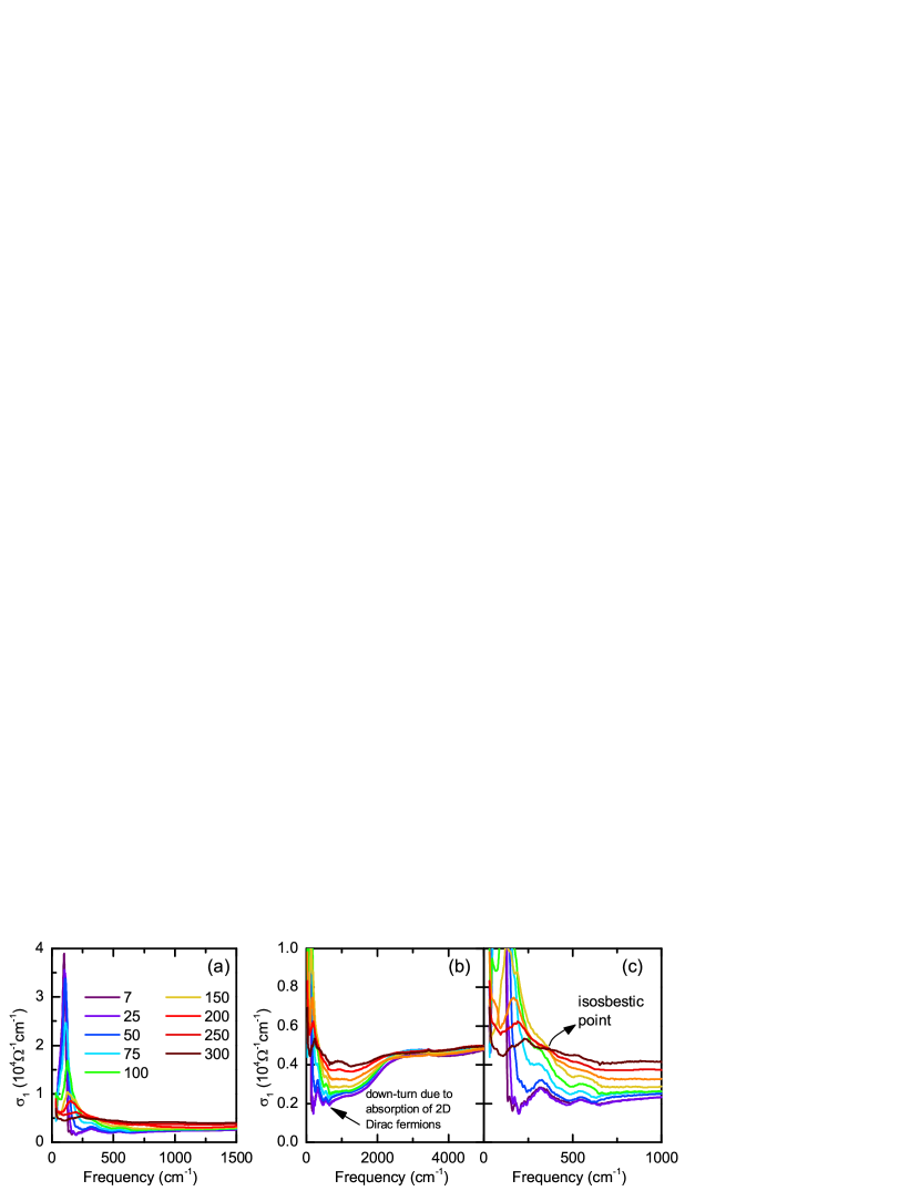
The optical conductivity is calculated via standard Kramers-Kronig analysis. Considering the highly metallic nature of the sample, we used a Hagen-Rubens extrapolation in the low-energy range. The obtained dc conductivity values agrees with transport measurements performed on the same sample [inset of Fig.1(b) of the main text]. For the high energy extrapolations, we used x-ray scattering functions Tanner (2015). The optical conductivity for selected temperatures have been given in linear scale in Fig. 4(a) and (b) for the different energy ranges.
The skin depth of the infrared radiation used in the measurements exceeds 30 nm for all temperatures and frequencies (in the far-infrared range, it is above 150 nm). Hence, our optical measurements reflect the bulk properties of Fe3Sn2.
.4 Localized carriers and Dirac fermions
In the real part of the optical conductivity, the strong absorption feature dominating the low energy spectra masks the Drude-like free carrier contribution, while the dc transport measurements corroborate the existence. In the measurement window employed here, we can only observe the small upturn of this Drude-component. Here we also examined other optical variables to ensure the existence of this sharp Drude-component, namely the imaginary part of the optical conductivity, , where one can see the signatures of the low-energy Drude-contribution more clearly Dressel and Grüner (2002).
In Fig. 5(a), the imaginary part of the optical conductivity is given. The zero crossing of this optical variable is defined as the screened plasma frequency. For Fe3Sn2, we estimated the screened plasma frequency as 1300 cm-1 and the temperature-dependence is negligibly small. The low-energy features reveals the signatures of the second plasma edge that can be associated with the free carrier response of the Dirac bands. As demonstrated in the inset of Fig. 5(a), in the case of Drude-like free carrier response with a high-energy absorption, the imaginary optical conductivity shows the zero-crossing at the screened plasma frequency followed by the broad maximum extrapolating to zero at . In the case of the localized carriers, the Drude-peak is shifting to the finite frequencies and in return low-energy response of is suppressed below zero.
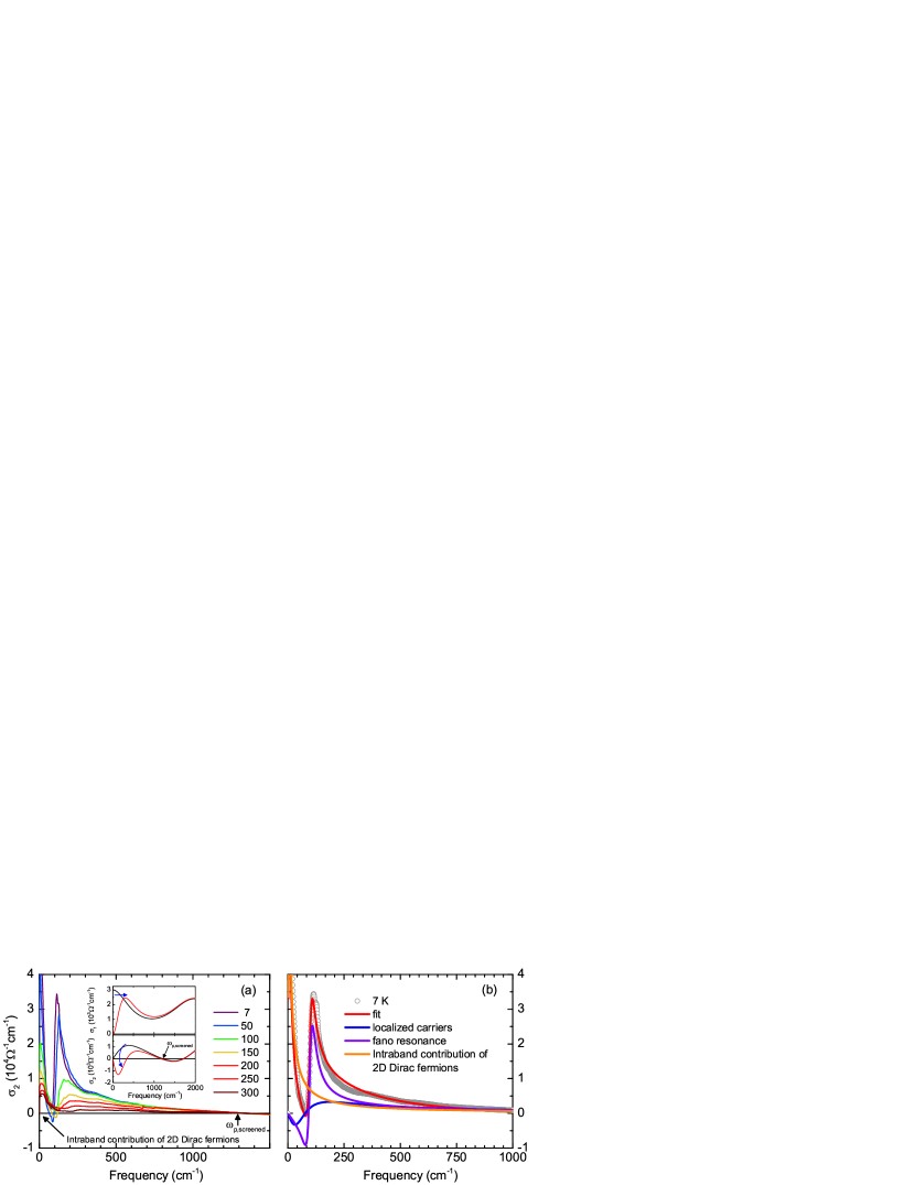
For the Fe3Sn2 system, we can reproduce the spectra with a similar analogy; however, the low-energy response reveals an additional zero-crossing below the energies described for the localized carriers along with the maximum as in the case of a regular Drude-component indicating the second contribution at lower energies. Below the spin-reorientation temperature a sharp Fano resonance appear in the spectra indicating a strong coupling to the electronic background.
We also want to point out that the zero-crossing of the imaginary optical conductivity is determined as the screened plasma frequency in the framework of the Drude interpretation, and in principle can be different for the Dirac fermions and/or localized carriers. On the other hand proposed models still consider it similar to the Drude approximation; therefore, here we used the same term.
.5 Decomposition of the optical conductivity
We determined that the optical conductivity spectra can be decomposed into two main parts, namely the response of the Dirac fermions and the localized carriers of the flat bands. We can further elaborate these contributions as the inter- and intra-band contributions of the 2D-Dirac fermions, conduction response of the localized carriers (modified Drude-component), and the temperature-independent van Hove singularities. At lower temperatures (below the spin-reorientation temperature), the modified Drude component alone cannot describe the strong low energy absorption. Instead, we defined this absorption with a Fano resonance Fano (1961), as described in Eq. 1, that is coupled to the localized carriers. In Fig. 6 a sample fit to the room temperature spectrum has been given. The fit to the low temperature spectrum with the strong absorption feature has been shown in the main text, Fig.2(a).
| (1) |
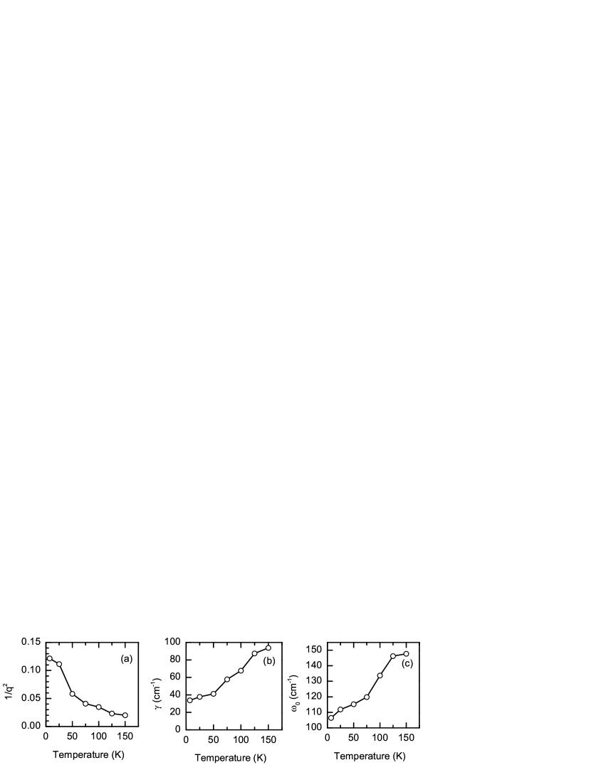
Here, is the vacuum impedance, , , and is the resonance frequency, line width, and the strength of the Fano mode, and is the dimensionless parameter that describes the asymmetry of the Fano resonance. Larger 1/ shows a stronger asymmetry, while for 1/=0, the regular Lorentzian line shape is recovered. Below the spin-reorientation temperature (around 150 K), this mode start to be prominent in the spectra and shows a strong temperature dependence. In Fig. 7, the fit parameter to this mode is given. The asymmetry of the mode (Fig. 7(a)) shows a very strong temperature dependence indicating a strong coupling to the electronic background, triggered with the spin reorientation. The Fano mode gets sharper and the resonance frequency shows a red shift with decreasing temperature; however, as demonstrated in Fig.3(c) of the main text, this shift in energy is much smaller than what is expected for the localization peak.
As several flat bands have been determined in the vicinity of the Fermi energy, several absorption features are also expected in the optical conductivity spectra, van Hove singularities arising due to the transitions between flat bands. Indeed several temperature-independent absorption modes are visible in the optical conductivity spectra, where the resonance frequency does not change, while the modes getting sharper. Although, we cannot discard the existence of more, we determined six of these van Hove singularities at resonance energies: 72, 141, 172, 223, 311, 542 cm-1. In Fig. 6 the decomposition of these bands for 7 K spectrum have been given along with the temperature evolution in Fig. 6 (b). Please note that the energy range where these van Hove singularities are dominant mostly covers the energies below the absorptions of 2D Dirac fermions, and the signature of the downturn due to step-like behavior is also clearly visible in the spectra (Fig. 4 (b)).
.6 Spectral Weight Analysis
In Fig.2 of the main text, a spectral weight (SW) analysis is shown for the different contributions to the optical conductivity. SW of the spectra have been calculated as
| (2) |
For the full spectra, the cut-off frequency have been chosen as 2 eV that reflects the whole measurement range. Although the overall SW transfer happens within 1 eV. The energy-dependent spectral weight for several temperatures have been given in Fig. 8 (a), demonstrating the conservation of the spectral weight and the energy range of the changes.
For the individual contributions, we first fit the whole spectrum with Drude (intraband contribution of Dirac fermions), Lorentzians (for the van Hove singularities), modified Drude component (for the localized carriers), and the Dirac contributions (reflecting the frequency-independent contributions). An example fit is given in Fig. 6 (a) The dc-conductivity estimated from the optical measurements have been kept as the dc-limit during the fits of the spectra. For the Dirac SW (interband and intraband), the low-lying absorption bands have been subtracted and the remaining spectra were integrated up to 1 eV. For the flat-band SW contribution, we followed the opposite trend, namely, we subtracted the Dirac contributions (intraband and interband) and integrated the spectra up to 1 eV. In Fig. 8 (a) and (b), the remaining spectra after the subtraction process used for the spectral weight analysis have been given. The SW conservation for the individual contributions holds as in the case of overall SW. The energy-dependent spectral weight for several temperatures have been given in Fig. 8, demonstrating the conservation of the spectral weight for the individual contributions and the energy range of the changes. One can also realize that the described Dirac points does not change significantly with temperature, while temperature smear out the step-like features.
.7 Optical conductivity of localized carriers
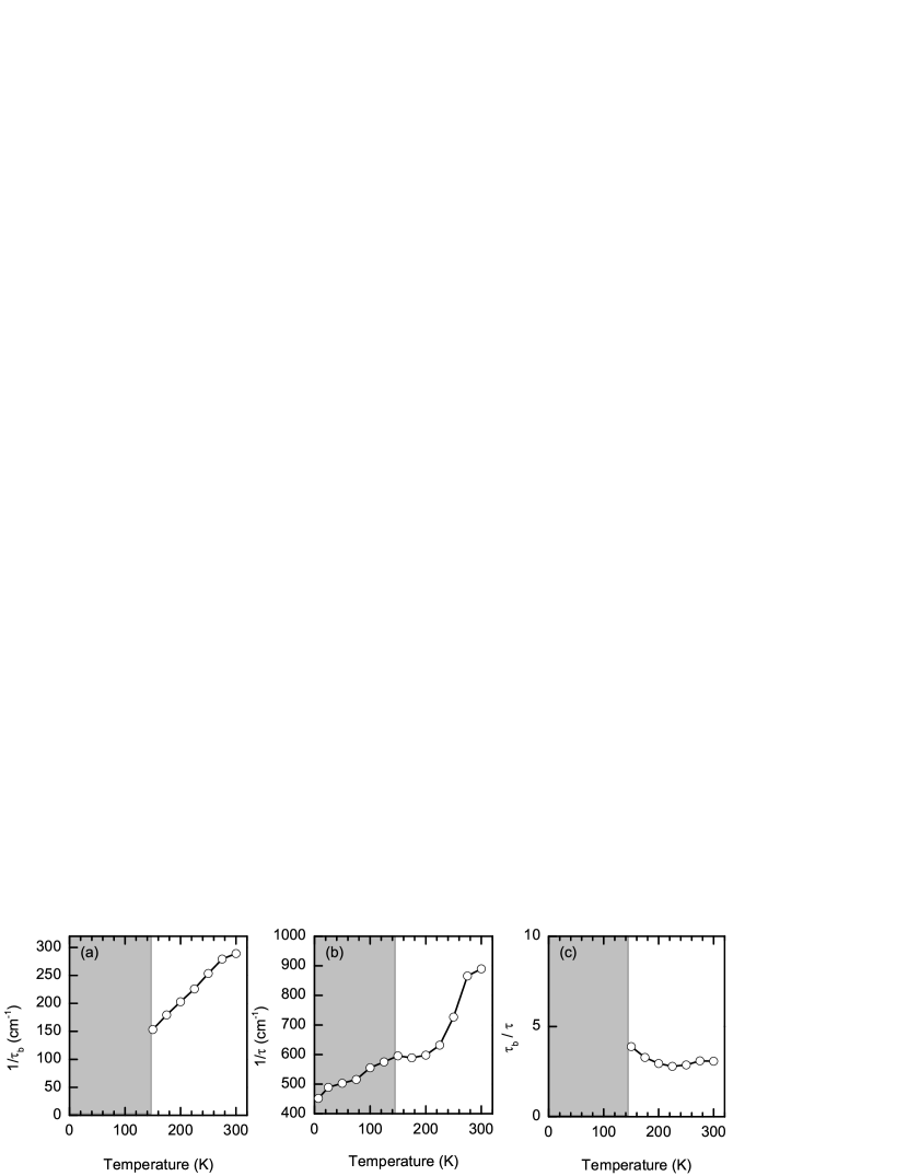
The optical conductivity that has been discussed in terms of localized carriers in the main text have been analysed with the model proposed by Fratini as described in Fratini et al. (2014, 2016); Fratini and Ciuchi (2020). Our description is restricted within the static limit, considering the limited low-energy measurement range. The inelastic scattering parameter within the static limit goes to infinity. So, within static limit, in Eq 3 for real part of the optical conductivity.
| (3) |
This equation describes the modified Drude component mentioned in the main text, where the position of the localization peak is determined by the backscattering rate of the electrons (1/) and the high-frequency tail is controlled by the elastic scattering rate (1/). In Fig. 9, the obtained fit parameters have been given as a function of temperature. Below the spin-reorientation temperature (marked with the gray area in Fig. 9), the backscattering of the electrons cannot be determined accurately due to the strong influence of the Fano resonance. Therefore, we remove it from the discussion, but the elastic scattering part can be determined from the imaginary part of the optical conductivity, as demonstrated in Fig. 5 (b).