Efficient Conversion of Nitrogen to Nitrogen-Vacancy Centers in Diamond Particles with High-Temperature Electron Irradiation
Abstract
Fluorescent nanodiamonds containing negatively-charged nitrogen-vacancy (NV-) centers are promising for a wide range of applications, such as for sensing, as fluorescence biomarkers, or to hyperpolarize nuclear spins. NV- centers are formed from substitutional nitrogen (P1 centers) defects and vacancies in the diamond lattice. Maximizing the concentration of NVs is most beneficial, which justifies the search for methods with a high yield of conversion from P1 to NV-. We report here the characterization of surface cleaned fluorescent micro- and nanodiamonds, obtained by irradiation of commercial diamond powder with high-energy (10 MeV) electrons and simultaneous annealing at 800 ∘C. Using this technique and increasing the irradiation dose, we demonstrate the creation of NV- with up to 25 % conversion yield. Finally, we monitor the creation of irradiation-induced spin-1 defects in microdiamond particles, which we associate with W16 and W33 centers, and investigate the effects of irradiation dose and particle size on the coherence time of NV-.
keywords:
Electron irradiation , P1 center , NV- center , conversion efficiency , Nanodiamonds1 Introduction
The negatively-charged Nitrogen-Vacancy (NV-) center, a point defect in diamond, has been established as a promising color center with unique properties, such as the ability to be polarized in the spin-state by illumination with green light, a long spin coherence time at room temperature, and bright fluorescence without photobleaching [1]. Owing to their optical and chemical properties, as well as exceptional biocompatibility [2], fluorescent nanodiamonds (FNDs) with high NV- concentration are promising candidates for the synthesis of fluorescence markers [3, 4], sensing probes [5, 6] and polarized MRI tracers [7, 8]. The brightness of FNDs depends on the average number of light-emitting color centers per particle and therefore requires a significant concentration of NV-.
The creation of NV- requires the presence of two types of impurities in the lattice, substitutional nitrogen defects (P1 centers), and vacancies. A single vacancy can recombine with a P1 center. Provided the presence of an electron donor, which can be another P1 center, such a recombination can lead to the formation of NV-. In conventional methods, the vacancies are created by irradiation at room temperature with high energy electrons [9, 10, 11], protons [12] or gamma rays [13]. At room temperature, however, the created vacancies remain at a fixed position in the diamond lattice. In order to induce the mobility of vacancies towards P1 centers, an additional step of annealing is required, which practically consists in heating up the sample above 800∘C. We label this method the room temperature (RT) irradiation technique.
A high NV- concentration can be achieved by irradiating nanodiamonds (NDs) with a high nitrogen content. However, single nitrogen defects in diamonds are also a major source for spin decoherence [14], photoluminescence quenching [15, 16, 17], and 13C nuclear spin relaxation [18, 19]. Therefore, a high residual P1 concentration reduces the nanodiamonds suitability for applications where the named properties are crucial. As a consequence, it is of major importance to optimize the NV- creation yield for a given nitrogen concentration, while, at the same time, minimizing the creation of other lattice defects.
Room temperature irradiation can be accompanied by the creation of unwanted paramagnetic defects due to vacancy aggregation [20]. In comparison, performing both irradiation and annealing simultaneously - a technique that we will label high temperature (HT) irradiation - provides a more homogeneous procedure in which created vacancies instantaneously become mobile and therefore immediately have the possibility to recombine with nitrogen atoms [21]. In this case, it is expected that the concentration of vacancies will remain continuously low during the full process, which can possibly allow reducing the formation of undesirable defects - e.g. divacancies [22] and vacancy clusters [23]- which induce additional spin decoherence [24]. In addition, in the case of bulk crystals, it has been reported a higher NV- creation after HT irradiation, compared to the case when RT irradiation is applied [21]. However, an investigation of possible benefits of this technique for the production of fluorescent nanodiamonds, in particular in terms of conversion efficiency from P1 to NV-, is missing.
In the present work, we examine the effect of electron irradiation on commercially available diamond powder of different sizes (Microdiamant AG, MSY: , , ), through the use of electron paramagnetic resonance (EPR) and the combination of an atomic force microscope (AFM) with a confocal microscope. We first compare, with these techniques, the RT and HT irradiation in terms of the created quantity of NV- and spin properties. We then investigate the NV- creation yield for and particles that underwent different doses of HT irradiation. Systematic analysis of the effect of increasing the irradiation dose is performed, demonstrating the possibility of reaching a conversion efficiency, defined as the ratio of the final NV- concentration over the initial P1 concentration, of up to 25%. Finally, coherence and relaxation properties of NV- centers, as well as the creation of additional spin-1 defects, are discussed.
2 Results and discussion
2.1 Evaluation of the nitrogen content in the starting material
The samples investigated in this work were obtained from diamond powder produced by the company Microdiamant AG, are of type Ib, and consist of different sizes: (Microdiamant, MSY 1.5-2.5), (Microdiamant, MSY 0-0.2) and (Microdiamant, MSY 0-0.05). These commercially available powders are produced through the High Pressure High Temperature (HPHT) technique, and subsequent milling is performed to obtain smaller size fractions below .
To characterize the effects of irradiation in terms of NV- formation, it is important to know the amount of nitrogen originally present in the material. Continuous wave electron paramagnetic resonance (CW EPR) spectroscopy has been established as a powerful tool for quantification of paramagnetic defects, such as P1 and NV- in diamond [25, 26]. In bulk type Ib crystals, this technique allows precise quantification of the nitrogen content, through the detection of P1 signal. P1 centers are usually quantified and characterized with CW EPR using their characteristic triplet spectrum associated with the hyperfine interaction of the unpaired electron with the 14N (, natural abundance 99.6 %) nuclear spin. Such a triplet can be conveniently distinguished from other contributions to the spectrum originating, e.g., from spin impurities with different features [27, 28]. Following such an analysis (detailed in Supporting Information, section “P1 Concentration with CW EPR”), the corresponding spin concentration can be obtained. The error on the determination of P1 spin concentrations with CW EPR was found to be about 15 % for the and 20 % for the and samples. The samples used for the present work originate from two different fabrication batches, and were measured with [P1] ppm and [P1] ppm respectively, a difference that could potentially be related to variations in the synthesis conditions. The and samples show respectively ppm and ppm of P1. Fainting P1 signal with decreasing particle size is consistent with data already reported in the literature for HPHT nanodiamonds obtained upon milling [28, 29]. One can hypothesize that electron acceptors or donors on the particle surface are responsible for conversion of a fraction of P1 in NDs to nitrogen with a different charge state, or, as it has been proposed, that part of P1 centers show a different (narrowed) spectrum in NDs due to strong exchange interaction with surface dangling bonds [30]. In the current work, we restrict ourselves to the quantification of the “core” P1 centers, that is P1 showing the characteristic hyperfine pattern due to 14N interaction. From the values given above, we expect that possible nitrogen that goes undetected with this method cannot play a dominant role in the NV- formation mechanism for the and particles. Whether the quantity of created NV- depends on such nitrogen impurities in smaller, e.g. 25 nm NDs, would need to be investigated elsewhere.
2.2 Room versus high temperature irradiation
To compare the yield of NV- creation after HT and RT irradiation, we have implemented both methods on diamond powder with particle sizes , and . We used in both cases high energy (10 MeV) electron irradiation, which allows to induce a homogeneous distribution of vacancies over a depth greater than [31], and thus offers the possibility to process large quantities of sample. The HT irradiated samples underwent irradiation and simultaneous heating to 800 °C (see Experimental section). For the RT samples, irradiation at room temperature was followed by annealing at 800 °C. Samples of a given size received identical electron doses ( for the sample, for the and samples). All samples underwent air oxidation as the final step (see Experimental section). The NV- concentration after irradiation was estimated with CW EPR for all samples, and, for the samples, also with a combined AFM-confocal microscope setup (see Experimental section). We first discuss the CW EPR technique, which allowed systematic characterization of the samples.
NV- centers, owing to their electron spin , can be characterized with EPR. It is important to note that the positions of NV- spectral lines in X-band depend strongly on the diamond orientation, as a consequence of the important zero-field splitting of NV-. Therefore, due to the random orientations of the NDs in powder samples, the EPR spectrum is broadly distributed over a magnetic field range of [27]. To prevent a too important error on the spin-counting estimate resulting from baseline drift during acquisition, the estimation of NV- concentration was performed considering a field region narrower than the full spectrum, as described in section “Spin-counting with CW EPR” in the Supporting Information. In the case of the samples, the estimation was carried out by considering a region centered on one intensive spectral line, appearing around a field of . This spectral feature corresponds to NV- centers oriented perpendicular to the magnetic field, which is the most common case. We found this method of NV- density determination to have an error of approximately 20 %. For the and samples, we rather performed double integration on the half-field transition of NV- [27, 26], which occurs at . Such a method allows reducing further the error on spin-counting, as described in section “NV- Concentration with CW EPR” of the Supporting Information. However, for these samples, a contribution from other spin-1 defects (non NV-) appears in the half-field region, which was estimated and subtracted (see section “Non-NV- Irradiation-Induced Defects”) to estimate the NV- concentration. The resulting error for the and samples was estimated to be 6 % and 7 %, respectively.
| Irradiation type | P1 conc. (ppm) | NV- conc., CW EPR (ppm) | NV- conc., AFM+confocal (ppm) | confocal ( |
|---|---|---|---|---|
| before irradiation | 5.2 | - | - | - |
| RT irradiated | 3.5 | 1.0 | 1.6 | 0.5 |
| irradiated | 1.9 | 2.7 | 2.8 | 0.5 |
| Irradiation type | P1 conc. (ppm) | NV- conc. (ppm) | ( | () |
|---|---|---|---|---|
| before irradiation | 27 | - | - | - |
| RT irradiated | 17 | 3.36 | 2.4 | 1.7 |
| irradiated | 18 | 3.44 | 2.7 | 2.0 |
| Irradiation type | P1 conc. (ppm) | NV- conc. (ppm) | ( | () |
|---|---|---|---|---|
| before irradiation | 74 | - | - | - |
| RT irradiated | 55 | 6.67 | 2.2 | 2.4 |
| irradiated | 50 | 7.41 | 2.2 | 2.5 |
The NV- concentrations obtained for the different samples (, and ) following RT and HT irradiation are given in Tables 1 to 3. The case of the sample is worth paying attention to, indeed, the CW EPR results reveal that the quantity of created NV- is more important in the HT irradiation case, in comparison with RT irradiation. To confirm this finding, we implemented an optical quantification method, previously described in Mindarava et al. [32]. This method consists in deducing the number of defects in NDs from the intensity of the photoluminescence (PL) signal in a combined AFM - confocal microscope. This analysis was performed on a set of measurements including 50 single ND particles for each irradiated sample, using light for illumination (see Experimental section for details). To estimate the concentration, the volume of each particle was estimated from their height, measured with the AFM tip, assuming a spherical particle shape. Using this method, for the sample we found that, on average, RT irradiation created three NV- centers per particle, while HT irradiation induced seven NV- centers per nanodiamond. The corresponding concentrations match the values obtained in CW EPR (Table 1). We remark that, in comparison with EPR, the optical method suffers from the combined effect of different error sources, including the uncertainty on the particle shape and the strong dependency of NV- fluorescence on the dipole orientation (the latter leads to fluctuations in the estimated NV- concentration even after averaging over several particles) [33]. Thus CW EPR allows a more precise absolute quantification of NV- spins than the optical method. In contrast, when considering the and sizes, we observe no significant difference related to changing the irradiation technique. We suggest that our observations could be described by considering the concentration of vacancies in the particles at a given time of irradiation, and the fact that due to diffusion at high temperature, vacancies might exit the crystal. Onoda et al. [34] determined the activation energy for vacancy migration to be eV, which relates to the vacancy diffusion coefficient through the formula with . The HT irradiation was performed with a dose rate . To accumulate a final dose of , a total irradiation time was needed. At , the diffusion coefficient is which yields a root mean square displacement . As a consequence, considering the dose applied, a vacancy created at the beginning of the irradiation process in the sample will likely exit the lattice (if it has not recombined meanwhile with a lattice defect). The probability of that event is significantly lower in the and case. Due to the possibility of the vacancy exiting the lattice in the case, we expect the recombination of two/more vacancies to form divacancies/vacancy clusters to be less frequent with HT irradiation, leading to a lower final concentration of such aggregates, as compared to RT irradiation. Such an effect could explain the higher NV- formation for high temperature irradiation, as divacancies are responsible for the conversion of NV- to the neutrally charged NV0 [35]. In addition, while the considerations above were made assuming the vacancies have diffusion properties as in the bulk, the fact that the vacancy migration energy can be lower close to a diamond surface (and thus the diffusion faster) [36], could be an additional explanatory factor for the observed size effect.
A side effect of the formation of NV- is the decrease in concentration of P1 spins. From the P1 concentrations given in Tables 1 to 3 (obtained with CW EPR) one can see that, as expected, the P1 concentration decreases in parallel to the NV- formation. We expect the creation of one NV- to take at maximum two P1 centers, corresponding to the case when one P1 center plays the role of nitrogen source and another P1 acts as an electron donor: 2P1+V0 NV0+P1 NV-+N+ (V0 is a single neutral vacancy). Remarkably, for the and samples, it can be seen from Tables 2 and 3, that the drop in P1 exceeds the NV- concentration concentration by more than a factor of two. This occurs because NV0 are created as well. Another nitrogen-containing defect, W33, is also formed as we discuss later on (see section “Non-NV- Irradiation-Induced Defects”).
The times of the and particles (Tables 2 and 3) were measured with Hahn echo using Pulsed EPR in X-band, on the 2900 G line. For the powder (Table 1), the Hahn echo coherence times were measured at zero magnetic field using a home-built confocal microscope setup (see Experimental section). The corresponding times correspond to an average over different single NV- centers. The evolution of the times with varying particle size can be described by considering spins both in the bulk (dominantly, P1) and on the surface as possible sources of decoherence. For and , the contribution from P1 needs to be taken into account (see discussion in section “NV- coherence and spin-lattice relaxation times”). In the NDs, decoherence is probably caused exclusively by surface spins, explaining the very short value [37] (for this size, the fact that similar values are obtained for room temperature and high temperature irradiated samples is probably a consequence of nearly identical surface states obtained following air oxidation). For the and samples, the spin-lattice relaxation times were also measured (using Pulsed EPR). We observe a slight shortening of for the size in comparison with , which could reflect the onset of surface-induced relaxation for NV- [38]. Overall, the coherence and spin-lattice relaxation times do not show a significant difference between the high and room temperature irradiated samples. The fact that very similar coherence times were observed for the samples following RT and HT irradiation suggests that for such small particles, the surface influence dominates over the effects related to bulk impurities.
Despite the difference between the room and high temperature approaches, only for the particles an increase in conversion efficiency upon HT irradiation was found. To explain this effect, we hypothesize that the possibility for the vacancies to diffuse out of the particles during irradiation works in favor of the HT irradiation technique. In the following sections, we discuss how the NV- creation and their spin properties depend on the dose of HT irradiation.
2.3 Effects of increasing the irradiation dose
An important parameter defining the efficiency of NV- formation is the dose of electron irradiation. Increasing the irradiation dose can increase the conversion efficiency from P1 to NV-. On the other hand, the irradiation dose must be kept under a certain limit as otherwise crystal damage becomes significant [20] and the lattice cannot recover its structure even after annealing [39]. Therefore, to improve NV- formation, the irradiation process has to be optimized.
To understand the effect of the irradiation dose on the formation of NV- defects, we have implemented high temperature irradiation with different doses on different fractions of the same sample batch. For this analysis, we selected (MSY2) and (MSY0.1) samples. The samples went through the same treatment as the HT irradiated samples described in section 2.2, but were subject to varying irradiation doses. The samples were irradiated with electron doses of 0.5, 1, 2, 3, 6 and -2, and are named correspondingly 0.5MSY2, 1MSY2, 2MSY2, 3MSY2, 6MSY2, 9MSY2. The samples were irradiated with doses of 0.5, 1, and -2 and are labeled 0.5MSY0.1, 1MSY0.1, 3MSY0.1.
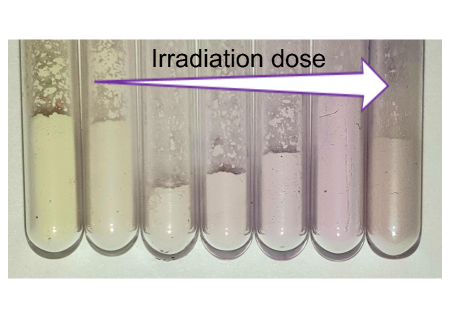
A fingerprint of the NV- concentration is the color of the sample. After cleaning the surface with air oxidation, the non-irradiated samples usually have yellow color, which reveals the presence of P1 centers in the diamond particles. With irradiation, as NV- centers are created, the color of the sample gradually changes to purple. The continuous alteration in color for the powder seen in Figure 1 reflects the increase in NV- concentration with increasing irradiation dose.
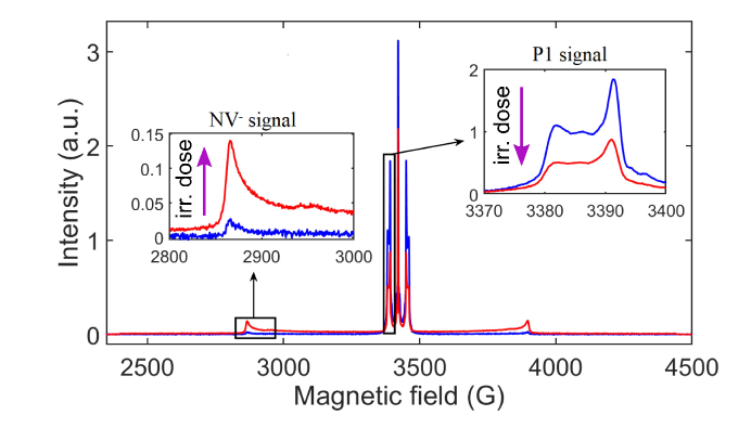
The evolution of P1 and NV- concentrations has been recorded with CW EPR, which gives the dependencies shown in Tables 4 and 5. The electron spin echo (ESE) EPR spectra shown in Figure 2, measured for two different irradiation doses, illustrate the efficient conversion from P1 to NV- induced by irradiation in the case of the powder. The intensity of the NV- spectrum raises upon increasing the irradiation dose with a simultaneous drop in P1 intensity, suggesting similar evolutions of the respective spin concentrations. We remark that the intensities of ESE-detected spectra are also affected by the decoherence processes (), however, this leads to a stable attenuation factor, depending only weakly on the irradiation dose (see its evaluation in Supporting Information section “Pulsed EPR spectrum”). Therefore, the changes in line intensities seen in both insets of Figure 2 reflect the conversion from P1 to NV- occurring with irradiation.
In addition, the P1 concentrations for the samples have been estimated using the effect of instantaneous diffusion (ID) with pulsed EPR (see section “Instantaneous diffusion” in Supporting Information). The method of ID is based on the dependence of on the flip angle induced by the refocusing pulse. As this method relies on the interaction between close spins [40, 41, 42], it provides a local information on the spin density (on the scale of a few nearest neighbors) and is therefore complementary to CW spin-counting. The ID method for P1 centers has been realized on the maximum of the low-field hyperfine pattern in the P1 spectrum. To analyze the ID data for P1, as described in the Supporting Information, one needs to take into account the inhomogeneous character of the excitation by the refocussing pulse, which leads to a non-uniform distribution of flip angles in the sample. The instantaneous diffusion method requires a concentration of the probed species high enough so that the ID effect can compete with other sources of NV- decoherence [42]. Therefore, the estimation of NV- concentration for the samples with a low NV- density was found not to be possible. For the samples with the highest conversion efficiencies, one can use this technique, as we demonstrated for the 6MSY2 sample (see section “Analysis of Instantaneous Diffusion for NV- Centers” in the Supporting Information), where it gives a result of 10.8 ppm, which is comparable with the result received with CW EPR.
| Sample name | Irradiation dose (-2 ) | P1 conc., CW (ppm) | P1 conc., ID (ppm) | NV- conc., CW (ppm) | ( | () |
|---|---|---|---|---|---|---|
| 0MSY2 | non-irradiated | 53 | 64 | - | - | - |
| 0.5MSY2 | 0.5 | 55 | 60 | 1.17 | 2.2 | 2.5 |
| 1MSY2 | 1 | 41 | 53 | 2.56 | 2.6 | 2.5 |
| 2MSY2 | 2 | 48 | 53 | 3.15 | 2.1 | 2.6 |
| 3MSY2 | 3 | 40 | 53 | 4.63 | 2.6 | 2.3 |
| 6MSY2 | 6 | 18 | 41 | 10.3 | 2.1 | 2.3 |
| 9MSY2 | 9 | 13 | 30 | 13.5 | 1.9 | 1.6 |
| Sample name | Irradiation dose (-2 ) | P1 conc., CW (ppm) | NV- conc., CW (ppm) | ( | ( |
|---|---|---|---|---|---|
| 0MSY0.1 | non-irradiated | 27 | - | - | - |
| 0.5MSY0.1 | 0.5 | 24 | 0.98 | 3.2 | 2.0 |
| 1MSY0.1 | 1 | 22 | 1.97 | 3.1 | 2.2 |
| 3MSY0.1 | 3 | 18 | 3.44 | 2.7 | 2.0 |
The results presented in the Tables 4 and 5 demonstrate a significant reduction in P1 concentration after irradiation, exceeding the concentration of created NV- by more than a factor of two. We interpret this as the consequence of several effects, as stated earlier, the creation of one NV- can take up to two P1 centers and, besides, other nitrogen-containing defects can be created. In this respect, we could evidence the creation of NV0, from the optical photoluminescence spectrum (see section “Photoluminescence spectra” in the Supporting Information) and W33 defects (see following section).
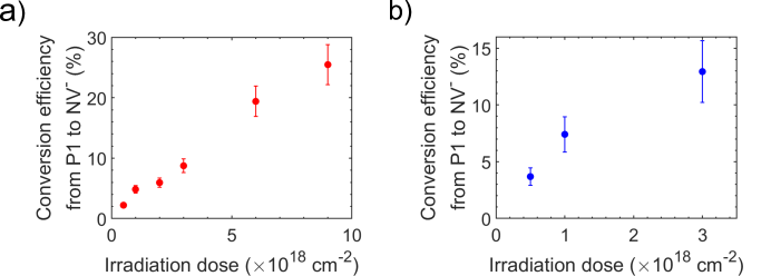
Figure 3 demonstrates the increase in P1 to NV- conversion efficiency with an increasing electron irradiation dose for the and diamond powder, reaching values of % for , at dose -2. For the for powder, we obtain a conversion efficiency of % , at dose -2. Interestingly, the conversion efficiency for the powder at the same dose is %, which is lower. As the P1 concentration for this sample is higher, our observations are consistent with the trend of decreasing conversion efficiency with increasing P1 content, which was already reported in [21].
To increase the NV- concentration further, one could increase the dose of irradiation. At high doses, one expects a saturation in NV- concentration, which we can not clearly observe in Figure 3. Further measurements, focusing on fluences above , would be needed to determine clearly the saturation parameters, in particular, the maximum attainable NV- concentration. We remark that the application of higher doses with HT irradiation would imply longer irradiation times and thus longer annealing times. This would increase the diffusion length of vacancies, which would then have the possibility to diffuse out of the crystal not only for the size, but also for bigger particles. For instance, with the parameters of our irradiation chamber, a dose would correspond to an annealing time of , which would result in a diffusion length (see section 2.2), meaning a significant fraction of vacancies would diffuse out of the lattice, considering particles. Following the analysis presented in section 2.2, this could lead to a better conversion efficiency in comparison with the RT technique, also for this particle size.
In addition, a side effect of high irradiation doses is the creation of non-NV- paramagnetic defects, as we discuss now.
2.4 Non-NV- irradiation-induced defects

The formation of paramagnetic defects aside of NV- is unavoidable during the irradiation process. Here we describe the appearance of such impurities following HT irradiation, for the and samples discussed in section 2.3
For these samples, EPR spectra in the half-field region reveal the presence of defects with appearing as additional lines (I and II) in the vicinity of the signal from NV-. For the samples, Figure 4 demonstrates the rise of the intensity of these additional lines, marked as I and II, with increasing irradiation dose. The positions of the lines for the I and II defects correspond to the expected positions for W33 and W16 centers, that were previously reported to appear as a consequence of room temperature irradiation [43]. A fit of the spectrum yields, for the samples, the concentrations given in Table 6. In the samples, the defect I (W33) also appears (Table 7), however, the defect II (W16) could not be detected. The concentrations were obtained after performing a fit of the CW spectrum including the contributions from NV-, I and II defects (see Supporting information, section “Simulation of the Half-Field Spectrum”), taking the parameters (spin Hamiltonian) for the simulation of W33 and W16 respectively from Nadolinny et al. [44] and Loubser et al. [25]. From Tables 6 and 7, one can see a linear increase of the W16 and W33 concentrations with the irradiation dose, suggesting that the structure of these centers involve one vacancy, or a split-vacancy with an interstitial atom. The structure of the W33 center in particular has been described by Nadolinny et al. [44] as a nitrogen-vacancy defect with a low symmetry, due to the presence of a positively charged nitrogen (N+) in its vicinity.
| Sample name | Irradiation dose (-2) | I center (W33) conc.(ppm) | II center (W16) conc.(ppm) |
|---|---|---|---|
| 0.5MSY2 | 0.5 | 0.10 | - |
| 1MSY2 | 1 | 0.23 | 0.10 |
| 2MSY2 | 2 | 0.24 | 0.11 |
| 3MSY2 | 3 | 0.42 | 0.15 |
| 6MSY2 | 6 | 1.14 | 0.57 |
| 9MSY2 | 9 | 1.93 | 0.87 |
| Sample name | Irradiation dose (-2 ) | I center (W33) conc.(ppm) |
|---|---|---|
| 0.5MSY0.1 | 0.5 | - |
| 1MSY0.1 | 1 | 0.08 |
| 3MSY0.1 | 3 | 0.34 |
2.5 NV- coherence and spin-lattice relaxation times
Estimation of the NV- coherence and spin-lattice relaxation time ( and ) for the and samples are given in Table 4 and 5, respectively.
One important source of NV- decoherence in non-irradiated bulk material or microdiamonds is the interaction with P1 centers [45], which can be supplanted by sources in the vicinity of the surface (e.g. dangling bonds) for small NDs [37, 46]. After irradiation, however, NV- centers and potentially other irradiation-induced defects (see previous section) can themselves be a source of decoherence. To distinguish the contributions from P1 centers and from other sources of NV- decoherence, we used the formula from Bauch et al. [45], that corresponds to a fit of the dependence of on the P1 concentration for single crystalline diamond with low NV- content. This dependence reads , as a function of P1 concentration, with [45]. From Fig. 5, we can draw several conclusions. First, looking at the samples with the lowest irradiation doses for particle sizes and (0.5MSY2 and 0.5MSY0.1), we observe that the discrepancy to the prediction is higher for the sample. We interpret this as a sign that the surface already plays a role in decoherence for particles, but has negligible effect in the case. We emphasize that such conclusion could only be made after simultaneously considering and P1 concentration, the latter being different between the and samples. Second, as the irradiation dose is increased, the measured data drifts away from the prediction, as the decrease in P1 concentration does not translate into a prolonged time. This demonstrates the growing influence of “non-P1” sources appearing as the irradiation dose is increased. Among these sources are NV- themselves, acting both through spectral and instantaneous diffusion [42]. Effects of NV--NV- interactions are naturally expected to become more important with increasing irradiation doses. As a confirmation of their role in decoherence, we could establish, for the 6MSY2 sample, that removing the effect of instantaneous diffusion between NV- yields an increase in from to (see section “Analysis of Instantaneous Diffusion for NV- Centers” in Supporting Information), which accounts for part of the deviation to the dashed line in Fig. 5a. Additionally, spectral diffusion, induced by NV- or by other defects such as the W16 and W33 center (see section “Non-NV- irradiation-induced defects”), can also play a role. Last, in the case, for which the seems to shorten as a result of irradiation, already at the lowest doses (which is not observed in the case), one cannot exclude the creation of new dangling bonds towards the surface. Further work would be needed to distinguish the respective contributions to decoherence, for the different particle sizes.
The NV- spin-lattice relaxation time, , measured for the samples (Table 4), may yield information on irradiation damage. We observe a drop in past the irradiation dose -2 (from 2.3 to 1.6 ms). Because of the particle size, we suppose any influence from surface defects can be excluded. As a hypothesis, the creation of fastly relaxing ( ns) paramagnetic impurities in the crystal under irradiation could explain the shortening of (owing to their very short time, such impurities are difficult to detect directly with EPR). Further studies performed with higher irradiation doses would be needed to confirm this trend.

3 Experimental section
3.1 Sample preparation
Commercially available micro- and nanodiamonds with a mean particle size of (Microdiamant, MSY 1.5-2.5, type Ib, HPHT), (Microdiamant, MSY 0-0.2, type Ib, HPHT) and (Microdiamant, MSY 0-0.05, type Ib, HPHT) have been used with size distributions between 1.5-, 0- and 0-, respectively. For the RT irradiated sample a 10 MeV electron accelerator (MB10-30MP-Mevex Corp., Stittsville Canada) operating under air atmosphere was used, which includes a permanent cooling to regulate the treatment temperature below 300 °C. After irradiation the sample was annealed in argon atmosphere at 800 °C for 5 hours.
The HT irradiation of the samples has been implemented within a ceramic holder placed in a quartz furnace under permanent argon flow. During warm up of the linear accelerator (also producing 10 MeV electrons) the quartz tube is flushed for approximately 30 minutes with argon. During irradiation, the argon flow is maintained at about 150 ml/min by means of a flow controller (GFC171 from Analyt), keeping the argon pressure close to . The treatment temperature was regulated by the dose per pulse and repetition frequency of the accelerator and was monitored by applying a thermocouple connected to the sample holder and regulated to be 800 °C. The HT irradiation dose rate is about -2 s-1.
Surface graphitization occured, either during HT irradiation or, in the case of the RT irradiated sample, during annealing. Therefore, after irradiation, all samples were subjected to air oxidation at 620 °C for 5 hours to remove the graphitic residues from the surface.
3.2 Optical measurements
For optical and AFM investigation of the ND samples (with HT and RT irradiation) of ND solution in demineralized water were spin-coated (5000 rpm for 40 seconds) on a plasma cleaned glass substrate. Microwaves were applied on NV- centers through a thick copper wire.
Fluorescence and NV- spin properties were measured with a home-built confocal microscope, where a laser was used for NV- excitation. The setup was controlled using the Qudi software package [47]. The fluorescence was collected through an oil-immersion objective (Olympus UPlanSApo 60x oil NA=1.35). After the bandpass filter with transmission between and the fluorescence was detected with an avalanche photodiode with single photon resolution (Excelitas Technologies). The pulse sequence for the time measurement was implemented using an Arbitrary Waveform Generator (Tektronix AWG70001A).
3.3 EPR measurements
X-band (9.6 GHz) CW and Pulse EPR measurements were implemented at room temperature on a Bruker Elexsys E580 EPR spectrometer with waveguide resonator (ER-4122MD4) and FlexLine resonator (ER-4118X-MD5), respectively. The spin-counting in CW has been performed with Bruker software (xEPR). Low microwave power was used in order to avoid saturation of the detected signal: < for measuring P1 and the “allowed” transition of NV- (at 2900 G), and < for the half-field transition of NV-. Experiments were performed with a decoupled cavity ().
The samples were measured inside of a quartz EPR tube from Wilmad-Labglass (707-SQ-100M) with an inner diameter of . Simulations of EPR spectra were performed using the EasySpin Matlab toolbox [48].
Details on the signal acquisition procedures and on the spectral simulation parameters are provided in Supporting Information.
4 Conclusions
A high NV- density is crucial for potential applications of NDs as fluorescent markers, to increase magnetometry sensitivity and for efficiency of techniques for 13C nuclear spin hyperpolarization. The results presented here demonstrate the possibility to reach a high formation yield of NV- defects in nano- and microdiamonds by implementing simultaneous electron irradiation and annealing. As we hypothesize, the possibility of the vacancies to diffuse out of the crystal during this ‘high temperature irradiation” process explains the higher NV- formation observed in the case of nanodiamonds. For bigger () particles, we demonstrated that a conversion efficiency of P1 to NV- of 25 % can be achieved, a figure that could potentially be increased by using higher irradiation doses. We observed, in and samples, the concomitant appearance of additional irradiation-induced spin-1 defects involving one vacancy, identified as W16 and W33 centers. Despite the creation of such defects, long NV- coherence and spin-lattice relaxation times prove that no severe irradiation damage has been caused. We expect that the presented irradiation technique will allow synthesis of fluorescent nanodiamonds with tailored NV- concentration, providing opportunity for their applications in nanoscale optical imaging, as magnetic sensors, and for nuclear spin hyperpolarization.
Acknowledgements
This work was supported by the DFG (CRC 1279), EU HYPERDIAMOND (Project ID 667192), VW Stiftung (No. 93432), BW Stiftung (No. BWINTSFIII-042), BMBF (Project No. 13N14438, 13GW0281C, 13N14808, 16KIS0832, 13N14810, 13N14990), ERC (Grant No. 319130), JSPS-KAKENHI (No. 17H02751). We thank Dr. Yan Liu for providing the confocal setup for optical measurements.
References
- [1] M. W. Doherty, N. B. Manson, P. Delaney, F. Jelezko, J. Wrachtrup, L. C. L. Hollenberg, The nitrogen-vacancy colour centre in diamond, Phys. Rep. 528 (1) (2013) 1–45. doi:10.1016/j.physrep.2013.02.001.
- [2] Y. Zhu, J. Li, W. Li, Y. Zhang, X. Yang, N. Chen, Y. Sun, Y. Zhao, C. Fan, Q. Huang, The biocompatibility of nanodiamonds and their application in drug delivery systems, Theranostics 2 (3) (2012) 302. doi:10.7150/thno.3627.
- [3] V. Vaijayanthimala, H.-C. Chang, Functionalized fluorescent nanodiamonds for biomedical applications, Nanomedicine 4 (1) (2009) 47–55. doi:10.2217/17435889.4.1.47.
- [4] K. Turcheniuk, V. N. Mochalin, Biomedical applications of nanodiamond (review), Nanotechnology 28 (25) (2017) 252001. doi:10.1088/1361-6528/aa6ae4.
- [5] Y. Wu, F. Jelezko, M. B. Plenio, T. Weil, Diamond quantum devices in biology, Angew. Chem., Int. Ed. 55 (23) (2016) 6586–6598. doi:10.1002/anie.201506556.
- [6] Y. Wu, T. Weil, Nanodiamonds for biological applications, Phys. Sci. Rev. 2 (6) (2017). doi:10.1515/psr-2016-0104.
- [7] D. E. J. Waddington, T. Boele, E. Rej, D. R. McCamey, N. J. C. King, T. Gaebel, D. J. Reilly, Phase-encoded hyperpolarized nanodiamond for magnetic resonance imaging, Sci. Rep. 9 (5950) (2017) 2045–2322. doi:10.1038/s41598-019-42373-w.
- [8] A. Ajoy, K. Liu, R. Nazaryan, X. Lv, P. R. Zangara, B. Safvati, G. Wang, D. Arnold, G. Li, A. Lin, et al., Orientation-independent room temperature optical 13c hyperpolarization in powdered diamond, Scien. Advan. 4 (5) (2018). doi:10.1126/sciadv.aar5492.
- [9] C. Laube, T. Oeckinghaus, J. Lehnert, J. Griebel, W. Knolle, A. Denisenko, A. Kahnt, J. Meijer, J. Wrachtrup, B. Abel, Controlling the fluorescence properties of nitrogen vacancy centers in nanodiamonds, Nanoscale 11 (2019) 1770–1783. doi:10.1039/C8NR07828A.
- [10] G. Dantelle, A. Slablab, L. Rondin, F. Lainé, F. Carrel, P. Bergonzo, S. Perruchas, T. Gacoin, F. Treussart, J. Roch, Efficient production of NV colour centres in nanodiamonds using high-energy electron irradiation, J. Lumin. 130 (9) (2010) 1655–1658. doi:10.1016/j.jlumin.2009.12.003.
- [11] C. Laube, Y. M. Riyad, A. Lotnyk, F. P. Lohmann, C. Kranert, R. Hermann, W. Knolle, T. Oeckinghaus, R. Reuter, A. Denisenko, A. Kahnt, B. Abel, Defined functionality and increased luminescence of nanodiamonds for sensing and diagnostic applications by targeted high temperature reactions and electron beam irradiation, Mater. Chem. Front. 1 (2017) 2527–2540. doi:10.1039/C7QM00241F.
- [12] J. Botsoa, T. Sauvage, M.-P. Adam, P. Desgardin, E. Leoni, B. Courtois, F. Treussart, M.-F. Barthe, Optimal conditions for center formation in type-1b diamond studied using photoluminescence and positron annihilation spectroscopies, Phys. Rev. B 84 (2011) 125209. doi:10.1103/PhysRevB.84.125209.
- [13] B. Campbell, A. Mainwood, Radiation damage of diamond by electron and gamma irradiation, Phys. Status Solidi A 181 (1) (2000) 99–107. doi:10.1002/1521-396X(200009)181:1<99::AID-PSSA99>3.0.CO;2-5.
- [14] J. F. Barry, J. M. Schloss, E. Bauch, M. J. Turner, C. A. Hart, L. M. Pham, R. L. Walsworth, Sensitivity optimization for NV-diamond magnetometry (2019). arXiv:1903.08176.
-
[15]
K. V. Bogdanov, M. V. Zhukovskaya, V. Y. Osipov, E. V. Ushakova, M. A. Baranov,
K. Takai, A. Rampersaud, A. V. Baranov,
Highly intensive emission of the
NV- centers in synthetic HPHT microdiamonds at low nitrogen doping,
APL Materials 6 (8) (2018) 086104.
arXiv:https://doi.org/10.1063/1.5045535, doi:10.1063/1.5045535.
URL https://doi.org/10.1063/1.5045535 - [16] A. I. Shames, V. Y. Osipov, K. V. Bogdanov, A. V. Baranov, M. V. Zhukovskaya, A. Dalis, S. S. Vagarali, A. Rampersaud, Does progressive nitrogen doping intensify negatively charged nitrogen vacancy emission from e-beam-irradiated ib type high-pressure-high-temperature diamonds, J. Phys. Chem. C 121 (9) (2017) 5232–5240. doi:10.1021/acs.jpcc.6b12827.
- [17] L.-J. Su, C.-Y. Fang, Y.-T. Chang, K.-M. Chen, Y.-C. Yu, J.-H. Hsu, H.-C. Chang, Creation of high density ensembles of nitrogen-vacancy centers in nitrogen-rich type ib nanodiamonds, Nanotechnology 24 (31) (2013) 315702. doi:10.1088/0957-4484/24/31/315702.
- [18] E. C. Reynhardt, G. L. High, Nuclear magnetic resonance studies of diamond, Progress in Nuclear Magnetic Resonance Spectroscopy 38 (1) (2001) 37–81. doi:https://doi.org/10.1016/S0079-6565(00)00025-X.
- [19] A. Ajoy, B. Safvati, R. Nazaryan, J. T. Oon, B. Han, P. Raghavan, R. Nirodi, A. Aguilar, K. Liu, X. Cai, et al., Hyperpolarized relaxometry based nuclear noise spectroscopy in hybrid diamond quantum registers, arXiv e-prints (2019) arXiv:1902.06204arXiv:1902.06204.
- [20] B. Campbell, W. Choudhury, A. Mainwood, M. Newton, G. Davies, Lattice damage caused by the irradiation of diamond, Nucl. Instrum. Methods Phys. Res., Sect. A 476 (3) (2002) 680 – 685, proc. of the 3rd Int. Conf. on Radiation Effects on Semiconductor Materials, Detectors and Devices. doi:https://doi.org/10.1016/S0168-9002(01)01664-3.
- [21] M. Capelli, A. Heffernan, T. Ohshima, H. Abe, J. Jeske, A. Hope, A. Greentree, P. Reineck, B. Gibson, Increased nitrogen-vacancy centre creation yield in diamond through electron beam irradiation at high temperature, Carbon 143 (2019) 714 – 719. doi:https://doi.org/10.1016/j.carbon.2018.11.0510.
- [22] B. Slepetz, M. Kertesz, Divacancies in diamond: a stepwise formation mechanism, Phys. Chem. Chem. Phys. 16 (2014) 1515–1521. doi:10.1039/C3CP53384K.
- [23] B. Slepetz, I. Laszlo, Y. Gogotsi, D. Hyde-Volpe, M. Kertesz, Characterization of large vacancy clusters in diamond from a generational algorithm using tight binding density functional theory, Phys. Chem. Chem. Phys. 12 (2010) 14017–14022. doi:10.1039/C0CP00523A.
- [24] F. Fávaro de Oliveira, D. Antonon, Y. Wang, P. Neumann, S. Ali Momenzadeh, T. Häußermann, A. Pasquarelli, A. Denisenko, J. Wrachtrup, Tailoring spin defects in diamond by lattice charging, Nat. Commun. 8 (2017) 15409. doi:10.1038/ncomms15409.
- [25] J. H. N. Loubser, J. A. van Wyk, Electron spin resonance in the study of diamond, Rep. Prog. Phys. 41 (8) (1978) 1201–1248. doi:10.1088/0034-4885/41/8/002.
- [26] O. A. Shenderova, A. I. Shames, N. A. Nunn, M. D. Torelli, I. Vlasov, A. Zaitsev, Review article: Synthesis, properties, and applications of fluorescent diamond particles, J. Vac. Sci. Technol., B 37 (3) (2019) 030802. doi:10.1116/1.5089898.
- [27] A. I. Shames, V. Y. Osipov, J. P. Boudou, A. M. Panich, H. J. von Bardeleben, F. Treussart, A. Y. Vul’, Magnetic resonance tracking of fluorescent nanodiamond fabrication, J. Phys. D: Appl. Phys. 48 (15) (2015) 155302. doi:10.1088/0022-3727/48/15/155302.
- [28] B. V. Yavkin, G. V. Mamin, M. R. Gafurov, S. B. Orlinskii, Size-dependent concentration of N0 paramagnetic centres in HPHT nanodiamonds, Magn. Reson. Solids 17 (2015) 15101.
- [29] T. Boele, D. E. J. Waddington, T. Gaebel, E. Rej, A. Hasija, L. J. Brown, D. R. McCamey, D. J. Reilly, Tailored nanodiamonds for hyperpolarized c13 MRI, Physical Review B 101 (15) (apr 2020). doi:10.1103/physrevb.101.155416.
-
[30]
G. G. Zegrya, D. M. Samosvat, V. Y. Osipov, A. Y. Vul’, A. I. Shames,
Size Effect in Electron Paramagnetic
Resonance Spectra of Impurity Centers in Diamond Nanoparticles, ArXiv
e-prints (2019).
arXiv:arXiv:1912.06330.
URL https://arxiv.org/abs/1912.06330 - [31] B. Campbell, A. Mainwood, Radiation damage of diamond by electron and gamma irradiation, Phys. Status Solidi A 181 (1) (2000) 99–107. doi:10.1002/1521-396X(200009)181:1<99::AID-PSSA99>3.0.CO;2-5.
-
[32]
Y. L. Mindarava, R. Blinder, Y. Liu, J. Scheuer, J. Lang, V. N. Agafonov, V. A.
Davydov, C. Laube, W. Knolle, B. Abel, B. Naydenov, F. Jelezko,
Synthesis
and coherent properties of 13c enriched sub-micron diamond particles with
nitrogen vacancy color centers, Carbon (2020).
doi:https://doi.org/10.1016/j.carbon.2020.04.071.
URL http://www.sciencedirect.com/science/article/pii/S0008622320304000 - [33] R. Epstein, F. Mendoza, Y. Kato, D. Awschalom, Anisotropic interactions of a single spin and dark-spin spectroscopy in diamond, Nature physics 1 (2) (2005) 94–98. doi:https://doi.org/10.1038/nphys141.
- [34] S. Onoda, K. Tatsumi, M. Haruyama, T. Teraji, J. Isoya, W. Kada, T. Ohshima, O. Hanaizumi, Diffusion of vacancies created by high-energy heavy ion strike into diamond, physica status solidi (a) 214 (11) (2017) 1700160. doi:10.1002/pssa.201700160.
-
[35]
P. Deák, B. Aradi, M. Kaviani, T. Frauenheim, A. Gali,
Formation of nv
centers in diamond: A theoretical study based on calculated transitions and
migration of nitrogen and vacancy related defects, Phys. Rev. B 89 (2014)
075203.
doi:10.1103/PhysRevB.89.075203.
URL https://link.aps.org/doi/10.1103/PhysRevB.89.075203 - [36] X. Hu, Y. Dai, R. Li, H. Shen, X. He, The diffusion of vacancies near a diamond (001) surface, Solid State Communications 122 (1-2) (2002) 45–48. doi:10.1016/s0038-1098(02)00069-8.
-
[37]
J. Tisler, G. Balasubramanian, B. Naydenov, R. Kolesov, B. Grotz, R. Reuter,
J.-P. Boudou, P. A. Curmi, M. Sennour, A. Thorel, M. Börsch,
K. Aulenbacher, R. Erdmann, P. R. Hemmer, F. Jelezko, J. Wrachtrup,
Fluorescence and spin properties of
defects in single digit nanodiamonds, ACS Nano 3 (7) (2009) 1959–1965.
doi:10.1021/nn9003617.
URL https://doi.org/10.1021/nn9003617 -
[38]
X. Song, J. Zhang, F. Feng, J. Wang, W. Zhang, L. Lou, W. Zhu, G. Wang,
A statistical correlation
investigation for the role of surface spins to the spin relaxation of
nitrogen vacancy centers, AIP Advances 4 (4) (2014) 047103.
doi:10.1063/1.4870938.
URL https://doi.org/10.1063/1.4870938 - [39] C. Uzan-Saguy, C. Cytermann, R. Brener, V. Richter, M. Shaanan, R. Kalish, Damage threshold for ion-beam induced graphitization of diamond, Appl. Phys. Lett. 67 (9) (1995) 1194–1196. doi:10.1063/1.115004.
- [40] S. Agnello, R. Boscaino, M. Cannas, F. Gelardi, Instantaneous diffusion effect on spin-echo decay: Experimental investigation by spectral selective excitation, Phys. Rev. B 64 (17) (2001) 174423. doi:10.1103/PhysRevB.64.174423.
- [41] M. Brustolon, A. Zoleo, A. Lund, Spin concentration in a possible esr dosimeter: An electron spin echo study on x-irradiated ammonium tartrate, J. Magn. Reson. 137 (2) (1999) 389 – 396. doi:https://doi.org/10.1006/jmre.1998.1671.
- [42] A. M. Tyryshkin, S. Tojo, J. J. L. Morton, H. Riemann, N. V. Abrosimov, P. Becker, H.-J. Pohl, T. Schenkel, M. L. W. Thewalt, K. M. Itoh, S. A. Lyon, Electron spin coherence exceeding seconds in high-purity silicon, Nat. Mater. 11 (2012) 143–147. doi:10.1038/nmat3182.
- [43] A. I. Shames, A. I. Smirnov, S. Milikisiyants, E. O. Danilov, N. Nunn, G. McGuire, M. D. Torelli, O. Shenderova, Fluence-dependent evolution of paramagnetic triplet centers in e-beam irradiated microcrystalline ib type hpht diamond, J. Phys. Chem. C 121 (40) (2017) 22335–22346. doi:10.1021/acs.jpcc.7b06514.
- [44] V. Nadolinny, A. Yelisseyev, J. Baker, D. Twitchen, M. Newton, B. Feigelson, O. Yuryeva, Mechanisms of nitrogen aggregation in nickel- and cobalt-containing synthetic diamonds, Diamond Relat. Mater. 9 (3) (2000) 883 – 886. doi:https://doi.org/10.1016/S0925-9635(99)00356-8.
- [45] E. Bauch, S. Singh, J. Lee, C. A. Hart, J. M. Schloss, M. J. Turner, J. F. Barry, L. Pham, N. Bar-Gill, S. F. Yelin, R. L. Walsworth, Decoherence of dipolar spin ensembles in diamond (2019). arXiv:1904.08763.
-
[46]
R. Tsukahara, M. Fujiwara, Y. Sera, Y. Nishimura, Y. Sugai, C. Jentgens,
Y. Teki, H. Hashimoto, S. Shikata,
Removing non-size-dependent
electron spin decoherence of nanodiamond quantum sensors by aerobic
oxidation, ACS Applied Nano Materials 2 (6) (2019) 3701–3710.
doi:10.1021/acsanm.9b00614.
URL https://doi.org/10.1021/acsanm.9b00614 - [47] J. M. Binder, A. Stark, N. Tomek, J. Scheuer, F. Frank, K. D. Jahnke, C. Müller, S. Schmitt, M. H. Metsch, T. Unden, T. Gehring, A. Huck, U. L. Andersen, L. J. Rogers, F. Jelezko, Qudi: A modular python suite for experiment control and data processing, SoftwareX 6 (2017) 85 – 90. doi:https://doi.org/10.1016/j.softx.2017.02.001.
- [48] S. Stoll, A. Schweiger, Easyspin, a comprehensive software package for spectral simulation and analysis in epr, J. Magn. Reson. 178 (1) (2006) 42–55. doi:https://doi.org/10.1016/j.jmr.2005.08.013.