Revealing the site selective local electronic structure of Co3O4 using resonant inelastic X-ray scattering
Abstract
We investigate mixed-valence oxide Co3O4 using Co resonant inelastic X-ray scattering (RIXS).
By setting resonant edges at Co2+ and Co3+ ions, the excitations on the two Co sites are probed selectively, providing detailed information on the local electronic structure of Co3O4.
The RIXS result reveals the 4T2 excited state of tetrahedral Co2+ site at 0.5 eV beyond the discriminative power of optical absorption spectroscopies.
Additionally, the 3T2g excited stated at 1.3 eV is uniquely identified for the octahedral Co3+ site.
Guided by cluster multiplet simulations, the ground-state character of the Co2+ and Co3+ site is determined to be high-spin 4A2(Td) and low-spin 1A1g(Oh), respectively.
This indicates that only the Co2+ site is magnetically active site at low-temperatures in Co3O4.
The ligand-to-metal charge transfer analysis suggests a formation of a strong covalent bonding between Co and O ions at the Co3+ site, while Co2+ is rather ionic.
I Introduction
Electronic properties in materials with strongly interacting electrons, such as transition-metal (TM) oxides, are governed by an inter-atomic hybridization (covalent bonding) building on the multiplet spectrum that is determined by Hund’s coupling, crystal-field splitting and spin-orbit coupling. The determination of the local electronic structure is a relatively straightforward task for localized materials, but it faces an experimental challenge for a mixed-valence TM compound, especially when the constituting TM ions have rich overlapping multiplet structure, which is the case for Co3O4.
Co3O4 crystalizes in a normal spinel structure (AB2O4) below 850 K Picard et al. (1980); Sparks et al. (2018). The Co ions in the A site with tetrahedral (Td) local environment are divalent (Co2+), while those in the B sites with octahedral (Oh)111The crystal structure analysis indicated that the Co3+ ions are located in a trigonal (pseudo Oh) local environment. However, our discussions follow the generally indication of Oh symmetry at the B site. local environment are trivalent (Co3+). An experimental determination on the multiplet character of two inequivalent Co sites is of fundamental importance to elucidate the magnetic coupling responsible for the antiferromagnetic order (below 40 K) Roth (1964); Mironova et al. (1994). The Co-site dependence of covalency is crucial for large exchange anisotropies achieved by a substitution of Ni or Fe for Co ions and its application to magneto-optical information storage Goodenough and Loeb (1955); Martens et al. (1985); Vaz et al. (2010). The covalency also influences charge capacity. Co3O4 shows a high charge capacity (700 mAh/g) and a good cycle performance on the nano-size negative-electrodes, which elevates Co3O4 to an influential compound for the lithium battery Poizot et al. (2000); Du et al. (2007). The catalytic activity of the Co2+ ions in Co3O4 is actively debated Kim et al. (2014); Wang et al. (2016); it is important for the oxygen evolution reaction step in the application of water oxidation Li and Shen (2014); Singh et al. (2007); Esswein et al. (2009); Jiao and Frei (2009); Xi et al. (2012); Zhang et al. (2018). The determination of the optical band gap is also relevant to the local electronic structure since a small band gap of Co3O4 in the visible region was found and allows the application in photovoltaic cells Singh et al. (2015); Qiao et al. (2013).
The electronic structure of Co3O4 has been characterized by the ultraviolet/visible (UV/Vis) Belova et al. (1983); Cook and van der Meer (1986); Miedzinska et al. (1987); Wang et al. (2004); Lima (2014) and near infrared (NIR) absorption Wood and Remeika (1967); Mironova et al. (1994). These absorption spectra show excitations at 0.82, 0.93, 1.64, and 2.81 eV Cook and van der Meer (1986), while diverse interpretations for these excitations are proposed Kim et al. (2014); Wang et al. (2004); Belova et al. (1983); Miedzinska et al. (1987); Mironova et al. (1994); Lima (2014), partly because the direct excitations are dipole forbidden that limits their discriminative power van Schooneveld et al. (2013); Liu et al. (2016). The tetrahedral symmetry of the Co2+ site effectively allows excitations, but the excitations lower than 0.82 eV are still an open question Mironova et al. (1994); Wood and Remeika (1967). Hibberd et al. performed Co -edge () x-ray absorption spectroscopy (XAS) Hibberd et al. (2015). With the aid of Co-based polyoxometalates (POMs), they revealed distinct absorption peaks for the Co valence (2 or 3) and local environment (Oh or Td) in Co -XAS spectra. However, due to possible overlap of the multiple Co-site signals and the lifetime broadening (200 meV), the reliability of the information on the local electronic structure is limited.
In this article, we study the Co-site resolved low-energy local excitations in Co3O4 using Co resonant inelastic x-ray scattering (RIXS). By setting the resonant photon energy to the distinct Co -XAS features of the Co2+ and Co3+ sites Hibberd et al. (2015), the site-selective local excitations are measured, which is referred to as site-resolved RIXS in recent literature van Schooneveld et al. (2013); Elnaggar et al. (2020); Winder et al. (2020); Lu et al. (2018). The (photon-in–photon-out) transitions in RIXS brings a broad sensitivity to excitations of the system, which provides a better determination on the local electronic structure including the orbital hybridization effects Magnuson et al. (2002); Wang et al. (2017); Hariki et al. (2018); Elnaggar et al. (2019). Recent improvement of the energy resolution reveals fine features due to small distortions van Schooneveld et al. (2013); Liu et al. (2016); Huang et al. (2017); Wang et al. (2019) and the spin-orbit coupling van Schooneveld et al. (2012). In combination with the theoretical simulations, Co RIXS provides accurate details about the electronic structures of both the ground state and the excited states with chemical site selectivity.
II Methodology
II.1 Experimental details
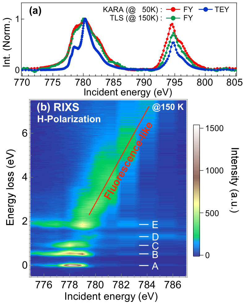
The experiments were performed on 99.9985% pure Co3O4 powder produced by Alfa Aesar. A cylindrical pellet Co3O4 with 10 mm in diameter and 0.5 mm in height was prepared for the measurements. Co XAS spectra were acquired at the soft X-ray WERA beamline of the Karlsruhe Research Accelerator (KARA) synchrotron in Germany. The instrumental resolution was calibrated to be 280 meV full width at half-maximum (FWHM) at the Co edge (780 eV). Both the total electron yield (TEY) and the fluorescence yield (FY) methods were employed. The Co RIXS measurements with linearly vertical () and horizontal () polarized incident X-rays were performed at the Taiwan Light Source (TLS) beamline 05A of National Synchrotron Radiation Research Center (NSRRC) in Taiwan Lai et al. (2014). The experimental energy resolution of the incident photon was 700 meV and the combined resolution of RIXS was 90 meV. Here we note that the RIXS resolution is much better than the incident photon resolution thanks to the position sensitive detector combined with the monochomator-spectrometer system based on the energy-compensation principle Lai et al. (2014); Fung et al. (2004). A grazing incident geometry () with the spectrometer at 90∘ was used. To compare the XAS spectra measured with different facilities and approaches, the background signals were subtracted from the spectra, as described in the Supplementary Material (SM). The subtracted spectra were normalized at the peak with the maximum intensity at the Co edge. We calibrated the photon energy of the RIXS beamline to the spectra acquired at the WERA beamline. The RIXS spectra measured with the -polarization were normalized for the exposure time. Then, the spectra with -polarization were normalized to -polarization ones according to the profile at high energies (above 2.5 eV). The measurements in the KARA-WERA and TLS-05A beamlines were carried out at 50 K and 150K, respectively.
II.2 Simulations
We analyze the experimental data using the cluster model which includes the Coulomb multiplet interaction, the crystal-field splitting, and the spin-orbit coupling on the X-ray excited Co site as well as a charge transfer between Co and O orbitals on the adjacent sites. The many-body Hamiltonian of the cluster model is solved using the Quanty program, which implements the configuration-interaction scheme de Groot (2005); Haverkort et al. (2012). To study the mixed-valence state of Co3O4, we use two cluster models simulating the spectra of the Co2+(Td) site and the Co3+(Oh) site. We take the initial parameter values of the cluster models from previous studies for cobaltate with a high-spin ground state (4B1 in D2d symmetry) and a low-spin ground state (1A1g in Oh symmetry) Liu et al. (2016); Tomiyasu et al. (2017); Wang et al. (2019). The parameter values are refined to represent the studied compound by a detailed comparison with the present high-resolution RIXS data and its polarization dependence, which will be discussed in Sec. III.2. To evaluate the used parameter values, we present the ones estimated by an -initio calculation for the real crystal structure of Co3O4. Starting with a density-functional calculation with local-density approximation (LDA), we construct a tight-binding model spanning Co and O bands by a Wannier projection. The parameter values (crystal-field and hopping parameters) are extracted in the tight-binding model.
III Results
III.1 Experimental results
Figure 1 shows the experimental Co XAS and RIXS results. The TEY spectrum shows a sharp feature at 778.5 eV and at 780.2 eV which are characteristic of the absorption by the Co2+ and the Co3+ sites in Co3O4, respectively Hibberd et al. (2015). The FY spectrum, on the other hand, is rather broad and the features are obscure. This is due to strong saturation and self-absorption effects for a bulk sample, which are more effective for features with strong absorption intensities. In the RIXS result (Fig. 1b), a fluorescence-like signal is observed and its excitation energy increases with incident photon energies as guided by a line. In addition, sharp features at 0.0, 0.5, 0.9, 1.2, and 1.9 eV (labelled by A to E in Fig 1b) are observed. The features A–C are resonantly enhanced at 778 eV incident photon energy, while the feature D is enhanced at 780 eV. The feature E is resonated at the energy slightly below 779 eV. Those features are attributed to local excitations of the Co2+ and/or Co3+ sites, as we will discuss later.
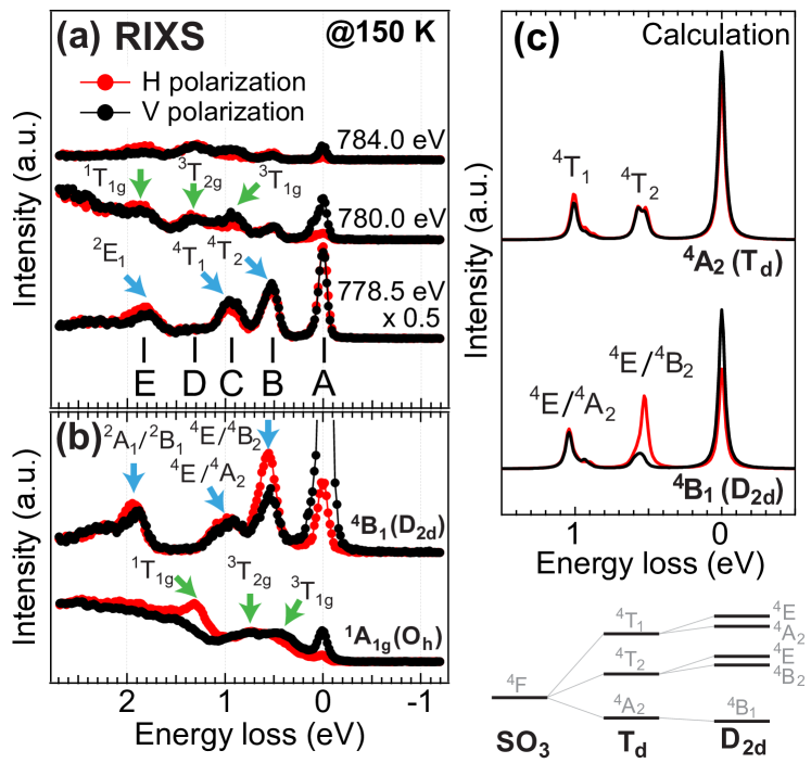
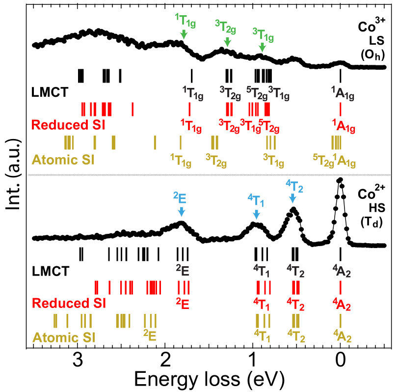
To gain more insight, figure 2a compares the - and -polarization spectra. At 780.0 eV, the feature C (at 0.9 eV) is enhanced by the -polarization, while the feature E (at 1.9 eV) shows opposite behaviour. This polarization dependence resembles the one of 3T1g and 1T1g excited states in LaCoO3 at 20 K, where the Co3+ ions have the 1A1g ground state, see Fig. 2a and 2b. However, the excitation energies differ largely in Co3O4 and LaCoO3, indicating that the crystal field splitting varies substantially between the two. In contrast, the feature B (at 0.5 eV) is identified as the 4T2 excited state on the Co2+(Td) site Mironova et al. (1994). As a reference, Fig. 2b shows Co RIXS in K5H[CoW12O40]xH2O Liu et al. (2016) with a divalent Co2+ ion and 4B1 (D2d) ground state in a distorted tetrahedral structure. In this reference compound, the distortion changes the ground state symmetry from the 4A2(Td) to the 4B1(D2d) that gives rise to a strong polarization dependence at 0.5 eV peak in the RIXS spectra. However, the feature B shows no dichroism in Co3O4, which suggests that the distortion is negligibly small on the Co2+ site and supports the 4A2(Td) symmetry of the ground state in the calculation.
III.2 Simulated results
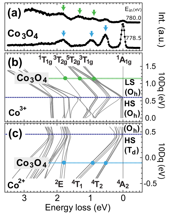
Guided by the observed low-energy features, we determine the low-energy local excitations on the two Co sites in Co3O4. In the cluster-model analysis, the crystal-field parameter () represents the energy splitting of Co 3 orbitals (() and () orbitals in Oh(Td) symmetry). Figures 3b and 3c show the excited states energies as a function of the for the Co3+ and Co2+ site, respectively. The RIXS probed at 780.0 eV shows three features at 0.9, 1.3, and 1.9 eV, indicated with the green arrows in Fig. 3a, which are attributed to the Co3+-site signals. As seen in Fig. 3b, the matching of the three features gives 1.15 eV at the Co3+ site. The value is far larger than the spin-state transition point ( 0.6 eV), suggesting that the ground state on the Co3+ site in Co3O4 is a robust low-spin singlet 1A1g. For the Co2+ site, the eV reproduces the energy position of the characteristic features at 0.5, 0.9, 1.9 eV in the RIXS data probed at 778.5 eV, compare Figs. 3a and 3c. The negative value indicates an inversion of the and manifolds in the Td symmetry.
| V | V | Udd | Upd | ||||
|---|---|---|---|---|---|---|---|
| Co | 0.10 | 0.55 | 4.5 | 1.0 | 2.0 | 4.5 | - |
| Co | 0.02 | 0.47 | 4.5 | 1.0 | 2.0 | 4.5 | 6.0 |
| Co | 1.15 | 1.90 | 1.5 | 3.12 | 1.8 | 6.5 | - |
| Co | 0.84 | 1.59 | 1.5 | 3.12 | 1.8 | 6.5 | 7.5 |
Table 1 summarizes the used parameter values. The charge transfer energy is an energy related to electron transfer from a ligand to the Co site and V/V are the values for electron hopping. The Udd and Upd values parameterize the Coulomb interaction, which are set to reference values Tomiyasu et al. (2017); Wang et al. (2017). Since the ligand-to-metal charge transfer also contributes to the energy splitting of Co 3 states, we provide the value which is calculated by the resultant energy splitting by including the charge transfer. To simulate a contraction of Co wave functions by the presence of the core hole Cramer et al. (1991), the value in the intermediate state is reduced from that in the ground state by 15%, see SM.
To evaluate the values in Table 1, the parameter values estimated by the LDA calculation are provided. For the Co2+() site, the estimated values are 0.10 eV for and 1.29 (1.82) eV for (). For the Co3+(Oh) site, the estimated values are 0.7 eV for and 3.03 (1.74) eV for () 222At the Co3+ site, the manifolds split into a singlet and doublet due to a small trigonal distortion. The value is estimated by averaging the hopping integrals over the two.. The value, which measures the trigonal distortion, is estimated as 0.05 eV. This value is much smaller than the required value ( 0.5 eV) for changing the ground state symmetry (singlet 1A1g) of the Co3+ site in Co3O4, but gives a minor correction to multiplet energies. Since the change affects minor on the spectra, we thus neglect it in our simulation and the site is referred to as Oh for simplicity. Overall the optimized values in the cluster-model simulation agree well with the -initio estimates. The small discrepancy in the value at the Co3+ site is likely due to an underestimate of the covalency in the LDA schemeHaverkort et al. (2012); Hariki et al. (2020).
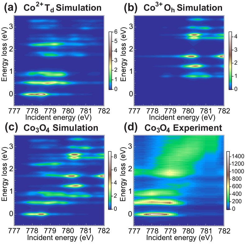
Figures 4a and 4b show the RIXS intensities calculated for the Co2+ and Co3+ sites, respectively, using the optimized parameters in Table 1. Since the low-spin (1A1g) ground state on the Co3+ site is angular isotropic, its intensity is strongly suppressed when the scattering angle is about 90∘ with the -polarization condition. Thus the intensity at zero energy loss (elastic line) is mainly due to the Co2+ site with the 4A2 ground state. Figure 4c shows the sum of the intensities of the two sites weighted by the ratio of 3:8 for the normalized (to total area) Co2+ and Co3+ spectra. The ratio simulates the combination of stoichiometry (1:2) and the number of holes (3:4). The incident energies of Co2+ and Co3+ spectra were adjusted by a shift of 1.8 eV to reproduce the experimental Co XAS (cf Fig. 5a). The cluster-model simulation above identifies the site-resolved excitation spectra in Co3O4. To reproduce the RIXS intensities, the saturation and the self-absorption effects need to be taken into account.
IV Discussion
IV.1 Site selectivity of partial fluorescence yield spectra
We extracted the partial fluorescence yield (PFY) spectra to see the local excitations in detail. Before the discussion on the PFY spectra, the simulated XAS result is presented in comparing with the experimental data. In Fig. 5a, the sum of the simulated XAS of Co2+ (blue) and Co3+ (green) sites well matches with the experimental TEY data (gray bars and red line). The contributions of two sites are separated in the incident energies, that enables to observe site-resolved local excitations by RIXS, as we shown in Fig. 4. Details of the local excitations can be stressed in PFY spectra, which are acquired for energy transfer of the excitations (A–E). The experimental data are obtained by fitting the RIXS intensities of these excitations (cf Fig. 4d), and the theoretical spectra are obtained by summing simulated intensities in a narrow energy window centered at these excitations in Fig. 4c. In the PFY spectra (Fig. 5b), the features A–B and D are unambiguously attributed to the Co2+-site and the Co3+-site excitations, respectively, while the features C and E show an overlap with both Co2+ and Co3+ regions. We stress that the overlap is essential since, according to our simulation in Fig. 4a and 4b, both two sites have excitations at around 0.9 and 1.9 eV. To discuss the two features, however, saturation and self-absorption effects needs to be taken into account properly.
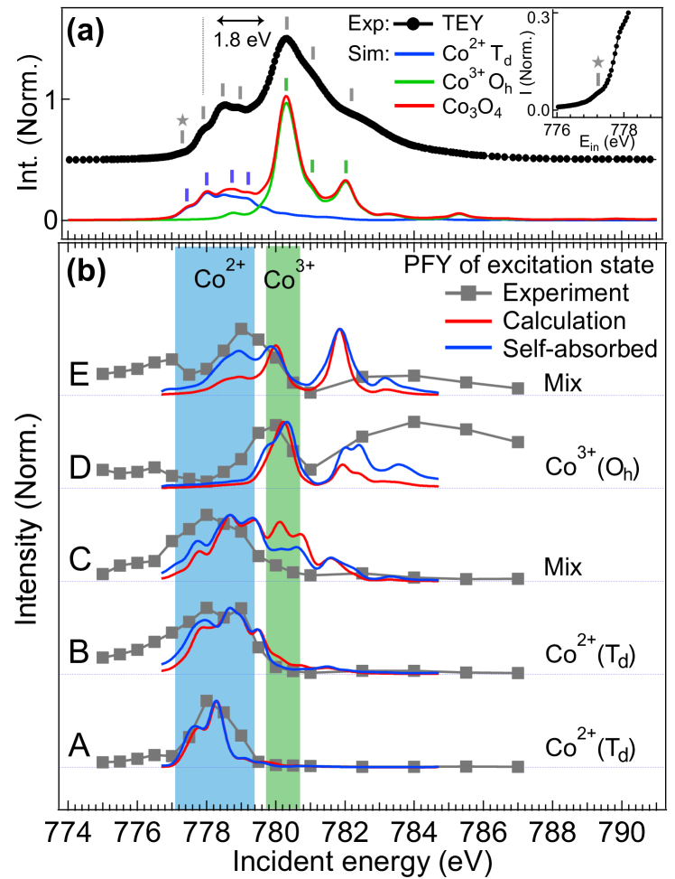
A systematic discrepancy is observed that the intensity at about 780 eV were always overestimated in the simulation, particularly, of the PFY spectra of the features C and E. This indicates that the spectral intensity with respect to the Co2+ site is more pronounced. To confirm the statement, we applied the saturation and self-absorption corrections as Achkar et al. (2011); Wang et al. (2020):
| (1) |
Here, , , and refer to the emission edge of element, the scattering angle, and the sample rotation angle, respectively. In this work, we employed the experimental geometry with and . The ) and ) are the absorption factor of photon-in and photon-out channel in the RIXS process, see further details about the background in the SM and the reference Wang et al. (2020). We multiply a self-absorption coefficient ()-1 to the simulated RIXS result (multiplication of ) and )). The formula implies that the saturation effect is more strong for a large absorption factor. Thus, the features at about 780 eV in PFY spectra are to be suppressed. Consequently, the PFY weights on the Co3+ region is largely suppressed for the feature C (cf. Fig. 5b). It also enhances the intensity at 782 eV of PFY spectra for the feature D and shows better agreement with the experiment. In the simulations, the energy broadening was assumed to be the same for both sites, which yields rather sharp features in the Co3+-site contribution compared to the experiment. A larger energy broadening for the Co3+ site is caused by the strong ligand-metal hybridization on the Co3+ site. To reproduce the broad XAS/PFY structure above the edge, the band formation of ligand 2 states must be taken into account, which is beyond the description of the cluster model used in this study.
The present analysis shown that the excited states at 0.5 eV and 1.3 eV are the unique features to identify the 4T2(Td) excited state on the Co2+ site and 3T1g(Oh) excited state on the Co3+ site. Furthermore, to the best of our knowledge, the 4T2 excited state of the Co2+ site in Co3O4 was only reported by Mironova et al. through infrared more than two decades ago Mironova et al. (1994). The result confirms that the Co3O4 is mainly composed by the magnetically active high-spin Co2+(Td) and the diamagnetic low-spin Co3+(Oh).
IV.2 Ligand-metal hybridization influence of different Co sites in Co3O4
We discuss the question whether the ligand-metal hybridization influences the local electronic structure based on the ligand-to-metal charge transfer model. Hibberd et al. have shown in their atomic-model analysis that the Slater integrals of the atomic Coulomb multiplet need to be reduced substantially to reproduce the Co XAS spectra, which implies that the ligand-metal hybridization is strong in Co3O4 Hibberd et al. (2015); Chen et al. (2011); Qiao et al. (2013). The sensitivity of RIXS to the excitations allows us to address the question, and furthermore study the site-dependence of the covalency in Co3O4. Figure 6 compares the energy diagrams obtained with three different models: (i) the atomic model with the bare (atomic) values of the Slater integrals, (ii) the one with reduced Slater integrals, and (iii) the cluster model including the ligand-to-metal charge transfer channel explicitly. Apparently, the atomic model (i) overestimates energies of observed excitations. For the Co2+ site, the atomic model (ii) with reduced Slater integrals (80% from the atomic values) yields good agreement with the experimental data. On the other hand, for the Co3+ (Oh) site, the Slater integrals are reduced to 55% (80%) from the atomic F (F) value to fit the experimental data. The unconventional reduction rates for the Co3+ (Oh) site imply that the screening effect via a ligand-to-metal charge transfer channel is not negligible. The cluster model including the ligand-to-metal charge transfer channel shows good agreement to the experimental results.
To obtain further information about the ligand-metal hybridization, Table 2 shows the configuration weights in the ground state of the cluster model using the optimized parameters in Table 1. The ligand-to-metal charge transfer channel mixes the ionic configuration (3) with ones with ligand holes (3 and ). In the Co3+ site, the ligand-hole configurations (3 and ) show large weights, which indicates that the Co3+ site is strongly hybridized with the ligand states. In contrast, the ligand-hole configuration (3) contributes only 20% to the ground state in the Co2+ site. This observation suggests that the Co3+ site is highly covalent, while the Co2+ site is rather ionic in Co3O4. The difference also affects the RIXS profile: the ionic Co2+ site shows sharp local excitations; the covalent Co3+ site exhibits a broad intense fluorescence-like feature (cf. Fig. 1).
The orbital covalency of the Co2+ (Co3+) cation can be analyzed using the approach described in the SM and literature Wang et al. (2017). The orbital covalencies of e and t2 orbitals on the tetrahedral Co2+ cation in Co3O4 are 100% and 80%. The e orbital is fully occupied and cannot participate in the ligand-metal hybridization, thus 100% orbital covalency is found. A high value of the cation orbital covalency for t2 orbital indicates that it less contributes to the ligand-hole configuration , which is consistent with the ionic character of the Co2+ site. For the Co3+ site, the orbital covalencies of eg and t2g orbitals are 50% and 100%. This indicates that the ligand-metal hybridization mainly influence to the eg orbital of the Co3+ ions. Although the Co3+ cation is the singlet ground state ( state in the atomic picture), the orbital forms a strong bonding with neighboring oxygen 2 orbitals, which involves the configuration with a ligand hole with eg symmetry, yielding the reduced value (50%) of the orbital covalency.
| weight of configurations | orbital covalency | ||||
| e(eg) | t2(t2g) | ||||
| Co2+() | 79 | 20 | 1 | 100 | 80 |
| Co3+() | 40 | 50 | 10 | 50 | 100 |
V Conclusion
We present the Co XAS and RIXS experimental results in comparison with cluster model simulations. The RIXS provides good chemical site selectivity to the local electronic structure, from which we are able to identify orbital covalencies of different ions in the compound. The polarization dependent analysis indicates the symmetry character of the excitations, which provides a solid guide to analyze the local electronic structure. By selecting characteristic excitations for the RIXS spectra, the PFY spectra have the ability to provide additional site dependent information. The result shows the 4T2 excited state of the tetrahedral Co2+ site at 0.5 eV, which is beyond the discriminative power of optical absorption. In addition, the 1A1g to 3T2g excitation of the octahedral Co3+ site at 1.3 eV can be uniquely identified. The ground state electronic structure of the Co2+ ions and the Co3+ ions are respectively high-spin 4A2(Td) and low-spin 1A1g(Oh), where the high-spin Co2+ must be the magnetically active site. Our result also shows strong ligand-metal hybridization on the Co3+ site, which indicates that the Co3+ site in Co3O4 is rather covalent. In contrast, the Co2+ site shows weak hybridization implying that Co2+ is more ionic. This chemical site selectivity will help the further understanding on the site-dependent catalytic activity and magnetic activity of the spinel cobalt oxides.
VI Acknowledgements
We gratefully acknowledge the synchrotron light source KARA and the KNMF at Karlsruhe, Germany, and the Taiwan Light Source at Hsinchu, Taiwan, for the provision of beamtime. The authors thank the technical staff for their help with the XAS and RIXS measurements. The experiments were supported by ERC advanced grant (grant agreement No. 340279-XRAYonACTIVE). D.J.H. was supported by the Ministry of Science and Technology of Taiwan under Grant No. 106-2112-M-213-008-MY3.
References
- Picard et al. (1980) J. P. Picard, G. Baud, J. P. Besse, and R. Chevalier, J. Less Common. Met. 75, 99 (1980).
- Sparks et al. (2018) T. D. Sparks, A. Gurlo, M. W. Gaultois, and D. R. Clarke, Phys. Rev. B 98, 024108 (2018).
- Note (1) The crystal structure analysis indicated that the Co3+ ions are located in a trigonal (pseudo Oh) local environment. However, our discussions follow the generally indication of Oh symmetry at the B site.
- Roth (1964) W. L. Roth, J. Phys. Chem. Solids 25, 1 (1964).
- Mironova et al. (1994) N. Mironova, V. Skvortsova, and U. Ulmanis, Solid State Commun. 91, 731 (1994).
- Goodenough and Loeb (1955) J. B. Goodenough and A. L. Loeb, Phys. Rev. 98, 391 (1955).
- Martens et al. (1985) J. W. D. Martens, W. L. Peeters, H. M. van Noort, and M. Erman, J. Phys. Chem. Commun. 46, 411 (1985).
- Vaz et al. (2010) C. A. F. Vaz, E. I. Altman, and V. E. Henrich, Phys. Rev. B 81, 104428 (2010).
- Poizot et al. (2000) P. Poizot, S. Laruelle, S. Grugeon, L. Dupont, and J.-M. Tarascon, Nature 407, 496 (2000).
- Du et al. (2007) N. Du, H. Zhang, B. Chen, J. Wu, X. Ma, Z. Liu, Y. Zhang, D. Yang, X. Huang, and J. Tu, Adv. Mater. 19, 4505 (2007).
- Kim et al. (2014) T. W. Kim, M. A. Woo, M. Regis, and K.-S. Choi, J. Phys. Chem. Lett. 138, 2370 (2014).
- Wang et al. (2016) H.-Y. Wang, S.-F. Hung, H.-Y. Chen, T.-S. Chan, H. M. Chen, and B. Liu, J. Am. Chem. Soc. 138, 36 (2016).
- Li and Shen (2014) Y. Li and W. Shen, Chem. Soc. Rev. 43, 1543 (2014).
- Singh et al. (2007) R. N. Singh, D. Mishra, Anindita, A. S. K. Sinha, and A. Singh, Electrochem. Commun. 9, 1369 (2007).
- Esswein et al. (2009) A. J. Esswein, M. J. McMurdo, P. N. Ross, A. T. Bell, and T. D. Tilley, J. Phys. Chem. C 113, 15068 (2009).
- Jiao and Frei (2009) F. Jiao and H. Frei, Angew. Chem. Int. Ed. 48, 1841 (2009).
- Xi et al. (2012) L. Xi, P. D. Tran, S. Y. Chiam, P. S. Bassi, W. F. Mak, H. K. Mulmudi, S. K. Batabyal, J. Barber, J. S. C. Loo, and L. H. Wong, J. Phys. Chem. C 116, 13884 (2012).
- Zhang et al. (2018) X. Zhang, Y.-S. Chen, P. V. Kamat, and S. Ptasinska, J. Phys. Chem. C 122, 13894 (2018).
- Singh et al. (2015) V. Singh, M. Kosa, K. Majhi, and D. T. Major, J. Chem. Theory Comput. 11, 64 (2015).
- Qiao et al. (2013) L. Qiao, H. Y. Xiao, H. M. Meyer, J. N. Sun, C. M. Rouleau, A. A. Puretzky, D. B. Geohegan, I. N. Ivanov, M. Yoon, W. J. Weber, and M. D. Biegalski, J. Mater. Chem. C 1, 4628 (2013).
- Belova et al. (1983) I. D. Belova, Y. E. Roginskaya, R. R. Shifrina, S. G. Gagarin, Y. V. Plekhanov, and Y. N. Venevtsev, Solid State Commun. 47, 577 (1983”).
- Cook and van der Meer (1986) J. G. Cook and M. P. van der Meer, Thin Solid Films 144, 65 (1986).
- Miedzinska et al. (1987) K. M. E. Miedzinska, B. R. Hollebone, and J. G. Cook, J. Phys. Chem. Solids 48, 649 (1987).
- Wang et al. (2004) X. Wang, X. Chen, L. Gao, H. Zheng, Z. Zhang, and Y. Qian, J. Phys. Chem. B 108, 16401 (2004).
- Lima (2014) A. F. Lima, J. Phys. Chem. Solids 75, 148 (2014).
- Wood and Remeika (1967) D. L. Wood and J. P. Remeika, J. Chem. Phys 46, 3595 (1967).
- van Schooneveld et al. (2013) M. M. van Schooneveld, R. W. Gosselink, T. M. Eggenhuisen, M. A. Samarai, C. Monney, K. J. Zhou, T. Schmitt, and F. M. F. de Groot, Angew. Chem. Int. Ed. 52, 1170 (2013).
- Liu et al. (2016) B. Liu, R.-P. Wang, E. N. Glass, C. L. Hill, T. Cuk, J. Okamoto, D. J. Huang, M. M. van Schooneveld, and F. M. F. de Groot, Inorg. Chem. 55, 10152 (2016).
- Hibberd et al. (2015) A. M. Hibberd, H. Q. Doan, E. N. Glass, F. M. F. de Groot, C. L. Hill, and T. Cuk, J. Phys. Chem. C 119, 4173 (2015).
- Elnaggar et al. (2020) H. Elnaggar, R. Wang, S. Lafuerza, E. Paris, A. C. Komarek, H. Guo, Y. Tseng, D. McNally, F. Frati, M. W. Haverkort, M. Sikora, T. Schmitt, and F. M. F. de Groot, Phys. Rev. B 101, 085107 (2020).
- Winder et al. (2020) M. Winder, A. Hariki, and J. Kuneš, “X-ray spectroscopy of rare-earth nickelate lunio3: Lda+dmft study,” (2020), arXiv:2004.01428 [cond-mat.str-el] .
- Lu et al. (2018) Y. Lu, D. Betto, K. Fürsich, H. Suzuki, H.-H. Kim, G. Cristiani, G. Logvenov, N. B. Brookes, E. Benckiser, M. W. Haverkort, G. Khaliullin, M. Le Tacon, M. Minola, and B. Keimer, Phys. Rev. X 8, 031014 (2018).
- Magnuson et al. (2002) M. Magnuson, S. Butorin, J.-H. Guo, and J. Nordgren, Phys. Rev. B 65, 205106 (2002).
- Wang et al. (2017) R.-P. Wang, B. Liu, R. J. Green, M. U. Delgado-Jaime, M. Ghiasi, T. Schmitt, M. M. van Schooneveld, and F. M. F. de Groot, J. Phys. Chem. C 121, 24919 (2017).
- Hariki et al. (2018) A. Hariki, M. Winder, and J. Kuneš, Phys. Rev. Lett. 121, 126403 (2018).
- Elnaggar et al. (2019) H. Elnaggar, R.-P. Wang, S. Lafuerza, E. Paris, Y. Tseng, D. McNally, A. Komarek, M. Haverkort, M. Sikora, T. Schmitt, and F. M. F. de Groot, ACS Appl. Mater. Inter. 11, 36213 (2019).
- Huang et al. (2017) H. Y. Huang, Z. Y. Chen, R.-P. Wang, F. M. F. de Groot, W. B. Wu, J. Okamoto, A. Chainani, A. Singh, Z.-Y. Li, J. S. Zhou, H.-T. Jeng, G. Y. Guo, J.-G. Park, L. H. Tjeng, C. T. Chen, and D. J. Huang, Nat. Commun. 8, 15929 (2017).
- Wang et al. (2019) R.-P. Wang, J. Geessinck, H. Elnaggar, Y. A. Birkhölzer, K. Tomiyasu, J. Okamoto, B. Liu, C.-H. Du, D.-J. Huang, G. Koster, and F. M. F. de Groot, Phys. Rev. B 100, 165148 (2019).
- van Schooneveld et al. (2012) M. M. van Schooneveld, R. Kurian, A. Juhin, K. Zhou, J. Schlappa, V. N. Strocov, T. Schmitt, and F. M. F. de Groot, J. Phys. Chem. C 116, 15218 (2012).
- Lai et al. (2014) C. H. Lai, H. S. Fung, W. B. Wu, H. Y. Huang, H. W. Fu, S. W. Lin, S. W. Huang, C. C. Chiu, D. J. Wang, L. J. Huang, T. C. Tseng, S. C. Chung, C. T. Chen, and D. J. Huang, J. Synchrotron Rad. 21, 325 (2014).
- Fung et al. (2004) H. S. Fung, C. T. Chen, L. J. Huang, C. H. Chang, S. C. Chung, D. J. Wang, T. C. Tseng, and K. L. Tsang, AIP Conf. Proc. 705, 655 (2004).
- de Groot (2005) F. M. F. de Groot, Coord. Chem. Rev. 249, 31 (2005).
- Haverkort et al. (2012) M. W. Haverkort, M. Zwierzycki, and O. K. Andersen, Phys. Rev. B 85, 165113 (2012).
- Tomiyasu et al. (2017) K. Tomiyasu, J. Okamoto, H. Y. Huang, Z. Y. Chen, E. P. Sinaga, W. B. Wu, Y. Y. Chu, A. Singh, R.-P. Wang, F. M. F. de Groot, A. Chainani, S. Ishihara, C. T. Chen, and D. J. Huang, Phys. Rev. Lett. 119, 196402 (2017).
- Cramer et al. (1991) S. P. Cramer, F. M. F. de Groot, Y. Ma, C. T. Chen, F. Sette, C. A. Kipke, D. M. Eichhorn, M. K. Chan, W. H. Armstrong, E. Libby, G. Christou, S. Brooker, V. McKee, O. C. Mullins, and J. C. Fuggle, J. Am. Chem. Soc. 113, 7937 (1991).
- Note (2) At the Co3+ site, the manifolds split into a singlet and doublet due to a small trigonal distortion. The value is estimated by averaging the hopping integrals over the two.
- Hariki et al. (2020) A. Hariki, M. Winder, T. Uozumi, and J. Kuneš, Phys. Rev. B 101, 115130 (2020).
- Achkar et al. (2011) A. J. Achkar, T. Z. Regier, H. Wadati, Y.-J. Kim, H. Zhang, and D. G. Hawthorn, Phys. Rev. B 83, 081106 (2011).
- Wang et al. (2020) R.-P. Wang, H. Elnaggar, C. J. Titus, K. Tomiyasu, J. Geessinck, G. Koster, F. Frati, J. Okamoto, D.-J. Huang, and F. M. F. de Groot, J. Synchrotron Rad. 27, online (2020).
- Chen et al. (2011) J. Chen, X. Wu, and A. Selloni, Phys. Rev. B 83, 245204 (2011).