Vibration-Enhanced Spin-Selective Transport of Electrons in DNA Double Helix
Abstract
The spin-selective transport through helical molecules has been a hot topic in condensed matter physics, because it develops a new research direction in spintronics, i.e., chiro-spintronics. Double-stranded DNA (dsDNA) molecules have been considered as promising candidates to study this topic, since the chiral-induced spin selectivity (CISS) effect in dsDNA was observed in experiment. Considering that the dsDNA molecules are usually flexible in mechanical properties, vibration may be one of important factors to influence the CISS effect. Here, we investigate the influences of electron-vibration interaction (EVI) on the spin-selective transport in dsDNA molecules. We uncover that the EVI not only enhances the CISS effect and the spin polarization () in dsDNA, but also induces a series of new spin-splitting transmission modes. More interesting, these vibration-induced transmission spectra tend to host the same values as those of the original spin-splitting transmission modes, making the spectra to display as a continuous platform even in the energy gap. Our work not only provides us a deep understanding into the influence of vibrations on the CISS effect in helical molecules, but also puts forwards a feasible route to detect the vibration-induced spin-polarized transport in low-dimensional molecular systems.
I \@slowromancapi@. INTRODUCTION
Recently, molecule spintronics has attracted remarkable research interests since the spin-filtering effect (SFE) and spin-polarization transport were observed in double-stranded DNA (dsDNA) molecules and other helical oligopeptides in experiments Göhler et al. (2011); Sanvito (2011); Naaman (2012); Kettner et al. (2015); Abendroth et al. (2019a, b); Mondal et al. (2015). The SFE in organic molecules not only provides us a way to manipulate the spin degrees of freedom of electrons, but also inspires the search for a new class of materials to build spintronic devices, as organic molecules usually preserve long spin relaxation time and may self-assemble on different substrates. Among the possible mechanisms, the helical-induced spin-orbital coupling (SOC) has been identified as the key factor in generating spin polarization in helical molecules Guo and Sun (2012a, b); Pan et al. (2016); Gutierrez et al. (2012); Kettner et al. (2018); Gutierrez et al. (2013); Geyer et al. (2017); Guo and Sun (2012a, 2014). Thus, the chiral-induced spin selectivity (CISS) has been recognized as an important research frontiers, giving birth to a new research direction in spintronics: chiro-spintronics or chiral-based spintronics Mondal et al. (2018). It is inspiring that based on the CISS effect, the spin-resolved currents can be generated and controlled at a molecular level, if chiral molecules are utilized as spin-specific transport media.
To develop chiro-spintronic devices, one of the crucial conditions is to enhance CISS effect or to achieve high spin-polarized transport in helical molecules. To this end, several effective ways such as applying an external gate voltage Guo and Sun (2012b) and using magnetic helix Sarkar and Maiti (2019) have been put forwards in theory. These proposed ways are tightly related with unique molecular structures, such as the double helixes in dsNDA molecules. Considering the fact that most helical molecules are usually flexible in mechanical property, lattice disordering and electron dephasing may easily occur in these spintronic devices, leading to the lose of information. This inspires us to explore new physical mechanism that may enhance the CISS effect and spin-polarized transport in dsDNA molecules. Moreover, just due to the aforementioned structural flexibility, the electron-vibration interaction (EVI) Karasch et al. (2018) may occur and play an important role in the spin-dependent transport through dsDNA.
In this work, we focus on the influence of the EVI on the CISS effect and the spin-polarized transport in a dsDNA molecule, which hosts the helical chain-induced SOC, the environment-induced dephasing process, the interchain and intrachain hopping integrals and the onsite vibration modes, by using the Landauer-Büttiker formula Guo and Sun (2012b); Pan et al. (2016); Fu et al. (2019). Our theoretical investigations uncover that the EVI may enhance the spin polarization in the dsDNA molecule, and also bring a series of new spin-splitting transmission modes in the transmission spectra. In some special structures, the vibration-induced additional resonance tunnelings lead to the spin-dependent transport in the band-gap regime of dsDNA molecules. Interestingly, these new spin-dependent transmission modes possess the same spin polarization as that in the original transmission modes. Moreover, the vibration-induced spin-dependent transport behaviors and the related spin polarization are rather robust against the the increasing dephasing. Although these theoretical results are obtained at zero temperature, they provide the fundamental understanding of the influences of EVI on the CISS effect and the spin-dependent transport in dsDNA molecules, and suggest a feasible route to detect the vibration-induced spin polarization in low-dimensional helical molecules.
The remainder of this paper is organized as follows. In Sec. \@slowromancapii@, we construct a dsNDA-based spintronic device, with a Hamiltonian model to describe the EVI, SOC, electronic hopping and dephasing process, and then introduce theoretical methods to study the spin-dependent transport. In Sec. \@slowromancapiii@, the spin-dependent transmission spectra and the related spin polarization in the dsDNA molecule are calculated, and the influences of the EVI on the CISS effect and spin-dependent transport are discussed in details. Finally, the main results are summarized in the last section.
II \@slowromancapii@. MODEL AND METHODS
II.1 A. Hamiltonian model of dsDNA-based device
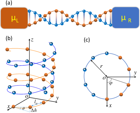
We construct a chiral chain-based dsDNA molecule coupled with two nonmagnetic leads, as illustrated schematically in Fig. 1(a). In the central dsDNA molecule, we consider the interchain and intrachain hopping integrals, the symmetry-induced SOC, the EVI, the onsite vibration modes and the environmental-induced dephasing in the spin-selective transport process. The model Hamiltonian of this dsDNA-based device can be described as Guo and Sun (2012a, b)
| (1) |
where is the Hamiltonian of usual two-leg model including the spin degree of freedom Guo and Sun (2012a), and can be described as = . Here, = describes the kinetic and potential energies of electrons of the molecule, and = ()() is the Hamiltonian of the SOC term; is the reduced Plank constant, is the speed of light, is the momentum operator, and = (, , ) are the Pauli matrices. By using the second quantization as described in Ref. Guo and Sun (2014), can be written as
| (2) |
Here, = (, ) and are the creation operator and the onsite energy at the lattice site in the dsDNA molecule, denoting the site in chain (= 1 or 2). and are the intrachain and interchain hopping integrals. is the SOC parameter with the expression = , where . As varies most rapidly in the near nuclear region, it is reasonable to consider its radical component only Guo and Sun (2012a). = ()() and = ()(). = () and =(). In the above expressions, and are the arc length and the twist angle between successive base-pairs respectively, as described in Figs. 1(b) and 1(c).
The Hamiltonian , where describes the electrons in the left and right nonmagnetic leads, and describes the electronic coupling between the dsDNA and the both leads with the strength , where is the creation operator for an electron in the left (right) lead, and represents the length of the dsDNA chain.
The EVI Hamiltonian can be described as Zimbovskaya and Nitzan (2018)
| (3) |
Here, () is the creation (annihilation) operator for the phonon mode. In the following analysis, we assume that the coupling parameters are set to be nonzero values only in the three cases: (i) for the onsite electrons in the dsDNA chain (i.e., = , = ), is reduced to ; (ii) for the intrachain nearest-hopping electrons (i.e., = , = ), is set as and (iii), for the interchain nearest-hopping electrons (i.e., = , = 1, = 2), is set as . It is reasonable to postulate that in the dsDNA molecules. In our calculations, is adopted.
The forth term in Eq. (1), , is the Hamiltonian of the Büttiker virtual leads and its coupling with each base of the dsDNA Büttiker (1986a, b); Kilgour and Segal (2015), simulating the phase-breaking processes due to the inelastic scattering with phonons and counterions Berlin et al. (2001, 2002). In the frame of the tight-binding model, is expressed as
| (4) |
where = (, ) and describe the creation operator and onsite energy of mode in Büttiker virtual leads, and is the coupling between the nucleobase and the virtual lead.
The last term, , represents the vibrational mode with the phonon frequency .
II.2 B. Lang and Firsov transformation
It is noted that we may eliminate the EVI term from the Hamiltonian (1) by employing the commonly used small polaron (Lang and Firsov) transformation Mitra et al. (2004); Hartle et al. (2008); Galperin et al. (2006a), which converts the Hamiltonian into the form = with = ()+()(). The transformed Hamiltonian reads as = , where the vibration term remains unchanged, while the electron part is reshaped into
| (5) |
It is clear that due to the EVI, the energy level of the dsDNA molecule is renormalized to . Assuming that EVI is sufficiently weak, i.e., , the coupling parameter can be renormalized in a similar way: = . Similarly, we can obtain = , = . The dressed tunneling matrix elements are transformed to , . Then, the Hamiltonian is transformed to , and is converted to be = , where and . Note that the phonon operator arises from the canonical transformation of the particle operator = Mitra et al. (2004); Braig and Flensberg (2003). For this model, we assume that the vibrational mode is coupled with a thermal phonon bath and that this coupling is strong enough so that the phonon maintains its thermal equilibrium state throughout the process. Therefore, the expected value of the phonon operator can be expressed as the following one Chen et al. (2005); Entin-Wohlman et al. (2006)
| (6) |
where denotes the equilibrium phonon population. Here, we consider the low temperature regime where , , so can be approximated by , which is independent of temperature. Thus, we can decouple the electron and phonon subsystems by replacing with its expectation value .
II.3 C. Spin-dependent transmission calculations
Using the Landauer-Bttiker formula Ryndyk et al. (2009, 2016); Fu and Yao (2011, 2010), the spin-dependent transmission coefficient of the dsDNA molecule from the th lead with spin to the th with spin can be calculated as
| (7) |
where are the retarded (advanced) Green’s functions for spin-up or spin-down electrons Mahan (2013). and are the linewidth functions describing the coupling between the leads and the dsDNA molecule, where is the incident electron energy (Fermi energy), and with the retarded (advanced) self-energy due to the coupling to the qth lead. For the real left/right lead, ; while for the virtual leads, , with the dephasing parameter and being the density of states of the leads. Note that as we calculate the spin-up (-down) transmission spectra from the real left lead () to the real right one (), the spin-up (-down) transmission coefficient is simplified as for convenience. Thus, the spin polarization in the dsDNA molecule is defined as = .
As interpreted above, when the operator is replaced by , the Hamiltonian can be decoupled from the vibration operator. The electronic Green’s functions on the Keldysh contour may be approximated as a product of the pure electronic term that can be computed based on the transformed Hamiltonian and the Franck-Condon factor Galperin et al. (2008, 2006b); Hartle and Thoss (2011),
| (8) | ||||
Similarly,
| (9) |
where the identification = . Here, the index represents the number of vibration phonons involved and are the coefficients, depending on the temperature and the strength of the EVI. At a finite temperature, can be expressed as
| (10) |
where = , , and is the modified Bessel function of the th order. At the zero temperature, can be simply read
| (11) |
For clarity, the electronic parameters are considered uniform along each helix of the dsDNA molecule. Based on the complementary base-pairing rule, the dsDNA molecule consists of four nucleobases, i.e., guanine (G), adenine (A), cytosine (C), and thymine (T). Because the structure and the atom number of these nuclear bases are different, the electronic parameters between the two DNA strands may be asymmetrical Voityuk et al. (2001); Senthilkumar et al. (2005); Hawke et al. (2010). For the dsDNA molecule considered here, is set to and , is taken as and = -0.08. To describe the asymmetry between the two helical chains, we employ an additional parameter , and set = , = with = 1.4. All these parameters are extracted from first-principles calculations Endres et al. (2004); Voityuk et al. (2001); Senthilkumar et al. (2005); Hawke et al. (2010) and the unit is eV. The SOC is estimated to = 0.01 eV, which is an order of magnitude smaller than the intrachain hopping integral. For the real leads, the parameters are fixed. The remaining parameters are taken as = 20, = 0.66 rad, and = , resembling the B-form dsDNA molecule, in which the helix makes a turn every 3.4 nm, and the distance between two neighboring base pairs is 0.34 nm Tang et al. (2019). Note that these parameters are used throughout this work, unless other values are explicitly mentioned.
III \@slowromancapiii@. RESULTS AND DISCUSSION
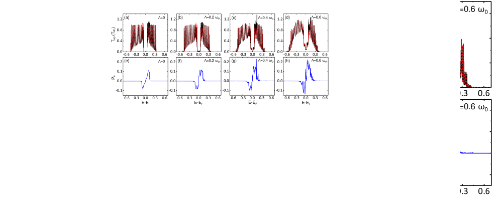
To illustrate the validity of our device model and theoretical method, we firstly calculated the spin-dependent transmission spectra of the dsDNA-based device without the EVI and dephasing process. The numerical results are drawn in Fig. 2(a), where a weak SOC is adopted as eV, and the other parameters are set as = 0 and = 0.05 eV. One may see that the conductance spectrum is consisted of two transmission bands, i.e., the highest occupied molecular orbital (HOMO) and the lowest unoccupied molecular orbital (LUMO), which are divided by an energy gap. For convenience, we call this energy gap the HOMO-LUMO gap. Many transmission peaks are found in both HOMO and LUMO bands, and their number is strictly equal to that of the base-pairs in dsDNA chain due to the quantum coherence effect. Meanwhile, the spin polarization occurs in the both HOMO and LUMO bands, especially near the edges of the HOMO-LUMO gap as drawn in Fig. 2(e). These properties are consistent with previous results Guo and Sun (2012b). As the EVI is taken into account in the dsDNA molecule in a range 0.2, the CISS effect and the corresponding spin-dependent transport behavior changes remarkably as illustrated in Figs. 2(b)-2(d). In particular, one can identify several interesting EVI-induced spin-resolved transport features as listed in the following:
(i) Some additional small transmission peaks appear in the central energy gap, indicating that the EVI assists the electronic conduction through the dsDNA in its HOMO-LUMO gap, while both spin-selective effect and electron-hole-type symmetry are hold in the transmission spectra. One may refer to Fig. S1 in Supplementary Material (SM) for more details see supplemental material at http://link.aps.org/supple mental/ for some vibration-induced spin-dpednent transmission spectra in the energy gap et al. .
(ii) The transmission peaks near the Fermi level in both HOMO and LUMO bands are enhanced as increases, indicating that the EVI contributes to charge transport through the dsDNA molecule, due to the vibration-induced additional conductance tunnelings.
(iii) The spin polarization is also enhanced with the increasing of the EVI as shown in Figs. 2(e)-2(h), supporting that EVI strengthens the CISS effect in the dsNDA molecule. This is because that the EVI enhances the electronic coherent effect, bringing more tunneling paths in the dsDNA molecule. This indicates that we may attain a new physical mechanism to enhance the CISS effect and the spin-dependent transport in dsDNA and related molecules.
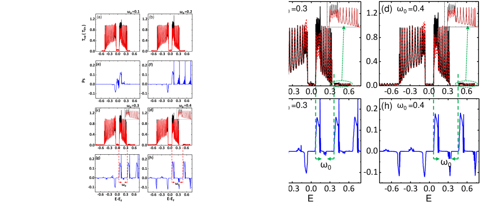
To elucidate the vibration-mediated spin-selective transport in the dsDNA molecule, we study the influence of the vibration frequency on the CISS effect in the dsDNA molecule. Figs. 3(a)-3(h) plot the spin-dependent transmission spectra and versus the Fermi level with increasing in the absence of the dephasing ( = 0). Similarly, one may find that some new resonance peaks in the HOMO-LUMO gap, driving the dsDNA molecule to a conductor. Nevertheless, their heights are much lesser than the peaks in the main transmission spectra. Moreover, both outsides of the HOMO and LUMO bands show additional small transmission peaks, remaining the spin-splitting characteristics regardless the change of vibration frequencies. It should be stressed that the new transmission modes in the high-energy regime nearly duplicate the transmission features of the LUMO band, as highlighted in the insets of Figs. 3(c) and 3(d). In comparison with the energy of the main transmission mode, the vibration-induced transmission mode is located just at , confirming that the new transmission modes originate from the vibration resonance. Consequently, as increases, the new transmission modes shift towards the high-energy direction while maintaining the all spin transport characteristics, as shown in Fig. 3(d). For the convenience in the following discussions, we call the main resonance mode for as the 0th transmission, and the new transmission mode at as the 1st transmission. Note that every peak in the 1st transmission mode serves as an extra conductance channel for electrons in the dsDNA molecule. From the general feature of the EVI, the vibration may induce a series of higher-order transmission modes, and the tunnelling peak in the modes should be localized at , with and . Nevertheless, due to tiny heights in these resonance peaks, it is difficult to observe them in the transmission spectra. In addition, it is noted that for , the vibration-induced 1st and higher-order transmission modes appear at , where denotes the energy related to the 0th transmission mode in the HOMO band.
Now we turn to examine the influence of vibration frequency on the spin polarization in the dsDNA molecule. In Figs. 3(e)-3(h), the corresponding versus with increasing are drawn. One may find that the spectra display several interesting characteristic properties: (i) As increases, mode related to the 0th transmission spectrum is enhanced, indicating that the vibration indeed enhances the CISS effect in chiral molecules. In fact, the EVI proposed here does not alter the SOC, nor break helical symmetry and the spin memory in dsDNA molecules, thus the origination of the spin splitting maintains well in the system. Moreover, the vibration brings several new resonance tunneling paths, much likes the situation that the increasing length of dsDNA chain produces more transmission peaks, which have already been confirmed by previous theoretical calculations and experimental observations Göhler et al. (2011); Guo and Sun (2012a). Consequently, is enhanced remarkably by the increase of vibration frequency in the present device models. (ii) With increasing , a series of new modes, such as the 1st, 2nd, and even 3rd mode, appear around , and meanwhile, the heights of nth () mode is enhanced to catch up to that of the 0th mode. As a result, all modes host the same shapes including their heights and widths, as illustrated in Figs. 3(g) and 3(h). This can be considered as a new finding in the field of chiro-spintronics, since it has not been reported so far. (iii) As increases to a large value, such as = 0.4 eV, some sharp valleys with appear and approach to the one in the 0th mode. This is due to the fact at every vibration-induced transmission spectrum reproduces the one in both HOMO and LUMO bands, and the resonance valleys are associated with the spin polarization in the HOMO band. For small values of , the vibration-induced transmission peaks related to these valleys are buried under the transmission spectra in the LUMO band.
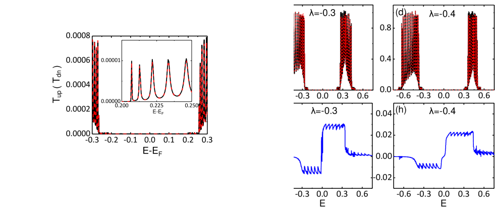
To illustrate the exotic finding that all vibration-induced spin-dependent transmission spectra possess the same modes, we consider further a particular dsDNA-based device model, in which the electronic hopping parameter in the dsDNA molecule is changed from -0.1 to -0.4 eV, while and are fixed as eV and , respectively. The corresponding spin-dependent transmission spectra are plotted in Figs. 4(a)-4(d). It is obvious that the increase of hopping leads to a larger band gap between the HOMO and LUMO bands, and meanwhile, more narrow conductance plateaus emerge in the transmission spectra. For the dsDNA molecule, electron can transport not only along the helical chain, but also within the base-pairs. As increases, the electronic localization in every base-pair is enhanced and charge transport along the main chains is frustrated. More interestingly, two asymmetrical platforms characterized by sawtooth shapes appear in the modes, as described in Figs. 4(e)-4(h). In particular, the appearance of the platform in the band gap indicates that there is filled with the vibration-induced spin-splitting tunneling peaks, although they are much difficult to observe in the transmission spectra. To support this derivation, in Fig. S2 in the SM see supplemental material at http://link.aps.org/supple mental/ for some vibration-induced spin-dpednent transmission spectra in the energy gap et al. , we redrawn the spin-dependent transmission spectra in the band-gap regime for more details. A series of vibration-produced spin-dependent transmission spectra are indeed filled in the band gap, while their transmission values decrease nearly by two orders of magnitude between two neighboring spectra towards the zero-energy direction. However, every new transmission spectrum hosts the same values, confirming further the aforementioned conclusion. Owning to the fact the frequency is too small set in the present device, the vibration-induced transmission spectra are difficult to display in the bang gap. Moreover, all spin-dependent transmission spectra contact with each other with the same modes, making the spectra to display as a wide platform. This property puts forwards a feasible route for us to detect the spin polarization produced by the vibrations in the chiral-molecule systems. In addition, as interpreted above, the increasing hinders the electrons to transport along the main helical chains, which reduces the SOC according to its Hamiltonian . As a result, the platform decreases obviously with increasing .
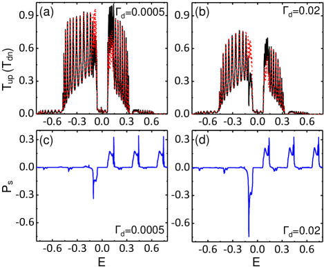
It is well known that the dephasing process occurs inevitably in dsDNA molecule in experiments, thus it is natural to ask whether the dephasing breaks the vibration-induced CISS effect and decreases the spin polarization in the dsDNA molecule. In Figs. 5(a) and 5(b), we plotted the spin-dependent transmission spectra versus for two values of the dephasing parameter in the presence of the EVI () and factors eV, eV and eV. Note that as increases to large values (two additional cases for values are supplemented in Fig. S4 see supplemental material at http://link.aps.org/supple mental/ for some vibration-induced spin-dpednent transmission spectra in the energy gap et al. ), the spin-dependent transmission bands and quickly decrease, because the dephasing process gives rise to the lose of electrons’ phase and spin memory. As a result, the coherence of the dsDNA molecule is reduced and meanwhile, the oscillation peaks in the transmission spectra decrease remarkably. However, the 1st and even higher-order vibration-induced spin-dependent transmission modes still remain in the transmission spectra, indicating that the decoherence effect on the vibration-induced transmission modes is much less than that on the main transmission spectra. Moreover, the modes related with the vibration-induced transmission spectra are barely influenced by the increase of , as illustrated in Figs. 5(c) and 5(d), which suggests the robustness of the vibration-induced spin-splitting transmissions in the dsDNA molecule. Moreover, numerical results show that the dephasing process may even enhance the spin polarization in the HOMO bands. To support the conclusions obtained above, we also considered two other dsDNA-based spintronics devices in the presence of dephasing (see Figs. S5 and S6 in the SM see supplemental material at http://link.aps.org/supple mental/ for some vibration-induced spin-dpednent transmission spectra in the energy gap et al. ), the same spin-dependent transport properties are achieved and the robustness of the vibration-induced spin-splitting transmission spectra are confirmed further.
IV \@slowromancapiv@. CONCLUSION
In summary, we investigate the influence of EVI on the CISS effect and the spin-dependent transport properties of the dsDNA molecule by considering helical symmetry-induced SOC, the dephasing process, and the interchain and intrachain hoppings with the Landauer-Büttiker formula. Our theoretical results show that the EVI not only enhances the spin polarization but also produces a series of new spin-dependent transmission channels through the dsDNA molecule. The vibration-induced spin-transmission spectra tend to retain the same spin-polarization mode as in the main spin-transmission spectra, making the spin-polarization spectra to display a series of platforms even in the band gap. Moreover, the vibration-induced spin-dependent transmissions are robust against the dephasing process, assisting the CISS and spin selective transport. These theoretical results provide new insights to understand the influence of the EVI on the CISS effect and the spin-polarized transport in dsDNA molecules. They also put forward a feasible route to enhance spin polarization induced by vibrations in low-dimensional molecular systems for applications.
V ACKNOWLEDGNEBTS
This work is supported by the Natural Science Foundation of China with Grants No. 11774104. Work at UCI was supported by DOE-BES (Grant No. DE-FG02-05ER46237). Computer simulations were partially performed at the U.S. Department of Energy Supercomputer Facility (NERSC).
References
- Göhler et al. (2011) B. Göhler, V. Hamelbeck, T. Z. Markus, M. Kettner, G. F. Hanne, Z. Vager, R. Naaman, and H. Zacharias, Science 331, 894 (2011).
- Sanvito (2011) S. Sanvito, Chem. Soc. Rev. 40, 3336 (2011).
- Naaman (2012) R. Naaman, J. Phys. Chem. Lett. 3, 2178 (2012).
- Kettner et al. (2015) M. Kettner, B. Göhler, H. Zacharias, D. Mishra, V. Kiran, R. Naaman, C. Fontanesi, D. H. Waldeck, S. Sek, J. Pawłowski, and J. Juhaniewicz, J. Phys. Chem. C 119, 14542 (2015).
- Abendroth et al. (2019a) J. M. Abendroth, K. M. Cheung, D. M. Stemer, M. S. El Hadri, C. Zhao, E. E. Fullerton, and P. S. Weiss, J. Am. Chem. Soc. 141, 3863 (2019a).
- Abendroth et al. (2019b) J. M. Abendroth, D. M. Stemer, B. P. Bloom, P. Roy, R. Naaman, D. H. Waldeck, P. S. Weiss, and P. C. Mondal, ACS Nano 13, 4928 (2019b).
- Mondal et al. (2015) P. C. Mondal, N. Kantor-Uriel, S. P. Mathew, C. Tassinari, F Fontanesi, and R. Naaman, Adv. Mater. 27, 1924 (2015).
- Guo and Sun (2012a) A.-M. Guo and Q.-F. Sun, Phys. Rev. Lett. 108, 218102 (2012a).
- Guo and Sun (2012b) A.-M. Guo and Q.-F. Sun, Phys. Rev. B 86, 035424 (2012b).
- Pan et al. (2016) T.-R. Pan, A.-M. Guo, and Q.-F. Sun, Phys. Rev. B 94, 235448 (2016).
- Gutierrez et al. (2012) R. Gutierrez, E. Díaz, R. Naaman, and G. Cuniberti, Phys. Rev. B 85, 081404 (2012).
- Kettner et al. (2018) M. Kettner, V. V. Maslyuk, D. Nuerenberg, J. Seibel, R. Gutierrez, G. Cuniberti, K.-H. Ernst, and H. Zacharias, J. Phys. Chem. Lett. 9, 2025 (2018).
- Gutierrez et al. (2013) R. Gutierrez, E. Diaz, C. Gaul, T. Brumme, F. Dominguez-Adame, and G. Cuniberti, J. Phys. Chem. C 117, 22276 (2013).
- Geyer et al. (2017) M. Geyer, R. Gutierrez, V. Mujica, and G. Cuniberti, J. Phys. Chem. C 123, 27230 (2017).
- Guo and Sun (2014) A.-M. Guo and Q.-F. Sun, Proc. Natl. Acad. Sci. USA 111, 11658 (2014).
- Mondal et al. (2018) P. C. Mondal, W. Mtangi, and C. Fontanesi, Small Methods 2, 1700313 (2018).
- Sarkar and Maiti (2019) S. Sarkar and S. K. Maiti, Phys. Rev. B 100, 205402 (2019).
- Karasch et al. (2018) P. Karasch, D. A. Ryndyk, and T. Frauenheim, Phys. Rev. B 97, 195401 (2018).
- Fu et al. (2019) H.-H. Fu, G.-F. Du, D.-D. Wu, Q.-B. Liu, and R. Wu, Phys. Rev. B 100, 085407 (2019).
- Zimbovskaya and Nitzan (2018) N. A. Zimbovskaya and A. Nitzan, J. Chem. Phys. 148, 024303 (2018).
- Büttiker (1986a) M. Büttiker, Phys. Rev. B 33, 3020 (1986a).
- Büttiker (1986b) M. Büttiker, IBM J. Res. Dev. 32, 63 (1986b).
- Kilgour and Segal (2015) M. Kilgour and D. Segal, J. Chem. Phys. 143, 024111 (2015).
- Berlin et al. (2001) Y. A. Berlin, A. L. Burin, and M. A. Ratner, J. Am. Chem. Soc. 123, 260 (2001).
- Berlin et al. (2002) Y. A. Berlin, A. L. Burin, and M. A. Ratner, Chem. Phys. 275, 61 (2002).
- Mitra et al. (2004) A. Mitra, I. Aleiner, and A. J. Millis, Phys. Rev. B 69, 245302 (2004).
- Hartle et al. (2008) R. Hartle, C. Benesch, and M. Thoss, Phys. Rev. B 77, 205314 (2008).
- Galperin et al. (2006a) M. Galperin, A. Nitzan, and M. A. Ratner, Phys. Rev. B 73, 045314 (2006a).
- Braig and Flensberg (2003) S. Braig and K. Flensberg, Phys. Rev. B 68, 205324 (2003).
- Chen et al. (2005) Z.-Z. Chen, R. Lü, and B.-F. Zhu, Phys. Rev. B 71, 165324 (2005).
- Entin-Wohlman et al. (2006) O. Entin-Wohlman, Y. Imry, and A. Aharony, Phys. Rev. B 81, 113408 (2006).
- Ryndyk et al. (2009) D. A. Ryndyk, R. Gutiérrez, B. Song, and G. Cuniberti, Energy Transfer Dynamics in Biomaterial Systems, edited by I. Burghardt, V. May, D. A. Micha, and E. R. Bittner (Springer Berlin Heidelberg, Berlin, Heidelberg, 2009) pp. 213–335.
- Ryndyk et al. (2016) D. A. Ryndyk et al., Theory of Quantum Transport at Nanoscale, Vol. 184 (Springer, 2016).
- Fu and Yao (2011) H.-H. Fu and K.-L. Yao, J. Chem. Phys. 134, 054903 (2011).
- Fu and Yao (2010) H.-H. Fu and K.-L. Yao, J. Appl. Phys. 108, 084510 (2010).
- Mahan (2013) G. D. Mahan, Many-particle physics (Springer Science & Business Media, 2013).
- Galperin et al. (2008) M. Galperin, M. A. Ratner, and A. Nitzan, Mol. Phys. 106, 397 (2008).
- Galperin et al. (2006b) M. Galperin, A. Nitzan, and M. A. Ratner, Phys. Rev. B 73, 045314 (2006b).
- Hartle and Thoss (2011) R. Hartle and M. Thoss, Phys. Rev. B 83, 115414 (2011).
- Voityuk et al. (2001) A. A. Voityuk, J. Jortner, M. Bixon, and N. Rösch, J. Chem. Phys. 114, 5614 (2001).
- Senthilkumar et al. (2005) K. Senthilkumar, F. C. Grozema, C. l. F. Guerra, F. M. Bickelhaupt, F. D. Lewis, Y. A. Berlin, M. A. Ratner, and L. D. A. Siebbeles, J. Am. Chem. Soc. 127, 14894 (2005), pMID: 16231945.
- Hawke et al. (2010) L. G. D. Hawke, G. Kalosakas, and C. Simserides, Euro. Phys. J. E 32, 291 (2010).
- Endres et al. (2004) R. G. Endres, D. L. Cox, and R. R. P. Singh, Rev. Mod. Phys. 76, 195 (2004).
- Tang et al. (2019) H.-Z. Tang, Q.-F. Sun, J.-J. Liu, and Y.-T. Zhang, Phys. Rev. B 99, 235427 (2019).
- (45) see supplemental material at http://link.aps.org/supple mental/ for some vibration-induced spin-dpednent transmission spectra in the energy gap, the influence of the disorder on the the vibration-induced spin-dependent transmision spectra, and the corresponding spin-polarization spectra in two different stuctures of the dsDNA moleucles, .