Epileptic Seizures Detection Using Deep Learning Techniques: A Review
Abstract
A variety of screening approaches have been proposed to diagnose epileptic seizures, using electroencephalography (EEG) and magnetic resonance imaging (MRI) modalities. Artificial intelligence encompasses a variety of areas, and one of its branches is deep learning (DL). Before the rise of DL, conventional machine learning algorithms involving feature extraction were performed. This limited their performance to the ability of those handcrafting the features. However, in DL, the extraction of features and classification are entirely automated. The advent of these techniques in many areas of medicine, such as in the diagnosis of epileptic seizures, has made significant advances. In this study, a comprehensive overview of works focused on automated epileptic seizure detection using DL techniques and neuroimaging modalities is presented. Various methods proposed to diagnose epileptic seizures automatically using EEG and MRI modalities are described. In addition, rehabilitation systems developed for epileptic seizures using DL have been analyzed, and a summary is provided. The rehabilitation tools include cloud computing techniques and hardware required for implementation of DL algorithms. The important challenges in accurate detection of automated epileptic seizures using DL with EEG and MRI modalities are discussed. The advantages and limitations in employing DL-based techniques for epileptic seizures diagnosis are presented. Finally, the most promising DL models proposed and possible future works on automated epileptic seizure detection are delineated.
Index Terms:
epileptic seizures, diagnosis, EEG, MRI, feature extraction, classification, deep learningI Introduction
Epilepsy is a non-communicable disease and one of the most common neurological disorders of humans, usually associated with sudden attacks [1]. Seizures are a swift and early abnormality in the electrical activity of the brain that disrupts the part of the whole body [2]. Epileptic seizures are affecting around 60 million people worldwide by varied kinds [3]. These attacks occasionally provoke cognitive disorders which can cause severe physical injury to the patient. Besides, people with epileptic seizures sometimes suffer emotional distress due to embarrassment and lack of appropriate social status. Hence, early detection of epileptic seizures can help the patients and improve their quality of life.
Various screening techniques have been developed to diagnose epileptic seizures, including MRI [4], EEG [5], Magnetoencephalography (MEG) [6] and Positron Emission Tomography (PET) [7]. The EEG signals are widely preferred as they are economical, portable, and show clear rhythms in the frequency domain [8]. The EEG provides the voltage variations produced by the ionic current of neurons in the brain, which indicate the brain’s bioelectric activity [9]. Diagnosing epilepsy with EEG signals is time-consuming and strenuous, as the epileptologist or neurologist needs to screen the EEG signals minutely. Also, there is a possibility of human error, and hence, developing a computer-based diagnosis may alleviate these problems.
Many machine learning algorithms have been developed using statistical, frequency domain and nonlinear parameters to detect epileptic seizures [10, 11, 12, 13, 14, 15]. In conventional machine learning techniques, the selection of features and classifiers is made by trial and error method. Also, one needs to have sound knowledge of signal processing and data mining to develop a robust model. Such models perform well for limited data. Nowadays, with the increase in the availability of data, machine learning techniques may not perform very well. Hence, the deep learning techniques, which are the state-of-art methods, have been employed.
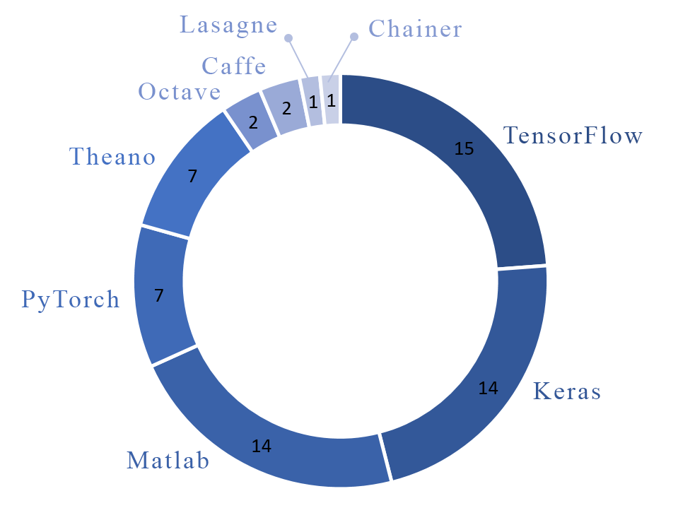
In traditional machine learning algorithms, most simulations were executed in the Matlab software environment, but the deep learning models are usually developed using Python programming language and its numerous open-source toolboxes. This change in the applied programming language helped the researchers to contribute to other works more comfortably and recreate the previously reached results more straightforward. Also created the accessibility of computation resource to everyone thanks to cloud computing, and lastly, made a more convenient path for the creation of application specified hardware for biomedical tasks. Figure 1 shows that, the Tensorflow and one of its high-level APIs, Keras, are widely used for epileptic seizure detection using deep learning in reviewed works due to their versatility and applicability.
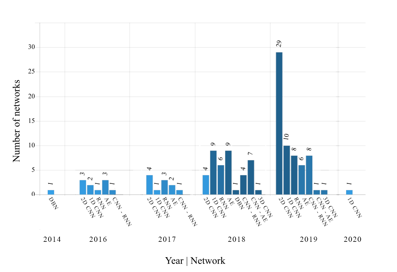
Since 2016, substantial research has embarked on the field of identifying epilepsy using deep learning models, such as Convolutional Neural Networks (CNN), Recurrent Neural Networks (RNN), Deep Belief Networks (DBN), Autoencoders (AE), CNN-RNN, and CNN-AE [16, 17, 18, 19]. The number of studies in this area using deep learning is growing by proposing new efficient models. Figure 2 provides the overview of number of studies conducted using various deep learning models from 2014 to 2020 in detecting epileptic seizures.
The main goals of this study are as follows:
-
•
Providing information on available EEG datasets.
-
•
Reviewing works done using various deep learning models for automated detection of epileptic seizures by various modalities.
-
•
Introducing future challenges on the detection of epileptic seizures.
-
•
Analyzing the best performing model for various modalities of data.
Epileptic seizures detection using deep learning is discussed in section II. Section III describes the non-EEG based epileptic seizure detection. Hardware used for epileptic seizures detection is provided in section IV. Discussion on the paper is outlined in section V. The challenges faced by employing deep learning methods for the epileptic seizure detection are summarized in section VI. Finally, the conclusion and future work are delineated in section VII.
II Epileptic Seizure Detection Based on Deep Learning Techniques
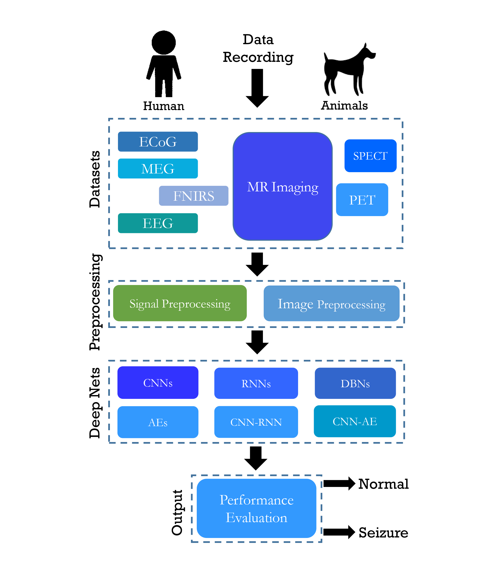
Figure 3 illustrates the working of a Computer-Aided Diagnosis System (CADS) for epileptic seizures using deep learning methods. The input to the deep learning model can be EEG, MEG, Electrocorticography (ECoG), functional Near-InfraRed Spectroscopy (fNIRS), PET, Single-Photon Emission Computed Tomography (SPECT), MRI. Then the signal is subjected to the preprocessing to remove the noise. Then these noise eliminated signals are used to develop the deep learning models. The performance of the model is evaluated using accuracy, sensitivity, and specificity. Additionally, a table combining all the works reviewed on epileptic seizure detection using deep learning is presented in the table form in Appendix A of the paper.
II-A Epileptic Datasets
Datasets play an important role in developing accurate and robust CADS. Multiple EEG datasets, namely, Freiburg [20], CHB-MIT [21], Kaggle [22], Bonn [23], Flint-Hills [13], Bern-Barcelona [24], Hauz Khas [13], and Zenodo [25] are available to develop the automated epileptic seizure detection systems. The signals from these datasets are recorded either intracranially or from the scalp of humans or animals. The supplementary information for each dataset is listed in table I.
| Dataset | Number of Patients | Number of Seizures | Recording | Total Duration (hour) | Sampling Frequency (Hz) |
| Flint-Hills [13] | 10 | 59 | Continues intracranial ling term ECoG | 1419 | 249 |
| Hauz Khas [13] | 10 | NA | Scalp EEG (sEEG) | NA | 200 |
| Freiburg [20] | 21 | 87 | Intracranial Electroencephalography (IEEG) | 708 | 256 |
| CHB-MIT [21] | 22 | 163 | sEEG | 844 | 256 |
| Kaggle [22] | 5 dogs | 48 | IEEG | 627 | 400 |
| 2 patients | 5 KHz | ||||
| Bonn [23] | 10 | NA | Surface and IEEG | 39 m | 173.61 |
| Barcelona [24] | 5 | 3750 | IEEG | 83 | 512 |
| Zenodo [25] | 79 neonatal | 460 | sEEG | 74 m | 256 |
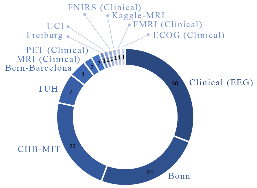
Figure 4 shows the number of times each dataset is used to detect epileptic seizures in reviewed researches. It is observable that the Bonn dataset is the most employed in reviewed research.
II-B Preprocessing
In a deep learning based CAD system, EEG signal preprocessing commonly involves three steps of noise removal, normalization, and signal preparation for deep learning network applications. In the noise removal step, finite impulse response (FIR) or infinite impulse response (IIR) filters are usually used to eliminate extra signal noise. Normalization is then performed using various schemes such as the z-score technique. Finally, different time domain, frequency, and time-frequency methods are employed to prepare the signals for the deployment of deep networks.
II-C Review of Deep Learning Techniques
In contrast to conventional neural networks, or so-called shallow networks, deep neural networks are structures with more than two hidden layers. Some recent deep nets have more than hundreds of layers [16]. This increase in the size of the networks results in a massive rise in the number of parameters of the network, requiring appropriate methods for learning, and also measures to avoid overfitting of the learned network. Convolutional networks use filters convolved with input patterns instead of multiplying a weight vector (matrix), which reduces the number of trainable parameters dramatically.
Furthermore, other methods are suggested to help the network to learn, as well [26]. Pooling layers reduce the size of the input pattern to the next convolutional layer. Batch normalization, dropout, early stopping, unsupervised or semi-unsupervised learning, and regularization techniques prevent the learned network from overfitting and increase the learning ability and speed. The AE and DBN are employed as unsupervised learning and then fine-tuned to avoid overfitting for limited labeled data. Long-Short-Time-Memory (LSTM) and Gated-Recurrent-Units (GRU) are recurrent neural networks capable of revealing the long term time dependencies of data samples.
II-C1 Convolutional Neural Networks (CNNs)
CNNs are one class of the most popular deep learning networks in which most of the researches in machine learning have been devoted to these networks [16]. They were initially presented for image processing applications but have recently been adopted to one and two-dimensional architectures for diagnosis and prediction of diseases using biological signals [27]. This class of deep learning networks is widely used for the detection of epileptic seizures using EEG signals. In two dimensional convolutional neural networks (2D-CNN), the one dimensional (1D) EEG signals are first transformed into two dimensions employing visualization methods such as spectrogram, higher order bispectrum, and wavelet transforms, then are applied to the input of the convolutional network. In 1D architectures, the EEG signals are applied in the form of one dimensional to the input of convolutional networks. In these networks, changes are made to the core architecture of 2D-CNN that are capable of processing the 1D-EEG signals. Therefore, since both 2D and one-dimensional convolutional neural networks (1D-CNNs) are used in the field of epileptic seizures detection, they are investigated separately.
2D - Convolutional Neural Networks
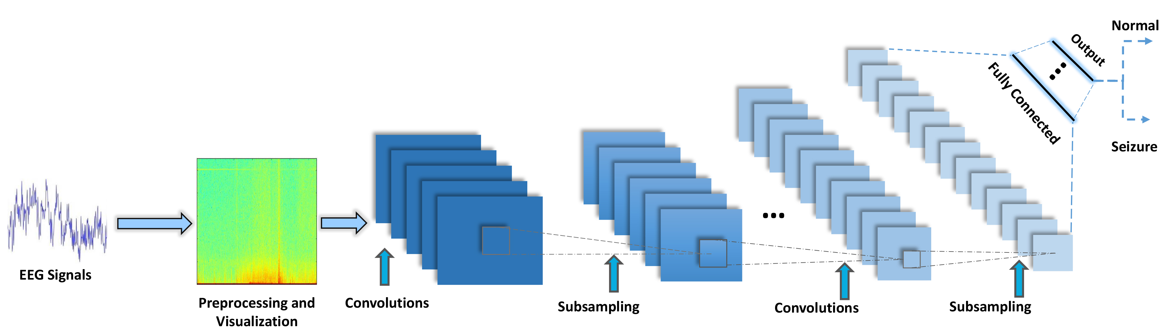
Nowadays, deep 2D networks are applied to resolve a wide range of computer vision obstacles such as image segmentation [28], medical image classification [29], and face recognition [30]. First, in 2012, Krizovsky et al. [31] suggested this network to solve image classification problems, and then quickly use similar networks for all different tasks such as medical image classification, in an effort to obviate the difficulties of previous networks and solve more intricate problems, increased remarkably. Figure 5 shows a general form of a 2D-CNN used for epileptic seizure detection. The application of 2D-CNN architectures is arguably the most important architecture of deep neural nets. Also, more information about visualization and preprocessing method can be found in Appendix A.
In one study [32], the SeizNet 16-layer convolutional network is introduced, with additional dropout layers and batch normalization (BN) behind each convolutional layer having a structure similar to VGG-Net. The researchers in [33] presented a new 2D-CNN model that can extract the spectral and temporal characteristics of EEG signals and used them to learn the general structure of seizures. Zuo et al. [34] developed the diagnosis of Higher Frequency Oscillations (HFO) epilepsy from 16-layer 2D-CNN and EEG signals. A deep learning framework called SeizureNet is proposed in [35] using convolution layers with dense connections. A novel deep learning model called the temporal graph convolutional network (TGCN) has been introduced by Covert et al. [36], comprising of five architectures with 14, 18, 22, 23 and 26 layers. Bouaziz et al. [37] split the EEG signals of CHB-MIT with 23 channels into 2-second time windows and then converted them into density images (spatial representation), which were fed as inputs to the CNN network.
Alexnet
FeiFei Li, Professor of Stanford University, created a dataset of labeled images of real-world objects and termed her project as ImageNet[38]. ImageNet organized a computer vision competition called ILSVRC annually to solve the image classification problems. Alex Krizhevsky revolutionized the image classification world with his algorithm, AlexNet, which won the 2012 ImageNet challenge and started the whole deep learning era [31]. AlexNet won the competition by achieving the top-5 test accuracy of 84.6%. Taqi et al. [39] used the AlexNet network to diagnose focal epileptic seizures. This proposed network used the feature extraction approach and eventually applied the softmax layer for classification purposes and achieved 100% accuracy. In another research, the AlexNet network was employed [40]. They transformed the 1D signal in to 2D image by passing through Signal2Image (S2I) module. The several methods used in this are signal as image, spectrogram, one layer 1D-CNN, and two-layer 1D-CNN.
VGG
A research team at Oxford proposed the Visual Geometry Group (VGG) CNN model in 2014 [41]. They configured various models, and one such model was called VGG-16 was submitted to ILSVRC 2014 competition. The model was known as VCG-16 because it comprised of 16 layers. It delivered an excellent performance in image detection and classification problems. Ahmedt-Aristizabal et al. [42] performed VGG-16 architecture to diagnose epilepsy from facial images. Their proposed approach attempted to extract and classify semiological patterns of facial states automatically. After recording the images, the proposed VGG architecture is trained primarily by well-known datasets, followed by various networks such as 1D-CNN and LSTM in the last few layers. In [40], the VGG network used one-dimensional and two-dimensional signals. To train the models, Adam’s optimizer and a cross-entropy error function were used. They used the batch size and number of epochs as 20 and 100 respectively. The idea of detecting epileptic seizures on the sEEG signal plots was examined by Emami et al. [43]. In the pre-processing step, the signals were segmented into different time windows and VGG-16 was used for classification using small (3×3) convolution filters to efficiently detect small EEG signal changes. This architecture was pre-trained by applying an ImageNet dataset to differentiate 1000 classes, and the last two layers had 4096 and 1000 dimensional vectors. They modified these last two layers to have 32 and 2 dimensions, respectively, to detect seizure and non-seizure classes.
GoogLeNet
GoogLeNet won the 2014 ImageNet competition with 93.3% top-5 test accuracy [44]. This 22-layer network was called GoogLeNet to honor Yann Lecun, who designed LeNet. Before the introduction of GoogLeNet, it was stated that by going deep, one could achieve better accuracy and results. Nevertheless, the google team proposed an architecture called inception, which achieved better performance by not going deep but by better design. It represented a robust design by using filters of different sizes on the same image. In the field of EEG signal processing to diagnose epileptic seizures, this architecture has recently received the attention of researchers. Taqi et al. [39] used this network in their preliminary researches to diagnose epileptic seizures. Their model was used to extract features from the Bern-Barcelona dataset and achieved excellent results.
ResNet
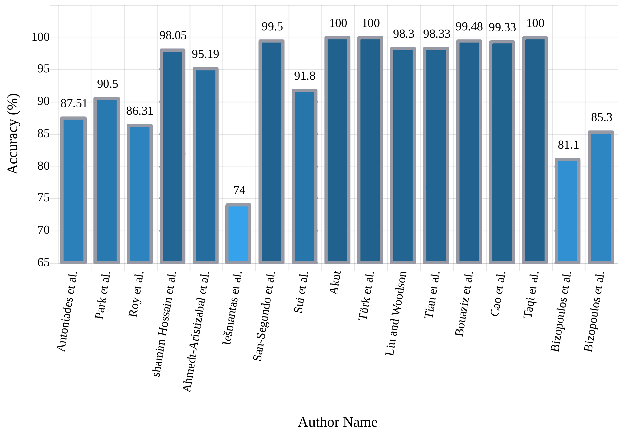
Microsoft’s ResNet won ImageNet challenge with 96.4% accuracy by applying a 152-layer network which utilized a Resnet module [45]. In this network, residual blocks were introduced, which were capable of training deep architecture by using identity skip connections which copied inputs of each layer to the next layer. The idea was to learn something different and new in the next layer. So far, not much research has been accomplished on the implementation of ResNet networks to diagnose epilepsy, but may grow significantly in coming days. Bizopoulos et al. [40] introduced two ResNet and DenseNet architectures to diagnose epileptic seizures and attained good results. They showed that S2I-DenseNet base model with an average of 70 epochs was sufficient to gain the best accuracy of 85.3%. A summary of the research accomplished in this section is reported in Table II. A comparison of accuracy obtained by each study is shown in Figure 6.
| Work | Networks | Number of Layers | Classifier | Accuracy% |
| [46] | 2D-CNN | 3 | Logistic Regression (LR) | 87.51 |
| 4 | ||||
| [47] | 2D-CNN | 9 | softmax | NA |
| [48] | Combination of 1D- CNN and 2D-CNN | 11 | sigmoid | 90.58 |
| [49] | 2D-CNN | 18 | softmax | NA |
| [50] | 2D-CNN/MLP hybrid | 11 | sigmoid | NA |
| [51] | 2D-CNN | 9 | softmax | 86.31 |
| [32] | SeizNet | 16 | NA | NA |
| [52] | 2D-CNN with 1D-CNN | 12 | softmax | NA |
| [33] | 2D-CNN | 9 | softmax | 98.05 |
| [34] | 2D-CNN | 16 | softmax | NA |
| [35] | SeizureNet | 133 | softmax | NA |
| [42] | 2D-CNN | VGG-16,8 | SVM | 95.19 |
| [53] | 2D-CNN | 6 | softmax | 74 |
| [54] | 2D-CNN | 12 | softmax and sigmoid | 99.50 |
| [55] | 2D-CNN | 16 | softmax | 91.80 |
| [36] | TGCN | 14 | sigmoid | NA |
| 18 | ||||
| 22 | ||||
| 22 | ||||
| 26 | ||||
| [56] | 2D-CNN | 23 | softmax | 100 |
| [57] | 2D-CNN | 5 | softmax | 100 |
| [58] | 2D-CNN | 14 | softmax | 2 classes 98.30 |
| 3 classes 90.10 | ||||
| [59] | 2D-CNN | 7 | MV-TSK-FS | 98.33 |
| 5 | ||||
| 3D-CNN | 8 | |||
| [37] | 2D-CNN | 8 | softmax | 99.48 |
| [60] | 2D-CNN | 23 | sigmoid | NA |
| 18 | RF | |||
| [61] | 2D-CNN | 7 | KELM | 99.33 |
| [39] | GoogleNet | Standard Networks | softmax | 100 |
| AlexNet | ||||
| LeNet | ||||
| [43] | 2D-CNN | VGG-16 | softmax | NA |
| [40] | Standard Networks | softmax | 85.30 | |
1D - Convolutional Neural Networks
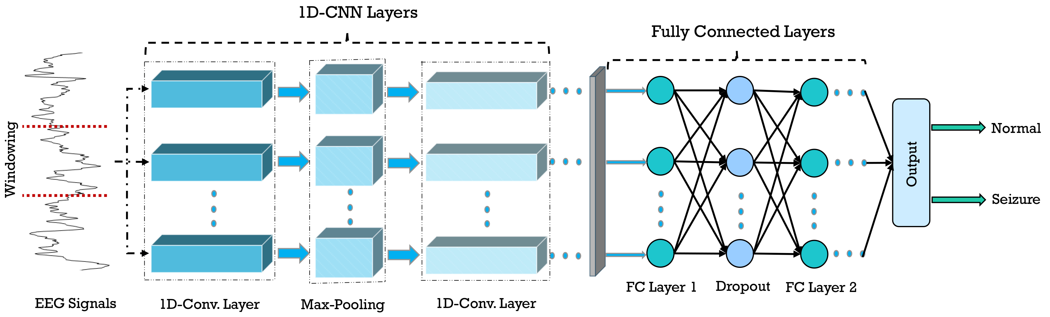
1D-CNNs are intrinsically suitable for processing of biological signals such as EEG for detection of epileptic seizures. These architectures present a more straightforward structure and a single pass of them is faster as compared to CNN with 2D architecture, due to lower number of parameters. The most important superiority of 1D to 2D architectures is the possibility of employing pooling and convolutional layers with a larger size. In addition to that, signals are 1D in nature, and using pre-processing methods to transform them to 2D may lead to information loss, yet all the data are preserved in 1D representation. Figure 7 shows a general form of a 1D-CNN used for epileptic seizure detection.
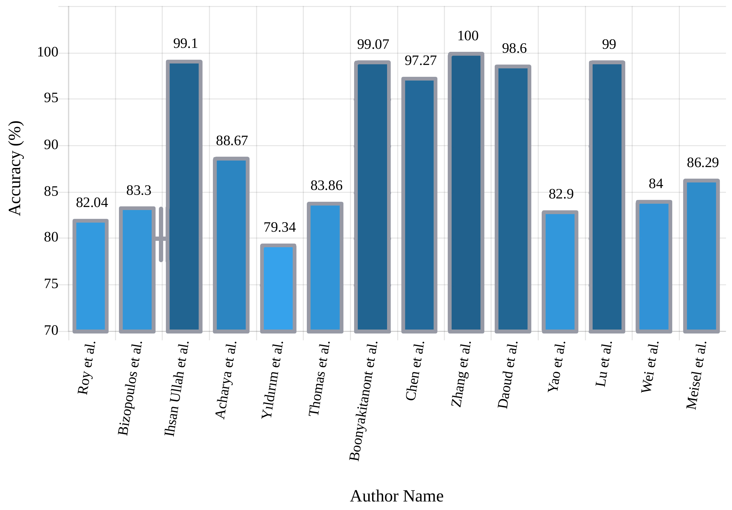
The authors in [40] pursued their experiments using one-dimensional LeNet, AlexNet, VGGnet, ResNet, and DenseNet architectures, and applied well-known 2D architectures in 1D space is the first study in this section. In [62], 1D-CNN was used for the feature extraction procedure. The researchers [63] used 1D-CNN in another research. In this work, the CHB-MIT dataset is adopted, and the signals from each channel are segmented into 4-second intervals; additionally, overlapping segments are also accepted to increase the data and accuracy. Combining CNNs with traditional feature extraction methods together was explored in [64]; they used the Empirical Mode Decomposition (EMD) method for feature extraction, and a CNN was applied to acquire high accuracy in the multi-class classification tasks. In [65], an integrated framework for the diagnosis of epileptic seizures is presented that combines the capability of interpreting probabilistic graphical models (PGMs) with advances in deep learning. The authors in [66] submitted a 1D-CNN architecture defined CNN-BP (stading for CNN bipolar). In this work, they used the data from patients monitored with combined foramen ovale (FO) electrodes and EEG surface electrodes. A new scheme to classify EEG signals based on temporal convolution neural networks (TCNN) is introduced by Zhang et al. [67]. Table III shows the summary of related works done using 1D CNNs. Figure 8 shows the sketch of accuracy (%) obtained by various authors using 1D-CNN models for seizure detection.
| Work | Networks | Number of Layers | Classifier | Accuracy% |
| [51] | 1D-CNN | 7 | softmax | 82.04 |
| [40] | 1D-CNN | VGGNet - 13 | 83.30 | |
| VGGNet - 19 | ||||
| Densenet - 161 | ||||
| [68] | P-1D-CNN | 14 | softmax | 99.10 |
| [69] | 1D-CNN | 13 | softmax | 88.67 |
| [70] | MPCNN | 11 | softmax | NA |
| [71] | 1D-FCNN | 11 | softmax | NA |
| [72] | 1D-CNN | 5 | binary LR | NA |
| [73] | 1D-CNN | 23 | softmax | 79.34 |
| [62] | 1D-CNN | 5 | softmax, SVM | 83.86 |
| [63] | 1D-CNN | 33 | NA | 99.07 |
| [74] | 1D-CNN | 4 | sigmoid | 97.27 |
| [67] | 1D-TCNN | NA | NA | 100 |
| [64] | 1D-CNN | 12 | softmax | 98.60 |
| [75] | 1D-CNN | 13 | NA | 82.90 |
| [76] | 1D-CNN with residual connections | 17 | softmax | 99.00 |
| 91.80 | ||||
| [65] | PGM-CNN | 10 | softmax | NA |
| [77] | 1D-CNN | 15 | softmax | 84 |
| [78] | 1D-CNN | 10 | sigmoid | 86.29 |
| [79] | 1D-CNN | 13 | softmax | NA |
| [66] | 1D-CNN-BP | 14 | sigmoid | NA |
| [80] | 1D-CNN | 9 | sigmoid | NA |
II-C2 Recurrent Neural Networks (RNNs)
Sequential data such as text, signals, and videos, show characteristics like variable and great length, which makes them not suitable for simple deep learning methods [26]. However, these data form a significant part of the information in the world, compelling the need for deep learning based schemes to process these types of data. RNNs are the solution suggested to overcome the mentioned challenges, and they are widely used for biomedical signal processing. Figure 9 shows a general form of RNN used for epileptic seizure detection. In the following section, an overview of popular RNN block structures is presented in addition to the reviewed papers.
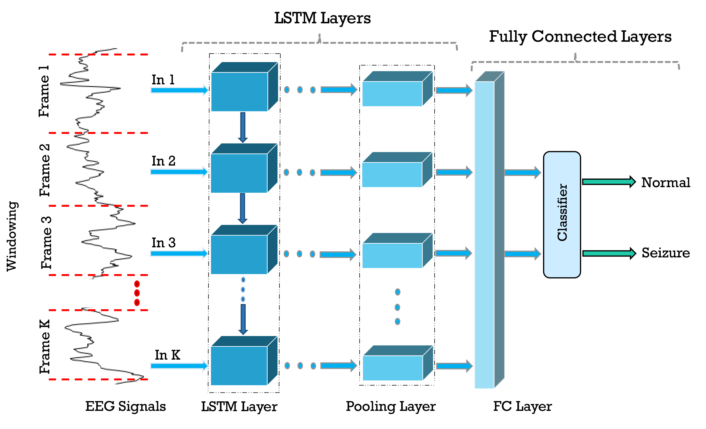
Long Short-Term Memory (LSTM)
The main problem of a simple recurrent neural network is short-term memory. RNN may leave out key information as it has a hard time transporting information from earlier time steps to the next steps in long sequence. Another drawback of RNN is the vanishing gradient problem [16, 17, 18, 19]. The problem arises because of the shrinking of gradients as it back-propagates. To solve the short-term memory problem, LSTM gates were created [16]. The flow of information can be regulated through gates. The gates can preserve the long sequence of necessary data, and throw away the undesired ones. The building block of LSTM is the cell state and its gates.
In this section, Golmohammadi et al. [50] evaluated two LSTM architectures with 3 and 4 layers together with the softmax classifier in their investigation and fetched up satisfactory results. In [74], a 3-layer LSTM deep network is used for feature extraction and classification. The last layer of this network is a sigmoid classification algorithm, and they achieved 96.82% accuracy. According to directed experiments in [80], they employed two LSTM and GRU architectures. The LSTM, GRU model architecture, comprised a layer of Reshape, four layers of LSTM / GRU with the activator, and one layer of Fully Connected (FC) with sigmoid activator. In another study, Yao et al. [81] practiced ten different and ameliorated Independently Recurrent Neural Network (IndRNN) architectures and achieved the best accuracy using Dense IndRNN with attention (DIndRNN) with 31 layers.
Gated Recurrent Unit (GRU)
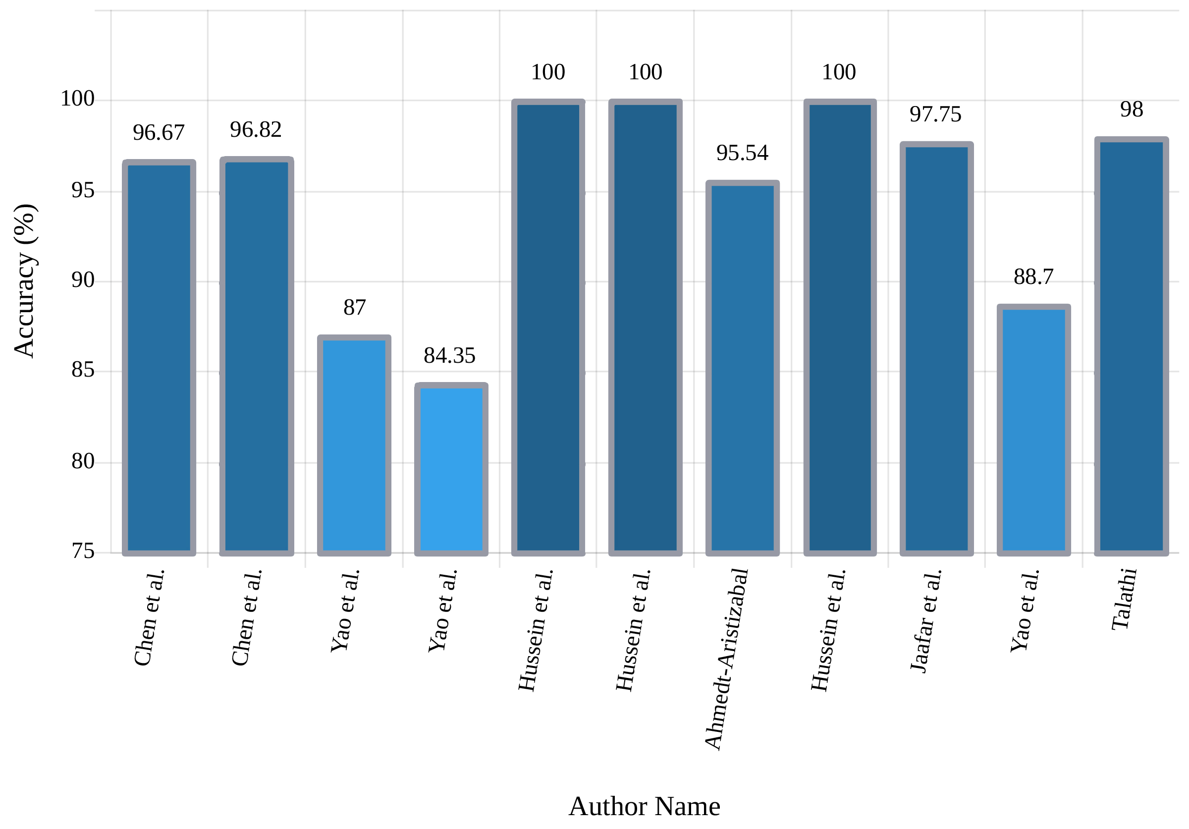
One variation of LSTM is GRU, which combines the input and forgets gates into one update gate [16, 17, 18, 19]. It merges the input and forgets gates and also makes some other modifications. The gating signals are decreased to two. One is the reset gate, and another is the updating gate. These two gates decide which information is necessary to pass to the output. In one experiment, Chen et al. [74] used a 3-layer GRU network with sigmoid classification and yielded 96.67% accuracy. A new GRU-based epileptic seizure detection system has been conducted by Talathi et al. [82]. In the proposed technique, in the pre-processing step, the input signals were split into time windows and calculated from each spectrogram window and then applied to a 4-layer GRU network with a softmax FC layer in the classification stage and achieved 98% accuracy. In another study, Roy et al. [83] employed a 5-layer GRU network and softmax classifier, achieved remarkable results. Table IV provides the summary of related works done using RNNs. Figure 10 shows the sketch of accuracy (%) obtained by various authors using RNN models for seizure detection.
| Work | Networks | Number of Layers | Classifier | Accuracy% |
| [50] | LSTM | 3 | sigmoid | NA |
| 4 | ||||
| [74] | GRU | 3 | sigmoid | 96.67 |
| [74] | LSTM | 3 | sigmoid | 96.82 |
| [75] | 15-IndRNN | 48 | NA | 87.00 |
| [75] | LSTM | 4 | NA | 84.35 |
| [80] | LSTM | 6 | sigmoid | NA |
| GRU | ||||
| [84] | RNN | NA | MLP with 2 layers (logistic sigmoid Classifier) | NA |
| [85] | LSTM | 4 | softmax | 100 |
| [86] | LSTM | 2 | sigmoid | 95.54 Validation |
| 5 | 91.25 Test | |||
| [87] | LSTM | 4 | softmax | 100 |
| [88] | LSTM | 3 | softmax | 97.75 |
| [81] | ADIndRNN-(3,3) | 31 | NA | 88.70 |
| [82] | GRU | 4 | LR | 98.00 |
| [83] | GRU | 5 | softmax | NA |
| [89] | LSTM | 4 | softmax | 100 |
II-C3 Autoencoders
Standard Autoencoders
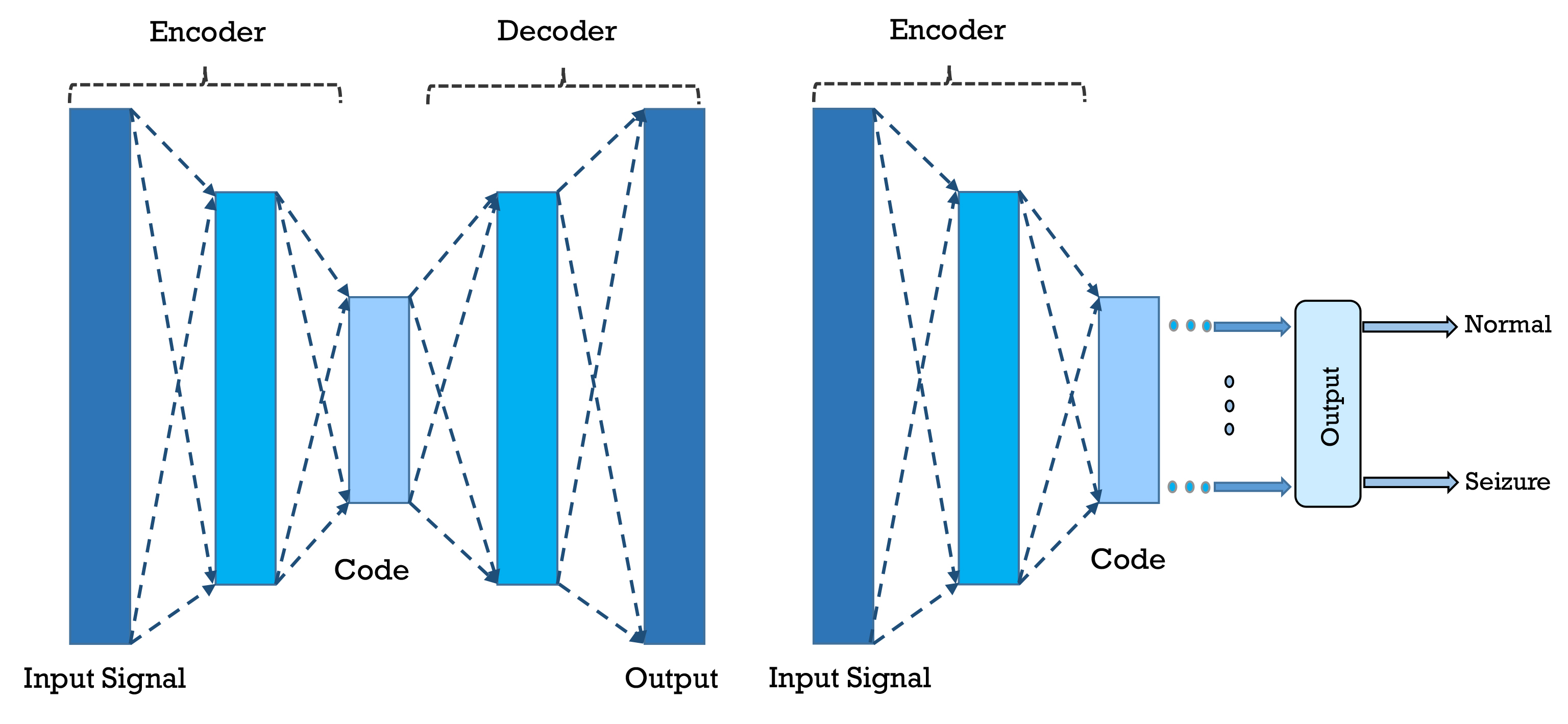
AE is an unsupervised neural network machine learning model for which the input is the same as output [16, 17, 18, 19]. Input is compressed to a latent-space representation, and then the output is obtained from the representation. So, in AE, the compression and decompression functions are coupled with the neural network. AE consists of three parts, i.e., encoder, code, and decoder. Autoencoder networks are the most commonly used for feature extraction or dimensionality reduction in brain signal processing. Figure 11 shows a general form of an AE used for epileptic seizure detection. As the first investigation in this section, Rajaguru et al. [90] separately surveyed the Multilayer Autoencoders (MAE) and Expectation-Maximization with Principal Component Analysis (EM-PCA) methods to diminish the representation dimensions and then employed the Genetic Algorithm (GA) for classification. Experiments indicated that the average classification accuracy of 93.78% is obtained when MAEs were applied for dimensionality reduction and combined with GA as classification. In another research, it was proposed to design an automated system based on AEs for the diagnosis of epilepsy using the EEG signal [91]. First, Harmonic Wavelet Packet Transform (HWPT) was used to decompose the signal to frequency sub-bands, and then fractal features, including Box-Counting (BC), Multi-Resolution BC (MRBC) and Katz Fractal Dimension (KFD) were extracted from each of the sub-bands.
Other Types of Autoencoders
To create a more robust representation, a number of scheme have been applied to autoencoders [26], such as Denoising AE (DAE) (which tries to recreate input from a corrupted form of it) [26], Stacked AE (SAE) (stacking few autoencoders on top of each other to go deeper) [26], and Sparse AutoEncoders(SpAE) (which attempts to harness from sparse representations) [26]. While these methods might pursue other objectives as well, for example, the DAE can be used to recover the corrupted input. However, the main goal in epileptic seizure detection systems is to prevent the hidden layer of autoencoder from merely learning the identity and finding more robust representation of the input. It is also common to combine two or more of these structures in an effort to get the best performance from them.
Works in this section begin with Golmohammadi et al. [50], who presented various deep networks, one of which is Stacked Denoising AE (SDAE). Their advised architecture in this section consists of three layers, and the final results demonstrate the proper performance of their approach. Qiu et al. [92] exerted the windowed signal and z-score normalization step of pre-processing EEG signals and imported pre-processed data into the Denoising Sparse AE (DSpAE) network. In their experiment, they achieved an outstanding performance of 100% accuracy. In [93], a high-performance automated EEG analysis system based on principles of machine learning and big data is presented, which consists of several parts. At first, the signal features are extracted by Linear Predictive Cepstral Coefficients (LPCC) coefficients, then three paths are applied for precise detection. The first pass is sequential decoding using Hidden Markov Models (HMM), the second pass is composed of both temporal and spatial context analysis based on deep learning, in the third pass, a probabilistic grammar is employed.
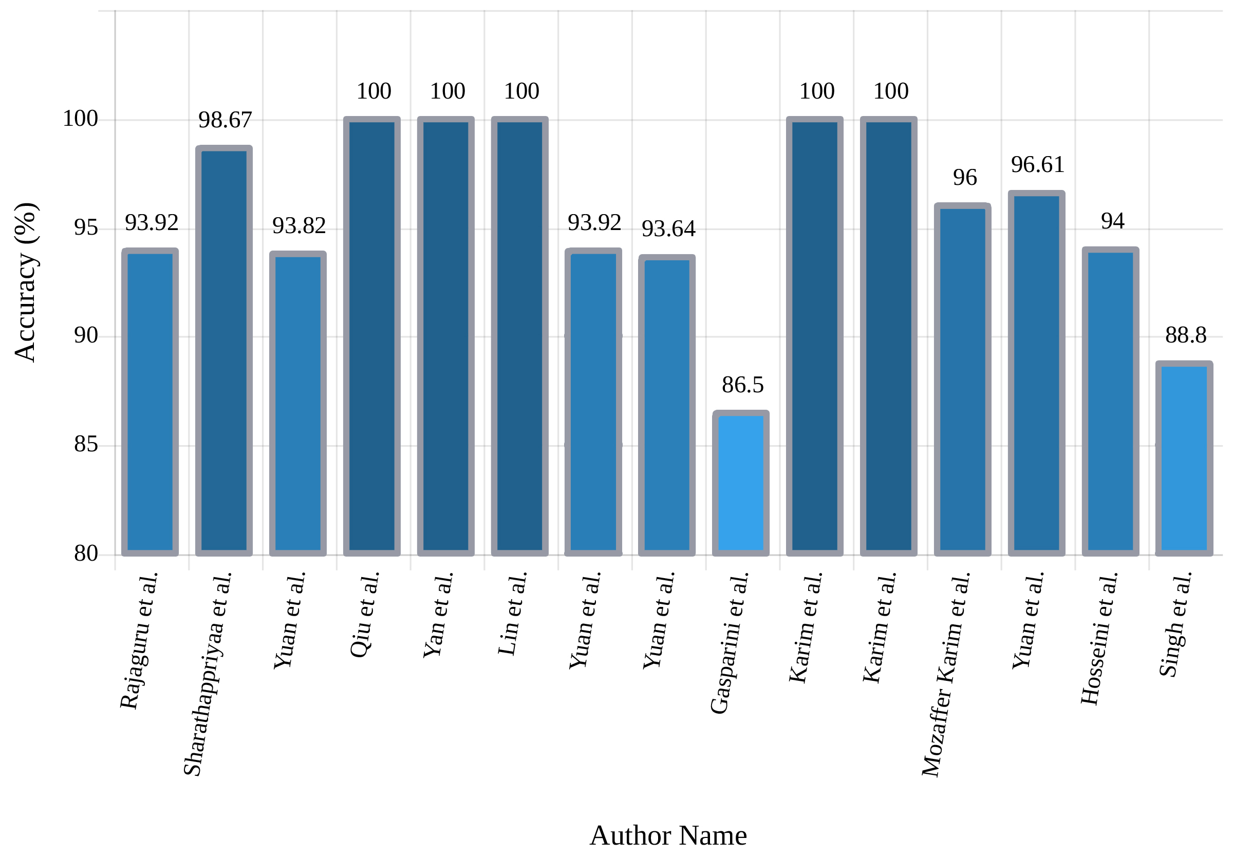
In another research, Yan et al. [94] proposed a feature extraction and classification method based on SpAE and Support Vector Machine (SVM). In this approach, first, the feature extraction of the input EEG signals is performed using SAE and, finally, the classification by SVM. Another SAE architecture was proposed by Yuan et al. [95], which is named Wave2Vec. In the pre-processing stage, the signals were first framed, and in the deep network segment, the SAE with softmax was applied and achieved 93.92% accuracy. Following the experiments of Yuan et al., in [96], different Stacked Sparse Denoising AE (SSpDAE) architectures have been tested and compared. In this work, feature extraction is accomplished by the SSpDAE network and finally classification by softmax. They obtained an accuracy of 93.64%. Table V provides the summary of related works done using AEs. Also, Figure 12 shows the comparison of the accuracies obtained by each of these investigations.
| Work | Networks | Number of Layers | Classifier | Accuracy% |
|---|---|---|---|---|
| [50] | SDAE | 3 | NA | NA |
| [90] | MAE | NA | GA | 93.92 |
| [91] | AE | 3 | softmax | 98.67 |
| [97] | AE | One layer | sigmoid | NA |
| [98] | SSpDAE | 2 hidden layers (intra channel) & 3 hidden layer (cross channel) + 2 hidden layer (FC)+ classifier | softmax | 93.82 |
| [92] | DSpAE | 3 | LR | 100 |
| [93] | SPSW-SDA | Each model has 3 hidden layers | LR | NA |
| 6W-SDA | ||||
| EYEM-SDA | ||||
| [94] | SpAE | single-layer SpAE | SVM | 100 |
| [99] | SSpAE | 3-hidden-layer SSpAE | softmax | 100 |
| [95] | Wave2Vec | NA | softmax | 93.92 |
| [95] | SSpDAE | 2 | softmax | 93.64 |
| [100] | SAE | 3 | softmax | 86.50 |
| [101] | SSpAE | 3 | softmax | 100 |
| [102] | Deep SpAE | 3 | softmax | 100 |
| [103] | SAE | 3 (2 AE+ classifier) | softmax | 96.00 |
| [96] | SAE | 3 | softmax | 96.61 |
| [104] | SSpAE | 3 (two sparse encoders as hidden layers+ classifier) | softmax | 94.00 |
| [105] | SAE | 3 | softmax | 88.80 |
II-C4 Deep Belief and Boltzmann Networks
Restricted Boltzmann Machines (RBM) is a variant of Deep Boltzmann Machines (DBM) and an undirected graphical model [16]. The unrestricted boltzmann machines may also have connections between the hidden units. Stacking the RBMs forms a DBN; RBM is the building block of DBN. DBNs are unsupervised probabilistic hybrid generative deep learning models comprising of latent and stochastic variables in multiple layers [16, 17]. Furthermore, a variation of DBN is called Convolutional DBN (CDBN), which could successfully scale the high dimensional model and uses the spatial information of the nearby pixels [16, 17]. Deep boltzmann machines are probabilistic, generative, unsupervised deep learning model which contains visible and multiple layers of hidden units [16, 17].
Xuyen et al. [106] used DBN to identify epileptic spikes in EEG data. The proposed architecture in their study consisted of three hidden layers and achieved an accuracy of 96.87%. In another study, Turner et al. [107] applied the DBN network to diagnose epilepsy and found promising results. More information about DBN architecture for epileptic seizures is shown in table VI.
II-C5 CNN - RNN
It is a highly efficient combination of deep learning networks to predict and diagnose epileptic seizures from EEG signals is the CNN-RNN architecture. Adding convolutional layers to RNN helps to find spatially nearby patterns more natural; meanwhile, RNN characteristic is more fitted for time-series processing. Starting with [50] as the first work in this section, they applied numerous pre-processing schemas; then, a modified CNN-LSTM architecture is proposed that consists of 13 layers and the sigmoid is used for the last layer. Finally, The proposed approach demonstrates high capability and performs competently.
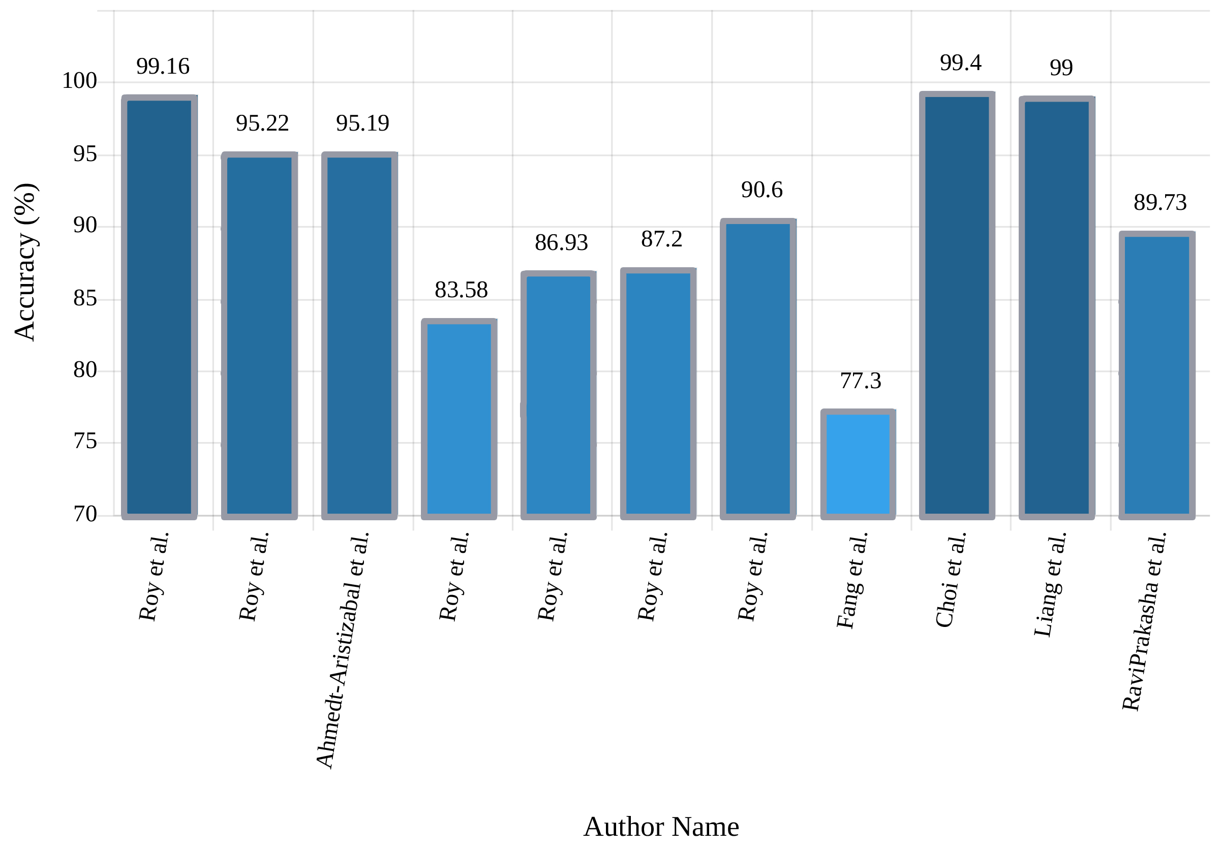
Roy et al. [51] used different CNN-RNN hybrid architectures to improve the experimental results. Their first network comprised a one-dimensional 7-layer CNN-GRU convolution architecture, and the second one is a three-dimensional (3D) CNN-GRU network. In another work, Roy et al. [83] concentrated on natural and abnormal brain activities and suggested four different deep learning architectures. The proposed ChronoNet model was developed using previous models. It achieved 90.60% and 86.57% training and test accuracies respectively.
Fang et al. [108] used the Inception-V3 network. At the outset, a preliminary training was used on this network. Then, to fine-tune this architecture, an RNN based network called Spatial Temporal GRU (ST-GRU) CNN was applied, and they demonstrated that their approach achieved 77.30% accuracy. Choi et al. [109] proposed a Multi-scale 3D-CNN with an RNN model for the detection of epileptic seizures. The CNN module output is applied as the input of the RNN module. The RNN module consists of a unilateral GRU layer that extracts the temporal feature of epileptic seizures and is finally classified using an FC layer. At the end of this section, generalized information from the CNN-RNN research is presented in Table VII and Figure 13, respectively.
| Work | Networks | Number of Layers | Classifier | Accuracy% |
| [50] | 2D-CNN biLSTM | 13 | sigmoid | NA |
| [51] | 1D CNN-GRU | 7 | softmax | 99.16 |
| [51] | TCNN-RNN | 10 | softmax | 95.22 |
| [42] | 2D CNN-LSTM | VGG-16 | sigmoid | 95.19 |
| [83] | C-RNN | 8 | softmax | 83.58 |
| [83] | IC-RNN | 14 | softmax | 86.93 |
| [83] | C-DRNN | 8 | softmax | 87.20 |
| [83] | ChronoNet | 14 | softmax | 90.60 |
| [110] | 2D CNN-LSTM | 8 | NA | NA |
| [108] | ST-GRU ConvNets | pre-trained Inception V3+ GRU + FC | NA | 77.30 |
| [109] | 3D-CNN biGRU | NA | NA | 99.40 |
| [111] | 2D CNN-LSTM | 18 | softmax | 99.00 |
| [112] | 1D CNN-LSTM | 7 | sigmoid | 89.73 |
| 8 |
II-C6 CNN - AEs
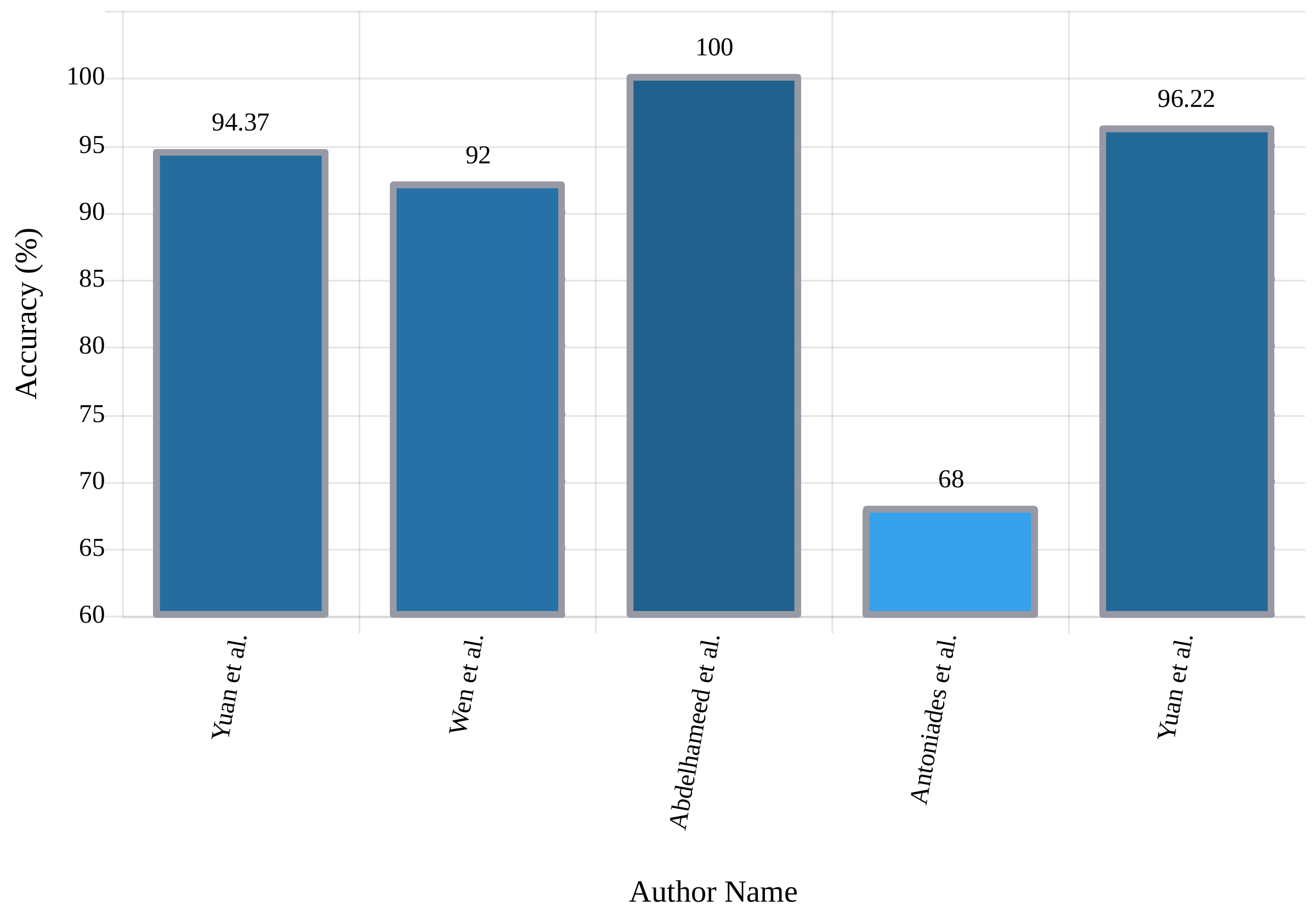
In addition to finding nearby patterns, convolutional layers can reduce the number of parameters in structures such as autoencoders. These two reasons make their combination suitable for many tasks like unsupervised feature extraction for epileptic seizure detection. In this section, a novel approach based on CNN-AE was presented by Yuan et al. [113]. At the feature extraction stage, two deep AE and 2D-CNN were used to extract the supervised and unsupervised features respectively. The unsupervised features were obtained directly from the input signals, and the supervised features were acquired from the spectrogram of the signals. Finally, the softmax classifier was utilized for classification and achieved 94.37% accuracy. In another investigation, Yuan et al. [114] proposed an approach called Deep Fusional Attention Network (DFAN) which can extract channel-aware representations from multi-channel EEG signals. They developed a fusional attention layer which utilized a fusional gate to fully integrate multi-view information to quantify the contribution of each biomedical channel dynamically. A multi-view convolution encoding layer, in combination with CNN, has also been used to train the integrated deep learning model. Table VIII provides the general information about the studies reviewed in the field of CNN-AE, and Figure 14 shows the accuracy of each research.
| Work | Networks | Number of Layers | Classifier | Accuracy% |
| [113] | CNN-AE | 10 | softmax | 94.37 |
| [115] | CNN-AE | 15 | Different Classifiers | 92.00 |
| [116] | 1D CNN-AE (feature extraction)+ MLP/LSTM/Bi LSTM (classification) | 16 + 3/1/1 | sigmoid | 100 2 classes |
| softmax | 99.33 3 classes | |||
| [117] | CNN-ASAE | 8 | LR | 66.00 |
| CNN-AAE | 7 | 68.00 | ||
| [114] | CNN-AE | NA | softmax | 96.22 |
III Non-EEG Based Epileptic Seizure Detection
III-A Medical Imaging Methods
Various deep learning models were developed to detect epileptic seizure using MRI, structural MRI (sMRI), functional MRI (fMRI), resting-state fMRI (rs-fMRI) and PET scans with or without EEG signals [118, 119, 120, 121, 122, 123, 124, 125]. These models outperformed the conventional models in terms of automatic detection and monitoring of the disease. However, due to the nature and difficulties of using imaging methods, these models are mostly practiced for localization of seizure and detection is not the main aim of these models.
The authors [118] proposed automatic localization and detection of Focal Cortical Dysplasia (FCD) from the MRI scan using a CNN. The FCD detection rate is only 50% despite the progress in the analytics of MRI scans. Gill et al. [119] proposed a CNN based algorithm with feature learning capability to detect FCD automatically. The researchers [120] designed DeepIED based on deep learning and EEG-fMRI scans for epilepsy patients, combining the general linear model with EEG-fMRI techniques to estimate the epileptogenic zone. Hosseini et al. [121] proposed an edge computing autonomic framework for evaluation, regulation, and monitoring of the epileptic brain. The epileptogenic network was estimated using rs-fMRI and EEG. Shiri et al. [125] presented a technique for direct attenuation correction of PET images applying emission data only via a CNN-AE. Nineteen radiomic features from 83 brain regions were evaluated for image quantification via Hammersmith atlas. At the end of this section, generalized information from the MRI researches for seizures epileptic detection is presented in Table IX.
| Work | Networks | Number of Layers | Classifier | Accuracy% |
| [118] | 2D-CNN | 30 | sigmoid | 82.50 |
| [119] | 2D-CNN | 11 | softmax | NA |
| [120] | ResNet | 31 | softmax | NA |
| Triplet | ||||
| [121] | 2D-CNN | NA | SVM | NA |
| [122] | 2D-CNN | 11 | softmax | 89.80 |
| 3D-CNN | 82.50 | |||
| [123] | 2D-CNN | NA | NA | NA |
| [124] | ResNet | 14 | sigmoid | 98.22 |
| VGGNet | ||||
| Inception-V3 | ||||
| SVGG-C3D | ||||
| [125] | Deep Direct Attenuation Correction (Deep-DAC) | 44 | tanh | NA |
III-B Other Detection Methods
Ravi Prakash et al. [112] introduced an algorithm based on deep learning for ECoG based Functional Mapping (ECoG-FM) for eloquent language cortex identification. However, the success rate of ECoG-FM is low as compared to Electro-cortical Stimulation Mapping (ESM). In another work, Rosas-Romero et al. [126] have used fNIRS to detect epileptic seizure and obtained better performance than using conventional EEG signals.
IV Hardware And Software Used For The Epileptic Seizure Detection
Fortunately, the privileged performance of deep learning algorithms has made them beneficial for commercial products. Nowadays various commercial products have been developed in the field of deep learning, one of which is deep learning applications and hardware for diagnosing epileptic seizures. In the first study investigated, the brain-computer interface (BCI) system was developed using an AE for epileptic seizure detection by Hosseini et al. [104]. In another study, Singh et al. [105] indicated a utilitarian product for the diagnosis of epileptic seizures, which comprised the user segment and the cloud segment. The block diagram of the proposed system presented by Singh et al. is shown in figure 15.
Kiral-Kornek et al. [127] demonstrated that deep learning in combination with neuromorphic hardware could help in developing a wearable, real-time, always-on, patient-specific seizure warning system with low power consumption and reliable long-term performance.
V Discussion
Anticipating and timely recognition of epileptic seizures is of the essence, as it directly influences the quality of life of patients with this disease and can enhance their confidence in all life’s stages. Numerous research has been fulfilled so far, concentrating on diagnosis epileptic seizures utilizing computer programs, but no efficient software programs or functional hardware have yet been implemented to recognize the disease; nevertheless, without an easy-to-use graphical interface, these studies might never be used in the real world. Until a few years ago, all the experimentation in this domain entailed traditional techniques, and due to practically the various deficiencies, they were not highly prosperous in assisting patients. As analyzed in this study, recent years of research into the diagnosis of epileptic seizures have led to the appearance of deep learning algorithms, and experts in the areas of artificial intelligence and signal processing reassure that these methods can sketch and lead to implementing concrete and functional tools. Table LABEL:tablelast in the Appendix yields more details of the research reviewed. It also shows what type of dataset, implementation tool, preprocessing, deep learning network, and evaluation method are utilized in the research.
As shown in this study, various deep learning structures are applied for epileptic seizure detection, yet none of them has total superiority over others, and the best structure should be chosen carefully based on the dataset and problem characteristics, such as the need for real-time detection or minimum acceptable accuracy. As shown earlier, the obtained accuracies for detecting epileptic seizures are fair, and works in this field need to concentrate more on using clinical datasets and predicting epileptic seizures.
VI Challenges
Currently, challenges holding researchers back can be summarized as follows; firstly, many datasets only contain selected segments of EEG signals, which is not suitable for real-world applications where detection must be done from real-time signals, and clinical datasets are usually not available publicly. Secondly, while the number of datasets in this field is quite a lot, yet they can not be combined easily due to different frequency sampling or other parameters, making the size of total usable data for a model to train small. Lastly, deep learning models require massive computational resources, and these resources are not accessible to everyone. The researchers need to work in this direction to interfere with the epileptic seizures with better models, helping the patients at any time, anywhere.
VII Conclusion and Future Works
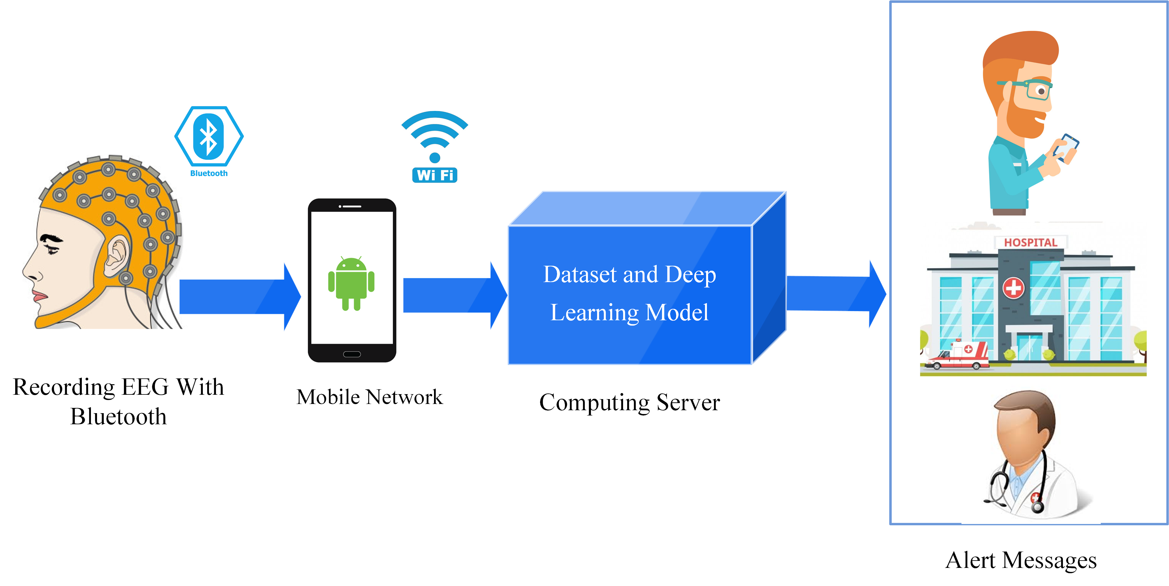
In this paper, a comprehensive review of researches in the field of epileptic seizure detection using numerous deep learning techniques such as CNNs, RNNs, and AEs is presented. Works studied in this research have used various screening methods, most importantly EEG and MRI. Finally, we have investigated deep learning based practical and applied hardware for diagnosing epileptic seizures. It is very encouraging that much of the future research will concentrate on hardware - practical applications to aid this kind of disease. The functional hardware has also been utilized to boost the performance of detection strategies. Furthermore, the models can be placed in the cloud by hospitals, so handheld applications, mobile or wearable devices, may be equipped with such models and the computations will be performed by cloud servers. The patients may also be benefited from predictive models for the epileptic seizure and take some measures to avert in a timely manner. Alert messages may be generated to the family, relatives, the concerned hospital, and doctor in the detection of epileptic seizures through the handheld devices or wearables, and thus the patient can be provided with proper treatment in time. Moreover, a cap with EEG electrodes in it can obtain the EEG signals and sent to the model kept in the cloud, so it can achieve real-time detection. Additionally, if we can detect interictal periods (early stage of seizure), early-stage immediately, the patient can take medication immediately and prevent seizure. Finally, this field of research requires more research by combining different screening methods for more precise and fast detection of epileptic seizures and also applying semi-supervised and unsupervised methods to further overcome the dataset size limits.
Appendix A
Table LABEL:tablelast shows details about all the works reviewed in this study.
| Work | Dataset | Tools | Preprocessing | Network | K-fold | Classifier | Accuracy% |
| [46] | Clinical | NA | Spectrogram | 2D-CNN | NA | LR | 87.51 |
| [47] | Clinical | MATLAB | Normalization | 2D-CNN | NA | softmax | NA |
| [48] | Clinical | NA | Filtering | 1D-CNN with 2D-CNN | NA | sigmoid | 90.50 |
| CHB-MIT | 85.60 | ||||||
| [49] | Clinical | Octave | Filtering, Re-referenced, Down Sampling | 2D-CNN | NA | softmax | NA |
| Keras | |||||||
| Theano | |||||||
| [50] | TUH EEG | NA | Filtering | CNN-RNN | NA | Different activation functions | NA |
| Clinical | |||||||
| [51] | TUH EEG | NA | Different methods | 1D-CNN-GRU | NA | softmax | 99.16 |
| [32] | Clinical | Keras | Down sampling, Z- normalization, augmentation | SeizNet | NA | NA | NA |
| [52] | Clinical | Python 3.6 | Z-Score normalization, STFT | 1D-CNN | NA | softmax | NA |
| PyTorch | 2D-CNN | ||||||
| [33] | CHB-MIT | PyTorch | Visualization | 2D-CNN | NA | softmax | 98.05 |
| [34] | Clinical | NA | Filtering, Visualization, Normalization | 2D-CNN | 10 | softmax | NA |
| [35] | TUH EEG | PyTorch | DivSpec | SeizureNet | 5 | softmax | NA |
| [42] | Clinical | Caffe | Different Methods | FRCNN with 2D-CNN | 5 | SVM | 95.19 |
| OpenCV | |||||||
| Keras | FRCNN with 2D-CNN-LSTM | sigmoid | |||||
| Theano | |||||||
| [53] | TUH EEG | TensorFlow | Feature Extraction | 2D-CNN | 10 | softmax | 74.00 |
| [54] | Bern Barcelona | Octave | Filtering, EMD, DWT, Fourier | 2D-CNN | 5 | sigmoid | 99.50 |
| Clinical | Keras | softmax | |||||
| [55] | Bern Barcelona | TensorFlow | STFT, Z-Score Normalization | 2D-CNN | 10 | softmax | 91.80 |
| [36] | Clinical | NA | STFT | TGCN | NA | sigmoid | NA |
| [56] | Bonn | NA | DWT | 2D-CNN | 10 | softmax | 100 |
| [57] | Bonn | Keras | CWT | 2D-CNN | 10 | softmax | 100 |
| [58] | Bonn | MATLAB | Filtering | 2D-CNN | NA | softmax | 99.60 |
| 90.10 | |||||||
| [59] | CHB-MIT | MATLAB | Over Sampling Method, FFT, WPD | 2D-CNN | 5 | MV-TSK-FS | 98.35 |
| TensorFlow | 3D-CNN | ||||||
| [37] | CHB-MIT | NA | Spatial Representation | 2D-CNN | NA | softmax | 99.48 |
| [60] | Clinical | MATLAB | Different Methods | 2D-CNN | 10 | sigmoid | NA |
| RF | |||||||
| [61] | CHB-MIT | NA | MAS | 2D-CNN | 5 | KELM | 99.33 |
| Clinical | |||||||
| [62] | Clinical | TensorFlow | Filtering, Down Sampling | 1D-CNN | 4 | softmax, SVM | 83.86 |
| [39] | Bern Barcelona | Caffe | NA | AlexNet | NA | softmax | 100 |
| GoogleNet | |||||||
| LeNet | |||||||
| [40] | UCI | PyTorch | Signal2Image | 2D one Layer CNN | NA | DenseNet | 85.30 |
| [43] | Clinical | Chainer | Filtering, Visualization | 2D-CNN | NA | softmax | NA |
| [68] | Bonn | TensorFlow | Data Augmentation | P-1D-CNN | 10 | Majority Voting | 99.10 |
| [69] | Bonn | MATLAB | Z-Score Normalization | 1D-CNN | 10 | softmax | 86.67 |
| [70] | CHB-MIT | NA | Filtering, Augmentation | MPCNN | NA | softmax | NA |
| [71] | Clinical | Keras | Down-Sampling, Filtering | 1D-FCNN | 5 | softmax | NA |
| [73] | TUH EEG | Keras | Normalization and Standardization | 1D-CNN | NA | softmax | 79.34 |
| [72] | Clinical | Theano | Filtering | 1D-CNN | NA | Binary LR | NA |
| Lasagne Library | |||||||
| [63] | CHB-MIT | NA | DWT, Feature Extraction, Normalization | 1D-CNN | 10 | NA | 99.07 |
| [74] | Bonn | NA | DWT, Normalization | 1D-CNN | 5 | sigmoid | 97.27 |
| [67] | Bonn | NA | Normalization | 1D-TCNN | NA | NA | 100 |
| [64] | Bonn | NA | EMD, MPF | 1D-CNN | 10 | softmax | 98.60 |
| [75] | CHB-MIT | NA | Windowing | IndRNN | 10 | NA | 87.00 |
| [76] | Bonn | TensorFlow | Filtering, Z-Score Normalization | 1D-CNN | NA | softmax | 99.00 |
| Bern Barcelona | 91.80 | ||||||
| [65] | CHB-MIT | PyTorch | Filtering | 1D-PCM-CNN | 5 | softmax | NA |
| Clinical | |||||||
| [77] | CHB-MIT | NA | MIDS, WGANs | 1D-CNN | NA | softmax | 84.00 |
| [78] | Clinical | NA | Down Sampling, PSD, FFT | 1D-CNN | 4 | sigmoid | 86.29 |
| [79] | CHB-MIT | TensorFlow | Filtering | 1D-CNN | 4 | softmax | NA |
| [66] | NA | Keras | Down Sampling, Filtering, Data Augmentation | CNN-BP | 5 | sigmoid | NA |
| TensorFlow | |||||||
| MATLAB | |||||||
| [80] | Clinical | NA | Filtering, DWT | 1D-CNN | NA | sigmoid | NA |
| LSTM | RF | ||||||
| GRU | SVM | ||||||
| [84] | CHB-MIT | MATLAB | Filtering, Montage Mapping | DRNN | NA | MLP | NA |
| [89] | Bonn | NA | Filtering | LSTM | NA | softmax | 100 |
| [85] | Bonn | MATLAB | Filtering | LSTM | 3 | softmax | 100 |
| Keras | 5 | ||||||
| TensorFlow | 10 | ||||||
| [86] | Bonn | Keras | Windowing | LSTM | 10 | sigmoid | 91.25 |
| [87] | Bonn | MATLAB | Filtering | LSTM | 3 | softmax | 100 |
| Keras | 5 | ||||||
| TensorFlow | 10 | ||||||
| [88] | Freiburg | Anaconda Navigator | Normalization, Filtering | LSTM | 5 | softmax | 97.75 |
| [81] | CHB-MIT | NA | Windowing | ADIndRNN | 10 | NA | 88.70 |
| Bonn | |||||||
| [82] | Bonn | Keras | Auto-Correlation | GRU | NA | LR | 98 |
| [83] | TUH EEG | NA | TCP | ChronoNet | NA | softmax | 90.60 |
| [90] | Clinical | NA | Windowing | AE with EM-PCA | NA | GA | 93.92 |
| [91] | Bonn | MATLAB | Filtering, HWPT, FD | AE | NA | softmax | 98.67 |
| [97] | Clinical | TensorFlow | Down Sampling, Filtering, Normalization | AE | NA | sigmoid | NA |
| [98] | CHB-MIT | NA | STFT | SSDA | NA | softmax | 93.82 |
| [92] | Bonn | MATLAB | Z-Score Normalization, Standardization | DSAE | NA | LR | 100 |
| [93] | TUH EEG | Open Source Toolkits | Different Methods | SDA | NA | LR | NA |
| Theano | |||||||
| [94] | Bonn | NA | Filtering | SAE | NA | SVM | 100 |
| [99] | Bonn | NA | Normalization | SSAE | NA | softmax | 100 |
| [95] | CHB-MIT | Theano | Scalogram | Wave2Vec | NA | softmax | 93.92 |
| [113] | CHB-MIT | PyTorch | Data Augmentation, STFT | CNN-AE | 5 | softmax | 94.37 |
| [100] | Clinical | NA | Filtering, CWT, Feature Extraction | SAE | NA | softmax | 86.50 |
| [101] | Bonn | NA | Taguchi Method | SSAE | NA | softmax | 100 |
| [102] | Clinical | NA | Dimension reduction, ESD | DeSAE | NA | softmax | 100 |
| [103] | Bonn | NA | DWT | SAE | NA | softmax | 96.00 |
| [96] | CHB-MIT | NA | Different Methods | mSSDA | NA | softmax | 96.61 |
| [104] | Clinical | MATLAB | PCA, I-ICA | SSAE | NA | softmax | 94 |
| [105] | Bonn | MATLAB | Windowing | SAE | NA | softmax | 88.80 |
| [106] | Clinical | MATLAB | DWT | DBN | NA | NA | 96.87 |
| [107] | Clinical | Theano | Normalization, Feature Extraction, Standardization | DBN | NA | LR | NA |
| SVM | |||||||
| KNN | |||||||
| [110] | CHB-MIT | NA | Image Based Representation | 2D CNN-LSTM | NA | NA | NA |
| [108] | Clinical | TensorFlow | Filtering | ST-GRU ConvNets | NA | NA | 77.30 |
| [109] | CHB-MIT | NA | STFT, 2D-mapping | 3D-CNN with Bi GRU | NA | NA | 99.40 |
| Clinical | |||||||
| [111] | CHB-MIT | NA | Visualization | 2D-CNN-LSTM | NA | softmax | 99.00 |
| [112] | clinical ECoG | NA | Filtering | 1D-CNN-LSTM | 5 | sigmoid | 89.73 |
| [115] | CHB-MIT | Scikit-Learn | Channel Selection | CNN-AE | 5 | Different Methods | 92.00 |
| Bonn | 10 | ||||||
| [116] | Bonn | NA | Windowing | 1D-CNN with Bi LSTM | NA | softmax | 99.33 |
| sigmoid | 100 | ||||||
| [117] | Clinical | Theano | Mapping | ASAE-CNN | NA | LR | 66.00 |
| AAE-CNN | 68.00 | ||||||
| [114] | CHB-MIT | PyTorch | STFT | CNN-AE | 5 | softmax | 96.22 |
| [118] | SCTIMST | FSL | Noise reduction with BM3D algorithm, Skull-stripping, Segmentation, Postprocessing | 2D-CNN | 5 | sigmoid | NA |
| Keras | |||||||
| TensorFlow | |||||||
| [119] | Clinical MRI | NA | Different Methods | 2D-CNN | 5 | softmax | NA |
| [120] | Clinical MRI | Brain Vision Analyzer | Filtering, ICA, BCG, GLM, MCS | ResNet | NA | softmax | NA |
| Triplet | |||||||
| [121] | ECoG Dataset | GIFT | Different Methods | 2D-CNN | NA | SVM | NA |
| Rs-FMRI Dataset | FSL | ||||||
| FreeSurfer | |||||||
| [122] | Clinical MRI | NA | Scaling Down | 3D-CNN | 5 | softmax | 89.80 |
| [123] | Clinical MRI | FSL | Connectivity Feature Extracion | 2D-CNN | NA | NA | NA |
| [124] | ImageNet | DPABI | ROI, Normalization, AAL, CNNI, Down-sampling, NNI (3D images) | 2D-ResNet | NA | sigmoid | 98.22 |
| Pulmonary nodules Kaggle | Python | 2D-VGGNET | |||||
| Keras | 2D-Inception V3 | ||||||
| Clinical PET | TensorFlow | 3D-SVGG-C3D | |||||
| [125] | Clinical PET | TensorFlow | OSEM, Data Augmentation Radionics Features | DAC | NA | tanh | NA |
References
- [1] N. Ghassemi, A. Shoeibi, M. Rouhani, and H. Hosseini-Nejad, “Epileptic seizures detection in eeg signals using tqwt and ensemble learning,” in 2019 9th International Conference on Computer and Knowledge Engineering (ICCKE). IEEE, 2019, pp. 403–408.
- [2] V. Gupta, A. Bhattacharyya, and R. B. Pachori, “Classification of seizure and non-seizure eeg signals based on emd-tqwt method,” in 2017 22nd International Conference on Digital Signal Processing (DSP). IEEE, 2017, pp. 1–5.
- [3] A. Bhattacharyya, R. B. Pachori, A. Upadhyay, and U. R. Acharya, “Tunable-q wavelet transform based multiscale entropy measure for automated classification of epileptic eeg signals,” Applied Sciences, vol. 7, no. 4, p. 385, 2017.
- [4] S. Kulaseharan, A. Aminpour, M. Ebrahimi, and E. Widjaja, “Identifying lesions in paediatric epilepsy using morphometric and textural analysis of magnetic resonance images,” NeuroImage: Clinical, vol. 21, p. 101663, 2019.
- [5] G. Zazzaro, S. Cuomo, A. Martone, R. V. Montaquila, G. Toraldo, and L. Pavone, “Eeg signal analysis for epileptic seizures detection by applying data mining techniques,” Internet of Things, p. 100048, 2019.
- [6] N. van Klink, A. Mooij, G. Huiskamp, C. Ferrier, K. Braun, A. Hillebrand, and M. Zijlmans, “Simultaneous meg and eeg to detect ripples in people with focal epilepsy,” Clinical Neurophysiology, vol. 130, no. 7, pp. 1175–1183, 2019.
- [7] N. Pianou and S. Chatziioannou, “Imaging with pet/ct in patients with epilepsy,” in Epilepsy Surgery and Intrinsic Brain Tumor Surgery. Springer, 2019, pp. 45–50.
- [8] A. Subasi, J. Kevric, and M. A. Canbaz, “Epileptic seizure detection using hybrid machine learning methods,” Neural Computing and Applications, vol. 31, no. 1, pp. 317–325, 2019.
- [9] R. Sharma and R. B. Pachori, “Classification of epileptic seizures in eeg signals based on phase space representation of intrinsic mode functions,” Expert Systems with Applications, vol. 42, no. 3, pp. 1106–1117, 2015.
- [10] E. Alickovic, J. Kevric, and A. Subasi, “Performance evaluation of empirical mode decomposition, discrete wavelet transform, and wavelet packed decomposition for automated epileptic seizure detection and prediction,” Biomedical signal processing and control, vol. 39, pp. 94–102, 2018.
- [11] M. Sharma, A. A. Bhurane, and U. R. Acharya, “Mmsfl-owfb: a novel class of orthogonal wavelet filters for epileptic seizure detection,” Knowledge-Based Systems, vol. 160, pp. 265–277, 2018.
- [12] M. Sharma, R. B. Pachori, and U. R. Acharya, “A new approach to characterize epileptic seizures using analytic time-frequency flexible wavelet transform and fractal dimension,” Pattern Recognition Letters, vol. 94, pp. 172–179, 2017.
- [13] E. B. Assi, D. K. Nguyen, S. Rihana, and M. Sawan, “Towards accurate prediction of epileptic seizures: A review,” Biomedical Signal Processing and Control, vol. 34, pp. 144–157, 2017.
- [14] U. R. Acharya, Y. Hagiwara, and H. Adeli, “Automated seizure prediction,” Epilepsy & Behavior, vol. 88, pp. 251–261, 2018.
- [15] U. R. Acharya, Y. Hagiwara, S. N. Deshpande, S. Suren, J. E. W. Koh, S. L. Oh, N. Arunkumar, E. J. Ciaccio, and C. M. Lim, “Characterization of focal eeg signals: a review,” Future Generation Computer Systems, vol. 91, pp. 290–299, 2019.
- [16] Y. LeCun, Y. Bengio, and G. Hinton, “Deep learning,” nature, vol. 521, no. 7553, pp. 436–444, 2015.
- [17] W. Pedrycz and S.-M. Chen, Development and Analysis of Deep Learning Architectures. Springer, 2020.
- [18] N. Srivastava and R. R. Salakhutdinov, “Multimodal learning with deep boltzmann machines,” in Advances in neural information processing systems, 2012, pp. 2222–2230.
- [19] D. Yu and L. Deng, “Deep learning and its applications to signal and information processing [exploratory dsp],” IEEE Signal Processing Magazine, vol. 28, no. 1, pp. 145–154, 2010.
- [20] M. Ihle, H. Feldwisch-Drentrup, C. A. Teixeira, A. Witon, B. Schelter, J. Timmer, and A. Schulze-Bonhage, “Epilepsiae–a european epilepsy database,” Computer methods and programs in biomedicine, vol. 106, no. 3, pp. 127–138, 2012.
- [21] A. H. Shoeb, “Application of machine learning to epileptic seizure onset detection and treatment,” Ph.D. dissertation, Massachusetts Institute of Technology, 2009.
- [22] A. E. Society. (2015) Seizure prediction challenge. [Online]. Available: https://www.kaggle.com/c/seizure-prediction
- [23] R. G. Andrzejak, K. Lehnertz, F. Mormann, C. Rieke, P. David, and C. E. Elger, “Indications of nonlinear deterministic and finite-dimensional structures in time series of brain electrical activity: Dependence on recording region and brain state,” Physical Review E, vol. 64, no. 6, p. 061907, 2001.
- [24] R. G. Andrzejak, K. Schindler, and C. Rummel, “Nonrandomness, nonlinear dependence, and nonstationarity of electroencephalographic recordings from epilepsy patients,” Physical Review E, vol. 86, no. 4, p. 046206, 2012.
- [25] N. Stevenson, K. Tapani, L. Lauronen, and S. Vanhatalo, “A dataset of neonatal eeg recordings with seizure annotations,” Scientific data, vol. 6, p. 190039, 2019.
- [26] I. Goodfellow, Y. Bengio, and A. Courville, Deep learning. MIT press, 2016.
- [27] O. Faust, Y. Hagiwara, T. J. Hong, O. S. Lih, and U. R. Acharya, “Deep learning for healthcare applications based on physiological signals: A review,” Computer methods and programs in biomedicine, vol. 161, pp. 1–13, 2018.
- [28] S. Pereira, A. Pinto, V. Alves, and C. A. Silva, “Brain tumor segmentation using convolutional neural networks in mri images,” IEEE transactions on medical imaging, vol. 35, no. 5, pp. 1240–1251, 2016.
- [29] N. Ghassemi, A. Shoeibi, and M. Rouhani, “Deep neural network with generative adversarial networks pre-training for brain tumor classification based on mr images,” Biomedical Signal Processing and Control, vol. 57, p. 101678, 2020.
- [30] I. Masi, Y. Wu, T. Hassner, and P. Natarajan, “Deep face recognition: A survey,” in 2018 31st SIBGRAPI conference on graphics, patterns and images (SIBGRAPI). IEEE, 2018, pp. 471–478.
- [31] A. Krizhevsky, I. Sutskever, and G. E. Hinton, “Imagenet classification with deep convolutional neural networks,” in Advances in neural information processing systems, 2012, pp. 1097–1105.
- [32] M. T. Avcu, Z. Zhang, and D. W. S. Chan, “Seizure detection using least eeg channels by deep convolutional neural network,” in ICASSP 2019-2019 IEEE International Conference on Acoustics, Speech and Signal Processing (ICASSP). IEEE, 2019, pp. 1120–1124.
- [33] M. S. Hossain, S. U. Amin, M. Alsulaiman, and G. Muhammad, “Applying deep learning for epilepsy seizure detection and brain mapping visualization,” ACM Transactions on Multimedia Computing, Communications, and Applications (TOMM), vol. 15, no. 1s, pp. 1–17, 2019.
- [34] R. Zuo, J. Wei, X. Li, C. Li, C. Zhao, Z. Ren, Y. Liang, X. Geng, C. Jiang, X. Yang et al., “Automated detection of high frequency oscillations in epilepsy based on a convolutional neural network,” Frontiers in computational neuroscience, vol. 13, p. 6, 2019.
- [35] U. Asif, S. Roy, J. Tang, and S. Harrer, “Seizurenet: a deep convolutional neural network for accurate seizure type classification and seizure detection,” arXiv preprint arXiv:1903.03232, 2019.
- [36] I. Covert, B. Krishnan, I. Najm, J. Zhan, M. Shore, J. Hixson, and M. J. Po, “Temporal graph convolutional networks for automatic seizure detection,” arXiv preprint arXiv:1905.01375, 2019.
- [37] B. Bouaziz, L. Chaari, H. Batatia, and A. Quintero-Rincón, “Epileptic seizure detection using a convolutional neural network,” in Digital Health Approach for Predictive, Preventive, Personalised and Participatory Medicine. Springer, 2019, pp. 79–86.
- [38] J. Deng, W. Dong, R. Socher, L.-J. Li, K. Li, and L. Fei-Fei, “Imagenet: A large-scale hierarchical image database,” in 2009 IEEE conference on computer vision and pattern recognition. Ieee, 2009, pp. 248–255.
- [39] A. M. Taqi, F. Al-Azzo, M. Mariofanna, and J. M. Al-Saadi, “Classification and discrimination of focal and non-focal eeg signals based on deep neural network,” in 2017 International Conference on Current Research in Computer Science and Information Technology (ICCIT). IEEE, 2017, pp. 86–92.
- [40] P. Bizopoulos, G. I. Lambrou, and D. Koutsouris, “Signal2image modules in deep neural networks for eeg classification,” in 2019 41st Annual International Conference of the IEEE Engineering in Medicine and Biology Society (EMBC). IEEE, 2019, pp. 702–705.
- [41] K. Simonyan and A. Zisserman, “Very deep convolutional networks for large-scale image recognition,” arXiv preprint arXiv:1409.1556, 2014.
- [42] D. Ahmedt-Aristizabal, C. Fookes, K. Nguyen, S. Denman, S. Sridharan, and S. Dionisio, “Deep facial analysis: A new phase i epilepsy evaluation using computer vision,” Epilepsy & Behavior, vol. 82, pp. 17–24, 2018.
- [43] A. Emami, N. Kunii, T. Matsuo, T. Shinozaki, K. Kawai, and H. Takahashi, “Seizure detection by convolutional neural network-based analysis of scalp electroencephalography plot images,” NeuroImage: Clinical, vol. 22, p. 101684, 2019.
- [44] C. Szegedy, W. Liu, Y. Jia, P. Sermanet, S. Reed, D. Anguelov, D. Erhan, V. Vanhoucke, and A. Rabinovich, “Going deeper with convolutions,” in Proceedings of the IEEE conference on computer vision and pattern recognition, 2015, pp. 1–9.
- [45] K. He, X. Zhang, S. Ren, and J. Sun, “Deep residual learning for image recognition,” in Proceedings of the IEEE conference on computer vision and pattern recognition, 2016, pp. 770–778.
- [46] A. Antoniades, L. Spyrou, C. C. Took, and S. Sanei, “Deep learning for epileptic intracranial eeg data,” in 2016 IEEE 26th International Workshop on Machine Learning for Signal Processing (MLSP). IEEE, 2016, pp. 1–6.
- [47] F. Achilles, F. Tombari, V. Belagiannis, A. M. Loesch, S. Noachtar, and N. Navab, “Convolutional neural networks for real-time epileptic seizure detection,” Computer Methods in Biomechanics and Biomedical Engineering: Imaging & Visualization, vol. 6, no. 3, pp. 264–269, 2018.
- [48] C. Park, G. Choi, J. Kim, S. Kim, T.-J. Kim, K. Min, K.-Y. Jung, and J. Chong, “Epileptic seizure detection for multi-channel eeg with deep convolutional neural network,” in 2018 International Conference on Electronics, Information, and Communication (ICEIC). IEEE, 2018, pp. 1–5.
- [49] M. C. Tjepkema-Cloostermans, R. C. de Carvalho, and M. J. van Putten, “Deep learning for detection of focal epileptiform discharges from scalp eeg recordings,” Clinical neurophysiology, vol. 129, no. 10, pp. 2191–2196, 2018.
- [50] M. Golmohammadi, S. Ziyabari, V. Shah, S. L. de Diego, I. Obeid, and J. Picone, “Deep architectures for automated seizure detection in scalp eegs,” arXiv preprint arXiv:1712.09776, 2017.
- [51] S. Roy, I. Kiral-Kornek, and S. Harrer, “Deep learning enabled automatic abnormal eeg identification,” in 2018 40th Annual International Conference of the IEEE Engineering in Medicine and Biology Society (EMBC). IEEE, 2018, pp. 2756–2759.
- [52] P. Nejedly, V. Kremen, V. Sladky, M. Nasseri, H. Guragain, P. Klimes, J. Cimbalnik, Y. Varatharajah, B. H. Brinkmann, and G. A. Worrell, “Deep-learning for seizure forecasting in canines with epilepsy,” Journal of neural engineering, vol. 16, no. 3, p. 036031, 2019.
- [53] T. Iesmantas and R. Alzbutas, “Convolutional neural network for detection and classification of seizures in clinical data,” arXiv preprint arXiv:1903.08864, 2019.
- [54] R. San-Segundo, M. Gil-Martín, L. F. D’Haro-Enríquez, and J. M. Pardo, “Classification of epileptic eeg recordings using signal transforms and convolutional neural networks,” Computers in biology and medicine, vol. 109, pp. 148–158, 2019.
- [55] L. Sui, X. Zhao, Q. Zhao, T. Tanaka, and J. Cao, “Localization of epileptic foci by using convolutional neural network based on ieeg,” in IFIP International Conference on Artificial Intelligence Applications and Innovations. Springer, 2019, pp. 331–339.
- [56] R. Akut, “Wavelet based deep learning approach for epilepsy detection,” Health information science and systems, vol. 7, no. 1, p. 8, 2019.
- [57] Ö. Türk and M. S. Özerdem, “Epilepsy detection by using scalogram based convolutional neural network from eeg signals,” Brain sciences, vol. 9, no. 5, p. 115, 2019.
- [58] J. Liu and B. Woodson, “Deep learning classification for epilepsy detection using a single channel electroencephalography (eeg),” in Proceedings of the 2019 3rd International Conference on Deep Learning Technologies, 2019, pp. 23–26.
- [59] X. Tian, Z. Deng, W. Ying, K.-S. Choi, D. Wu, B. Qin, J. Wang, H. Shen, and S. Wang, “Deep multi-view feature learning for eeg-based epileptic seizure detection,” IEEE Transactions on Neural Systems and Rehabilitation Engineering, vol. 27, no. 10, pp. 1962–1972, 2019.
- [60] A. H. Ansari, P. J. Cherian, A. Caicedo, G. Naulaers, M. De Vos, and S. Van Huffel, “Neonatal seizure detection using deep convolutional neural networks,” International journal of neural systems, vol. 29, no. 04, p. 1850011, 2019.
- [61] J. Cao, J. Zhu, W. Hu, and A. Kummert, “Epileptic signal classification with deep eeg features by stacked cnns,” IEEE Transactions on Cognitive and Developmental Systems, 2019.
- [62] J. Thomas, L. Comoretto, J. Jin, J. Dauwels, S. S. Cash, and M. B. Westover, “Eeg classification via convolutional neural network-based interictal epileptiform event detection,” in 2018 40th Annual International Conference of the IEEE Engineering in Medicine and Biology Society (EMBC). IEEE, 2018, pp. 3148–3151.
- [63] P. Boonyakitanont, A. Lek-uthai, K. Chomtho, and J. Songsiri, “A comparison of deep neural networks for seizure detection in eeg signals,” bioRxiv, p. 702654, 2019.
- [64] H. G. Daoud, A. M. Abdelhameed, and M. Bayoumi, “Automatic epileptic seizure detection based on empirical mode decomposition and deep neural network,” in 2018 IEEE 14th International Colloquium on Signal Processing & Its Applications (CSPA). IEEE, 2018, pp. 182–186.
- [65] J. Craley, E. Johnson, and A. Venkataraman, “Integrating convolutional neural networks and probabilistic graphical modeling for epileptic seizure detection in multichannel eeg,” in International Conference on Information Processing in Medical Imaging. Springer, 2019, pp. 291–303.
- [66] M. A. Jaoude, J. Jing, H. Sun, C. S. Jacobs, K. R. Pellerin, M. B. Westover, S. S. Cash, and A. D. Lam, “Detection of mesial temporal lobe epileptiform discharges on intracranial electrodes using deep learning,” Clinical Neurophysiology, vol. 131, no. 1, pp. 133–141, 2020.
- [67] J. Zhang, H. Wu, W. Su, X. Wang, M. Yang, and J. Wu, “A new approach for classification of epilepsy eeg signals based on temporal convolutional neural networks,” in 2018 11th International Symposium on Computational Intelligence and Design (ISCID), vol. 2. IEEE, 2018, pp. 80–84.
- [68] I. Ullah, M. Hussain, H. Aboalsamh et al., “An automated system for epilepsy detection using eeg brain signals based on deep learning approach,” Expert Systems with Applications, vol. 107, pp. 61–71, 2018.
- [69] U. R. Acharya, S. L. Oh, Y. Hagiwara, J. H. Tan, and H. Adeli, “Deep convolutional neural network for the automated detection and diagnosis of seizure using eeg signals,” Computers in biology and medicine, vol. 100, pp. 270–278, 2018.
- [70] A. Page, C. Shea, and T. Mohsenin, “Wearable seizure detection using convolutional neural networks with transfer learning,” in 2016 IEEE International Symposium on Circuits and Systems (ISCAS). IEEE, 2016, pp. 1086–1089.
- [71] A. O’Shea, G. Lightbody, G. Boylan, and A. Temko, “Neonatal seizure detection using convolutional neural networks,” in 2017 IEEE 27th International Workshop on Machine Learning for Signal Processing (MLSP). IEEE, 2017, pp. 1–6.
- [72] A. R. Johansen, J. Jin, T. Maszczyk, J. Dauwels, S. S. Cash, and M. B. Westover, “Epileptiform spike detection via convolutional neural networks,” in 2016 IEEE International Conference on Acoustics, Speech and Signal Processing (ICASSP). IEEE, 2016, pp. 754–758.
- [73] Ö. Yıldırım, U. B. Baloglu, and U. R. Acharya, “A deep convolutional neural network model for automated identification of abnormal eeg signals,” Neural Computing and Applications, pp. 1–12, 2018.
- [74] X. Chen, J. Ji, T. Ji, and P. Li, “Cost-sensitive deep active learning for epileptic seizure detection,” in Proceedings of the 2018 ACM International Conference on Bioinformatics, Computational Biology, and Health Informatics, 2018, pp. 226–235.
- [75] X. Yao, Q. Cheng, and G.-Q. Zhang, “A novel independent rnn approach to classification of seizures against non-seizures,” arXiv preprint arXiv:1903.09326, 2019.
- [76] D. Lu and J. Triesch, “Residual deep convolutional neural network for eeg signal classification in epilepsy,” arXiv preprint arXiv:1903.08100, 2019.
- [77] Z. Wei, J. Zou, J. Zhang, and J. Xu, “Automatic epileptic eeg detection using convolutional neural network with improvements in time-domain,” Biomedical Signal Processing and Control, vol. 53, p. 101551, 2019.
- [78] C. Meisel, R. E. Atrache, M. Jackson, and T. Loddenkemper, “Deep learning from wristband sensor data: towards wearable, non-invasive seizure forecasting,” arXiv preprint arXiv:1906.00511, 2019.
- [79] R. Yuvaraj, J. Thomas, T. Kluge, and J. Dauwels, “A deep learning scheme for automatic seizure detection from long-term scalp eeg,” in 2018 52nd Asilomar Conference on Signals, Systems, and Computers. IEEE, 2018, pp. 368–372.
- [80] K. Fukumori, H. T. T. Nguyen, N. Yoshida, and T. Tanaka, “Fully data-driven convolutional filters with deep learning models for epileptic spike detection,” in ICASSP 2019-2019 IEEE International Conference on Acoustics, Speech and Signal Processing (ICASSP). IEEE, 2019, pp. 2772–2776.
- [81] X. Yao, Q. Cheng, and G.-Q. Zhang, “Automated classification of seizures against nonseizures: A deep learning approach,” arXiv preprint arXiv:1906.02745, 2019.
- [82] S. S. Talathi, “Deep recurrent neural networks for seizure detection and early seizure detection systems,” arXiv preprint arXiv:1706.03283, 2017.
- [83] S. Roy, I. Kiral-Kornek, and S. Harrer, “Chrononet: a deep recurrent neural network for abnormal eeg identification,” in Conference on Artificial Intelligence in Medicine in Europe. Springer, 2019, pp. 47–56.
- [84] L. Vidyaratne, A. Glandon, M. Alam, and K. M. Iftekharuddin, “Deep recurrent neural network for seizure detection,” in 2016 International Joint Conference on Neural Networks (IJCNN). IEEE, 2016, pp. 1202–1207.
- [85] R. Hussein, H. Palangi, R. Ward, and Z. J. Wang, “Epileptic seizure detection: a deep learning approach,” arXiv preprint arXiv:1803.09848, 2018.
- [86] D. Ahmedt-Aristizabal, C. Fookes, K. Nguyen, and S. Sridharan, “Deep classification of epileptic signals,” in 2018 40th Annual International Conference of the IEEE Engineering in Medicine and Biology Society (EMBC). IEEE, 2018, pp. 332–335.
- [87] R. Hussein, H. Palangi, R. K. Ward, and Z. J. Wang, “Optimized deep neural network architecture for robust detection of epileptic seizures using eeg signals,” Clinical Neurophysiology, vol. 130, no. 1, pp. 25–37, 2019.
- [88] S. T. Jaafar and M. Mohammadi, “Epileptic seizure detection using deep learning approach,” UHD Journal of Science and Technology, vol. 3, no. 2, pp. 41–50, 2019.
- [89] R. Hussein, H. Palangi, Z. J. Wang, and R. Ward, “Robust detection of epileptic seizures using deep neural networks,” in 2018 IEEE International Conference on Acoustics, Speech and Signal Processing (ICASSP). IEEE, 2018, pp. 2546–2550.
- [90] H. Rajaguru and S. K. Prabhakar, “Multilayer autoencoders and em-pca with genetic algorithm for epilepsy classification from eeg,” in 2018 Second International Conference on Electronics, Communication and Aerospace Technology (ICECA). IEEE, 2018, pp. 353–358.
- [91] V. Sharathappriyaa, S. Gautham, and R. Lavanya, “Auto-encoder based automated epilepsy diagnosis,” in 2018 International Conference on Advances in Computing, Communications and Informatics (ICACCI). IEEE, 2018, pp. 976–982.
- [92] Y. Qiu, W. Zhou, N. Yu, and P. Du, “Denoising sparse autoencoder-based ictal eeg classification,” IEEE Transactions on Neural Systems and Rehabilitation Engineering, vol. 26, no. 9, pp. 1717–1726, 2018.
- [93] M. Golmohammadi, A. H. Harati Nejad Torbati, S. Lopez de Diego, I. Obeid, and J. Picone, “Automatic analysis of eegs using big data and hybrid deep learning architectures,” Frontiers in human neuroscience, vol. 13, p. 76, 2019.
- [94] B. Yan, Y. Wang, Y. Li, Y. Gong, L. Guan, and S. Yu, “An eeg signal classification method based on sparse auto-encoders and support vector machine,” in 2016 IEEE/CIC International Conference on Communications in China (ICCC). IEEE, 2016, pp. 1–6.
- [95] Y. Yuan, G. Xun, Q. Suo, K. Jia, and A. Zhang, “Wave2vec: Deep representation learning for clinical temporal data,” Neurocomputing, vol. 324, pp. 31–42, 2019.
- [96] Y. Yuan, G. Xun, F. Ma, Q. Suo, H. Xue, K. Jia, and A. Zhang, “A novel channel-aware attention framework for multi-channel eeg seizure detection via multi-view deep learning,” in 2018 IEEE EMBS International Conference on Biomedical & Health Informatics (BHI). IEEE, 2018, pp. 206–209.
- [97] A. Emami, N. Kunii, T. Matsuo, T. Shinozaki, K. Kawai, and H. Takahashi, “Autoencoding of long-term scalp electroencephalogram to detect epileptic seizure for diagnosis support system,” Computers in biology and medicine, vol. 110, pp. 227–233, 2019.
- [98] Y. Yuan, G. Xun, K. Jia, and A. Zhang, “A multi-view deep learning method for epileptic seizure detection using short-time fourier transform,” in Proceedings of the 8th ACM International Conference on Bioinformatics, Computational Biology, and Health Informatics, 2017, pp. 213–222.
- [99] Q. Lin, S.-q. Ye, X.-m. Huang, S.-y. Li, M.-z. Zhang, Y. Xue, and W.-S. Chen, “Classification of epileptic eeg signals with stacked sparse autoencoder based on deep learning,” in International Conference on Intelligent Computing. Springer, 2016, pp. 802–810.
- [100] S. Gasparini, M. Campolo, C. Ieracitano, N. Mammone, E. Ferlazzo, C. Sueri, G. G. Tripodi, U. Aguglia, and F. C. Morabito, “Information theoretic-based interpretation of a deep neural network approach in diagnosing psychogenic non-epileptic seizures,” Entropy, vol. 20, no. 2, p. 43, 2018.
- [101] A. M. Karim, M. S. Güzel, M. R. Tolun, H. Kaya, and F. V. Çelebi, “A new generalized deep learning framework combining sparse autoencoder and taguchi method for novel data classification and processing,” Mathematical Problems in Engineering, vol. 2018, 2018.
- [102] A. M. Karim, M. S. Güzel, M. R. Tolun, H. Kaya, and F. V. Çelebi, “A new framework using deep auto-encoder and energy spectral density for medical waveform data classification and processing,” Biocybernetics and Biomedical Engineering, vol. 39, no. 1, pp. 148–159, 2019.
- [103] A. M. Karim, Ö. Karal, and F. Çelebi, “A new automatic epilepsy serious detection method by using deep learning based on discrete wavelet transform,” no, vol. 4, pp. 15–18, 2018.
- [104] M.-P. Hosseini, H. Soltanian-Zadeh, K. Elisevich, and D. Pompili, “Cloud-based deep learning of big eeg data for epileptic seizure prediction,” in 2016 IEEE global conference on signal and information processing (GlobalSIP). IEEE, 2016, pp. 1151–1155.
- [105] K. Singh and J. Malhotra, “Stacked autoencoders based deep learning approach for automatic epileptic seizure detection,” in 2018 First International Conference on Secure Cyber Computing and Communication (ICSCCC). IEEE, 2018, pp. 249–254.
- [106] T. X. Le, T. T. Le, V. V. Dinh, Q. L. Tran, L. T. Nguyen, and D. T. Nguyen, “Deep learning for epileptic spike detection,” VNU Journal of Science: Computer Science and Communication Engineering, vol. 33, no. 2, pp. 1–13, 2017.
- [107] J. Turner, A. Page, T. Mohsenin, and T. Oates, “Deep belief networks used on high resolution multichannel electroencephalography data for seizure detection,” in 2014 AAAI Spring Symposium Series, 2014.
- [108] Z. Fang, H. Leung, and C. S. Choy, “Spatial temporal gru convnets for vision-based real time epileptic seizure detection,” in 2018 IEEE 15th International Symposium on Biomedical Imaging (ISBI 2018). IEEE, 2018, pp. 1026–1029.
- [109] G. Choi, C. Park, J. Kim, K. Cho, T.-J. Kim, H. Bae, K. Min, K.-Y. Jung, and J. Chong, “A novel multi-scale 3d cnn with deep neural network for epileptic seizure detection,” in 2019 IEEE International Conference on Consumer Electronics (ICCE). IEEE, 2019, pp. 1–2.
- [110] P. Thodoroff, J. Pineau, and A. Lim, “Learning robust features using deep learning for automatic seizure detection,” in Machine learning for healthcare conference, 2016, pp. 178–190.
- [111] W. Liang, H. Pei, Q. Cai, and Y. Wang, “Scalp eeg epileptogenic zone recognition and localization based on long-term recurrent convolutional network,” Neurocomputing, 2019.
- [112] H. RaviPrakash, M. Korostenskaja, E. M. Castillo, K. H. Lee, C. M. Salinas, J. Baumgartner, S. M. Anwar, C. Spampinato, and U. Bagci, “Deep learning provides exceptional accuracy to ecog-based functional language mapping for epilepsy surgery,” bioRxiv, p. 497644, 2019.
- [113] Y. Yuan, G. Xun, K. Jia, and A. Zhang, “A multi-view deep learning framework for eeg seizure detection,” IEEE journal of biomedical and health informatics, vol. 23, no. 1, pp. 83–94, 2018.
- [114] Y. Yuan and K. Jia, “Fusionatt: Deep fusional attention networks for multi-channel biomedical signals,” Sensors, vol. 19, no. 11, p. 2429, 2019.
- [115] T. Wen and Z. Zhang, “Deep convolution neural network and autoencoders-based unsupervised feature learning of eeg signals,” IEEE Access, vol. 6, pp. 25 399–25 410, 2018.
- [116] A. M. Abdelhameed, H. G. Daoud, and M. Bayoumi, “Epileptic seizure detection using deep convolutional autoencoder,” in 2018 IEEE International Workshop on Signal Processing Systems (SiPS). IEEE, 2018, pp. 223–228.
- [117] A. Antoniades, L. Spyrou, D. Martin-Lopez, A. Valentin, G. Alarcon, S. Sanei, and C. C. Took, “Deep neural architectures for mapping scalp to intracranial eeg,” International journal of neural systems, vol. 28, no. 08, p. 1850009, 2018.
- [118] K. B. Dev, P. S. Jogi, S. Niyas, S. Vinayagamani, C. Kesavadas, and J. Rajan, “Automatic detection and localization of focal cortical dysplasia lesions in mri using fully convolutional neural network,” Biomedical Signal Processing and Control, vol. 52, pp. 218–225, 2019.
- [119] R. S. Gill, S.-J. Hong, F. Fadaie, B. Caldairou, B. C. Bernhardt, C. Barba, A. Brandt, V. C. Coelho, L. d’Incerti, M. Lenge et al., “Deep convolutional networks for automated detection of epileptogenic brain malformations,” in International Conference on Medical Image Computing and Computer-Assisted Intervention. Springer, 2018, pp. 490–497.
- [120] Y. Hao, H. M. Khoo, N. von Ellenrieder, N. Zazubovits, and J. Gotman, “Deepied: An epileptic discharge detector for eeg-fmri based on deep learning,” NeuroImage: Clinical, vol. 17, pp. 962–975, 2018.
- [121] M.-P. Hosseini, T. X. Tran, D. Pompili, K. Elisevich, and H. Soltanian-Zadeh, “Deep learning with edge computing for localization of epileptogenicity using multimodal rs-fmri and eeg big data,” in 2017 IEEE International Conference on Autonomic Computing (ICAC). IEEE, 2017, pp. 83–92.
- [122] M. Yan, L. Liu, S. Chen, and Y. Pan, “A deep learning method for prediction of benign epilepsy with centrotemporal spikes,” in International Symposium on Bioinformatics Research and Applications. Springer, 2018, pp. 253–258.
- [123] E. Gleichgerrcht, B. Munsell, S. Bhatia, W. A. Vandergrift III, C. Rorden, C. McDonald, J. Edwards, R. Kuzniecky, and L. Bonilha, “Deep learning applied to whole-brain connectome to determine seizure control after epilepsy surgery,” Epilepsia, vol. 59, no. 9, pp. 1643–1654, 2018.
- [124] H. Jiang, F. Gao, X. Duan, Z. Bai, Z. Wang, X. Ma, and Y.-W. Chen, “Transfer learning and fusion model for classification of epileptic pet images,” in Innovation in Medicine and Healthcare Systems, and Multimedia. Springer, 2019, pp. 71–79.
- [125] I. Shiri, P. Ghafarian, P. Geramifar, K. H.-Y. Leung, M. Ghelichoghli, M. Oveisi, A. Rahmim, and M. R. Ay, “Direct attenuation correction of brain pet images using only emission data via a deep convolutional encoder-decoder (deep-dac),” European radiology, vol. 29, no. 12, pp. 6867–6879, 2019.
- [126] R. Rosas-Romero, E. Guevara, K. Peng, D. K. Nguyen, F. Lesage, P. Pouliot, and W.-E. Lima-Saad, “Prediction of epileptic seizures with convolutional neural networks and functional near-infrared spectroscopy signals,” Computers in biology and medicine, vol. 111, p. 103355, 2019.
- [127] I. Kiral-Kornek, S. Roy, E. Nurse, B. Mashford, P. Karoly, T. Carroll, D. Payne, S. Saha, S. Baldassano, T. O’Brien et al., “Epileptic seizure prediction using big data and deep learning: toward a mobile system,” EBioMedicine, vol. 27, pp. 103–111, 2018.