Exciton-exciton interaction beyond the hydrogenic picture in a MoSe2 monolayer in the strong light-matter coupling regime
Abstract
In transition metal dichalcogenides layers of atomic scale thickness, the electron-hole Coulomb interaction potential is strongly influenced by the sharp discontinuity of the dielectric function across the layer plane. This feature results in peculiar non-hydrogenic excitonic states, in which exciton-mediated optical nonlinearities are predicted to be enhanced as compared to their hydrogenic counterpart. To demonstrate this enhancement, we performed optical transmission spectroscopy of a MoSe2 monolayer placed in the strong coupling regime with the mode of an optical microcavity, and analyzed the results quantitatively with a nonlinear input-output theory. We find an enhancement of both the exciton-exciton interaction and of the excitonic fermionic saturation with respect to realistic values expected in the hydrogenic picture. Such results demonstrate that unconventional excitons in MoSe2 are highly favourable for the implementation of large exciton-mediated optical nonlinearities, potentially working up to room temperature.
The realization of solid-state photonic nanostructures featuring a large third-order optical nonlinearity is a high stake objective. Arrays of coupled nonlinear optical microcavities for instance, would constitute a powerful simulator of nonequilibrium quantum many-body physics Tomadin:2010a ; Noh:2016 , in which phenomena such as a fractional quantum Hall states Angelakis:2008b ; Umcalilar:2012 ; hafezi:2013 , fermionized states Carusotto:2009 , nontrivial topological phenomena Tang:2016 , and a variety of nonequilibrium quantum phase transitions Tomadin:2010b ; Jin:2013 ; Jin:2014 ; Raferty:2014 have been predicted. This nonlinearity is also currently a key mechanism in optical communication and computation, as photonic logic gates are mostly built upon it Andalib:2009 ; Liu:2011 ; Mabuchi:2011 ; Espinosa:2013 ; Salman:2015 . Increasing further the nonlinearity lowers the energy required to switch the gate, up to a point where the operation works in the quantum regime Flamini:2019 as required in future quantum computing and communication devices based on photons Obrien:2009 .
In this context, excitonic states, i.e. bound electron-hole pairs in semiconductor nanostructures, are ideally suited. Their dipole moment provides both strong interaction with light, and a large third order optical nonlinearity due to Coulomb interaction (of magnitude ) between excitons Ciuti:1998 , and to the fermionic saturation (of magnitude ) of the involved electrons and holes Rochat:2000 .
Photonic waveguides and micropillar microcavities containing quantum dots constitute a successful exploitation of the excitonic nonlinearity Lodahl:2015a . The quantum confinement of the excitonic states at the nanometer scale enhances the fermionic saturation, resulting in a giant nonlinearity Auffeves:2007 . In this system, the conversion of a classical input into various kinds of quantum states of light at cryogenic temperatures has been demonstrated Faraon:2008a ; Reinhard:2012a ; Kim:2013a ; Javadi:2015a ; Santis:2017a . However, scaling up such a system is challenging, due to the way semiconductor quantum dots are fabricated Senellart:2017a . Another strategy known as polariton quantum blockade Verger:2006 , gets rid of this difficulty at the expense of a weaker nonlinearity. A semiconductor quantum well (QW) is embedded in the spacer of an optical microcavity such that its excitonic transition is in the strong coupling regime with the cavity resonance Weisbuch:1992a . The resulting exciton-polariton states inherit the excitonic nonlinearity, and optimize at the same time the coupling with light Carusotto:2013 . The onset of the polariton blockade regime has been demonstrated recently in GaAs-based microcavities at cryogenic temperatures Munoz:2019a ; Delteil:2019a .
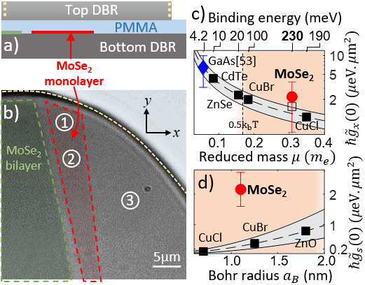
In order to achieve a robust and practical implementation of exciton-mediated optical nonlinearities, like e.g. for room temperature operation, Arsenide-based semiconductor materials feature a too weak excitonic binding energy . is larger in high-bandgap semiconductors, but only at the expense of lower and (see Fig.1.c-d). This trade-off is governed by the hydrogenic character of the excitonic states in conventional materials, in which and depend only on , and the exciton Bohr radius. More explicitly, Ciuti:1998 , and Rochat:2000 , where is the Rabi splitting in the strong coupling regime. In fact, since , where is the exciton reduced mass, depends only on the electron and hole effective masses and .
In this context, monolayers of semiconductor transition metal dichalchogenides (TMDCs) Wang:2018a offer a unique opportunity to break this constrained trade-off. Owing to their atomic-scale thickness, the dielectric constant exhibits a sharp discontinuity across the material plane. The resulting in-plane dependence of the effective Coulomb interaction between electrons and holes is strongly modified, and the resulting excitonic states are non-hydrogenic Berkelbach:2013 ; Chernikov:2014a . Since and are fixed by the spatial characteristic of the Coulomb interaction and of the excitonic wavefunction, we expect both to deviate from the hydrogenic exciton (HE) picture. A 30% enhancement of is actually predicted in TMDCs as compared to hydrogenic excitons of identical reduced mass Shahn:2017a . Signatures of non-negligible excitonic nonlinearities have been observed already in other TMDCs Barachati_2019 , in charged Emmanuele_2020 and excited states of excitons Scuri:2018 , and in polaron-polaritons Tan:2020 .
In this work, we take advantage of the giant oscillator strength of TMDCs excitons Poellmann:2015 ; Robert:2016a ; Moody:2015 ; Korn:2011a to put a MoSe2 monolayer in the strong coupling regime with the resonance of a microcavity Dufferwiel:2015a ; Sidler:2017a ; Lundt:2019a , and carry out spatially-resolved optical transmission spectroscopy with pulsed laser light, as a function of the intensity. The obtained spectra exhibit signatures of a nonlinear response, from which we derive a quantitative estimate of , , and the polarization dependence of the latter. The monolithic microcavity that we investigate is shown in Fig.1.(a,b) with its main features highlighted (See Lundt:2019a ; SI for details). Its quality factor and Rabi splitting amount to and meV respectively. The latter is derived from the anti-crossing of the polariton modes that we observe in a transmission measurement upon sweeping temperature. Details of this characterization can be found in SI .
We then move on to the nonlinear transmission measurements. We use a pulsed Ti:sapphire laser that delivers fs pulses with a spectrum of tunable mean energy meV and a bandwidth meV. Its purpose is the ultrafast creation of a dense polariton population resonantly, without overheating the sample, and to perform a broadband transmission spectrum measurement which is able to capture both the lower and upper polaritons. Heating from residual absorption is further suppressed by chopping the laser into a on-off duty-cycle. The beam is prepared into a Gaussian mode which is focused on the microcavity surface into a m waist size spot. We use a quarter-wave plate to tune the laser polarization among the states , where , is the linear polarization basis oriented as shown in Fig.1.(b), and is the wave plate rotation angle with respect to . In the first part of this work we use -polarized light (). The time-integrated transmitted light intensity is collected with a microscope objective, and imaged at the entrance focal plane of a 300 grooves/mm grating spectrometer. By doing so, we obtain space and frequency-resolved transmission spectra . Since our aim is to provide a quantitative estimate of the interactions, we also need to know the electromagnetic energy in each pulse. To do so, the time-averaged laser power is measured at the input cryostat window, just before the laser light impinges the cavity backside using a thermal-head powermeter. is thus measured from pJ, which is well below the onset of the nonlinear regime, up to several hundreds of pJ, which is well above.
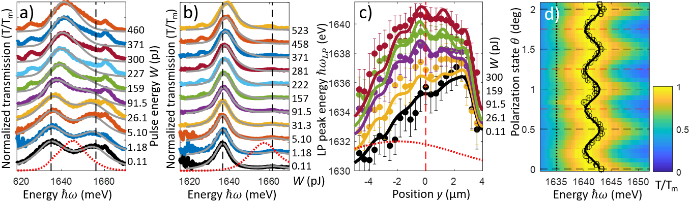
Two such measurements, realized in area (1) and (2), at temperatures K and K, respectively, are shown in Fig.2.a and Fig.2.b. The excitonic fraction of the polariton field in each case is and , respectively. We indeed exploit the fact that the excitonic transition energy is temperature-dependent to control the detuning between the bare cavity (frequency ) and the excitonic level (frequency ) SI , and hence the excitonic fraction () of the lower (upper) polariton states Carusotto:2013 . The effective polariton-polariton interaction constant is thus varied as it depends on Rochat:2000 ; SI . The laser pulse spectral overlap with the polariton modes is also different in the two experiments. We take advantage of these variations to test the robustness of our quantitative estimate of and , as they should not depend on these parameters. The plotted transmission spectra are normalized to their maximum for clearer representation. In the linear regime (bottom spectra, pJ), we observe both the upper and lower polariton resonances, with a mostly equal weight at K, and with a dominant lower polariton peak at , consistently with their respective photonic fraction. Two smaller peaks are also visible in these spectra at meV and meV that we traced back, by real space analysis, to bare cavity resonances situated within the small gap separating the MoSe2 monolayer from the bilayer. Upon increasing , the polaritonic resonances exhibit a clear and consistent trend: at moderate , the lower polariton peak blueshifts, while the upper polariton essentially does not. This behaviour difference is key to distinguish between the contributions of and to the nonlinearity. Indeed, while Coulomb interaction contributes to blueshift both lower and upper polaritons, the saturation causes a reduction of the effective Rabi spitting, and thus shifts the lower and upper polaritons in opposite directions SI . The trend we observe thus indicates that the saturation contributes significantly to the nonlinearity, consistently with recent reports Gu:2020 .
We interpret these spectra quantitatively, by theoretical simulation of the polariton field ultrafast evolution, including the shape of the laser pulse in time and space. Specifically, we derive a mean-field input/output theory in the exciton-photon basis SI , including exciton-exciton interactions and saturation effects. Owing to the exciton state properties, has two contributions: and corresponding to the interactions between parallel and opposite spin excitons, that couple to co- and cross-circularly polarized light. After transformation into the polarization basis, the equation of motion for the exciton and photon fields read:
| (1) | |||||
| (2) | |||||
where is the -polarized incident laser pulse field density, is the bare cavity resonance frequency at vanishing in-plane wavevector , its effective mass, and is the potential describing the spatial dependence , is the excitonic transition frequency, of which we can neglect the kinetic contribution and is the Rabi splitting, is the excitonic non-radiative relaxation rate, is the cavity radiative decay rate, and () are the cavity coupling rate on the laser input side (of the transmission side). Finally, , and are the saturation and exciton-exciton interaction constants in the -polarization basis, given by SI
| (3) | |||||
| (4) |
Note that in Eq.(1-2) we have neglected the contribution of the cross-polarized components of the exciton and photon fields since the laser excites only one component, and the interactions terms provide only density-mediated couplings which vanish if one of the two fields is zero. We also checked experimentally that the polariton modes exhibit no birefringence SI .
In order to fully account for the time profile of the excitation pulse, and of the Gaussian shape of the spot in real space, we solved this model numerically. The experimental parameters entering the model are the microcavity and laser characteristics, which are known accurately. The interaction constants and , are thus the only free parameters. We first apply this model to the spectra shown in Fig.2.(a,b) (in the polarization state). and are thus derived with their uncertainty by numerical optimization of the fit between the model and the measurements SI . This analysis yield eV.m2 and eV.m2 for the experiment at K shown in Fig.2.a. The experiment at K consistently yields eV.m2, and eV.m2, albeit with a much larger uncertainty due to the fact that the upper polariton contribution to the spectra is small, and hence prevents determining accurately the relative contribution of and . We also derive the excitonic densities (half-width-at-half-maximum in time and space) that increases from cm-2 (pJ) to cm-2 (pJ). Note that at high , the saturation effect is large and our model is expected to overestimate it in this regime Rochat:2000 ; Combescot:2008 ; Kyriienko_2019 ; Emmanuele_2020 . This is indeed the trend that we observe in the last four spectra in Fig.2.b, in which the theory predicts a slightly smaller Rabi splitting than in the experiment. Yet, except for this feature, the spectral shape and peak energies evolution for increasing are in very good agreement with the experiment.
We cross-checked this quantitative analysis by looking at another footprint of the nonlinearity: the nontrivial spatially-dependent transmission spectrum that results from the interplay between the Gaussian shape of the spot and the nonlinearity. Fig.2.c shows the lower polariton transmission peak energy , plotted versus , where is the position along a diameter of the laser spot, and is the laser spot intensity maximum position. The lowest spectrum (black) is obtained in the linear regime (pJ) and thus shows the lower polariton potential , from which we derive . For increasing the nonlinearity changes this shape as the blueshift depends on the local density and excitonic fraction. We can fit this behaviour quantitatively with our model, and a good agreement is obtained for eV.m2, and eV.m2. The large uncertainty reflects the fact that the upper polariton contribution is weak in the dataset, and the relative contributions of and are hard to distinguish. Yet, the result is consistent with the spectral analysis.
We verified that the nonlinearities that we measure in this work come from the monolayer and not from any other materials within the structure. We thus measured in area 3, which is a bare cavity free from MoSe2. The area exhibits a sharp cavity mode, that does not shift (meV) up to the highest applied pulse energy (nJ), as is shown in detail in SI .
In Fig.1.c and Fig.1.d, we plotted the theoretical HE interaction constants (in which we assumed that ) versus , and versus (dashed lines). is introduced in order for the theory to agree quantitatively with the measurement in Estrecho et al. Estrecho:2019 , where eV.m2 is found for a planar microcavity with GaAs quantum wells SI . This deviation might arise from the strict 2D approximation of the excitonic wavefunction in the theory, which is likely inaccurate in realistic quantum wells Estrecho:2019 . Using excitonic reduced masses from the literature SI , a few materials are highlighted (squares) along these theoretical curve. In Fig.1.c, the bulk exciton binding energies are also indicated for each material on the top axis as reference SI . The measurements obtained from the analysis of Fig.2.a are shown as a red circle in Fig.1.c-d. Our measured is found to moderately exceed HE’s theory, and is fully compatible with the enhancement (hollow square in Fig.1.c) predicted in Shahn:2017a , while exceeds HE’s theory by a large factor . A possible origin of this larger deviation is already visible in the HE picture, in which depends directly on the dielectric function square (via ), while essentially does not.
We finally characterized the spin anisotropy of the nonlinearity at K, during the same experimental run as that shown in Fig.2.a, by measuring the transmission spectrum versus . The results are shown in Fig.2.d: upon increasing from (linear polarization) to (circular polarization) at a fixed pJ, the spectrum exhibits a global redhsift of meV. Using our model and Eqs.(3,4) SI , this behaviour implies that is about twice larger than , and positive. In TMDC monolayers Shahn:2017a , like in conventional materials, the Coulomb interaction between polaritons is in principle dominated by exchange interaction Ciuti:1998 , for which is expected to be negative and small as compared to Glazov:2009 ; Vladimirova:2010 . Our result differs from this picture, and is thus highly non-trivial. Its precise interpretation requires a fully dedicated investigation that exceeds the scope of the present work.
A possible explanation could be the involvement of an intermediate state, like spin-2 dark excitons Glazov:2009 , or biexcitons Carusotto:2010a ; Takemura:2017 . In such a mechanism, is enhanced and takes a positive sign when the two-polaritons state is close, and on the high energy side of the intermediate state. In a MoSe2 monolayer, the dark exciton state is a few meV above the bright one Robert:2020 , such that the upper polariton state, nominally meV above the bright exciton, could benefit from this resonance at the peak intensity, when the saturation brings it closer. A resonance with the biexciton state is expected meV Hao:2017 ; Bleu:2020 below the bright exciton, which is meV above the nominal energy of the lower polariton, and thus also favourable at the peak intensity. Finally, at such large , higher order many-body correlations and the composite nature of excitons might start to contribute, such that our estimate of might be too inaccurate. Yet, owing to the robustness that the model has demonstrated in reasonably capturing the measurements in Fig.2.(a-c), we expect that this estimate is at least qualitatively correct; namely, that is positive and comparable in magnitude to and .
In summary, we have shown that a MoSe2 monolayer in the strong coupling regime displays enhanced exciton-mediated optical nonlinearity as compared to comparable HE excitons, in particular via the excitonic saturation mechanism. We also observe a non-trivial spin anisotropy of the interaction which deserves future investigation. Our results demonstrate that non-hydrogenic exciton in MoSe2, and potentially in other TMDC materials, offer new perspectives for the engineering of exciton-mediated optical nonlinearities.
Acknowledgements.
PS and AV contributed equally to this work. The authors acknowledge fruitful discussions with D. Basko, O. Kyriienko and D. Ferrand. P.S., M.R. and J.R. are supported by the French National Research Agency in the framework of the Investissements d’Avenir program (ANR-15-IDEX-02) and by the research grant ANR-16-CE30-0021. S.T. acknowledges support from NSF DMR 1838443 and ARO Materials STIR program. M.K., N.L., S.H., and C.S. acknowledges support by the State of Bavaria. C.S. acknowledges support by the European Research Commission (ERC, Project unLiMIt-2D, 697228). T.V. acknowledges the ARC Centre of Excellence for Engineered Quantum Systems (CE170100009). A.V. acknowledges the European Union Horizon 2020 research and innovation programme under the Marie Skłodowska-Curie grant agreement No 754303.References
- (1) A. Tomadin, and R. Fazio, JOSA B 27 A130 (2010)
- (2) C. Noh, and D.G. Angelakis, Rep. Prog. Phys. 80, 016401 (2016)
- (3) J. Cho, D. G. Angelakis, and S. Bose, Phys. Rev. Lett. 101, 246809 (2008)
- (4) R. O. Umucalilar, and I. Carusotto, Phys. Rev. Lett. 108, 206809 (2012)
- (5) M. Hafezi, M. D. Lukin, and J. M. Taylor, New Journal of Physics 15, 063001 (2013)
- (6) I. Carusotto, D. Gerace, H. E. Tureci, S. De Liberato, C. Ciuti, and A. Imamoglu, Phys. Rev. Lett. 103, 033601 (2009)
- (7) J. Tangpanitanon, V. M. Bastidas, S. Al-Assam, P. Roushan, D. Jaksch, and D. G. Angelakis, Phys. Rev. Lett. 117, 213603 (2016)
- (8) A. Tomadin, V. Giovannetti, R. Fazio, D. Gerace, I. Carusotto, H. E. Türeci, and A. Imamoglu Phys. Rev. A 81, 061801(R) (2010)
- (9) J. Jin, D. Rossini, R. Fazio, M. Leib,and M. J. Hartmann, Phys. Rev. Lett. 110, 163605 (2013)
- (10) J. Jin, D. Rossini, M. Leib, M. J. Hartmann, and R. Fazio, Phys. Rev. A 90, 023827 (2014)
- (11) J. Raftery, D. Sadri, S. Schmidt, H. E. Türeci, and A. A. Houck, Phys. Rev. X 4, 031043 (2014)
- (12) P. Andalib, and N. Granpayeh, JOSA B 26 10-16 (2009)
- (13) Ye Liu, Fei Qin, Zi-Ming Meng, Fei Zhou, Qing-He Mao, and Zhi-Yuan Li, Optics Express 19, 1945-1953 (2011)
- (14) H. Mabuchi, Appl. Phys. Lett. 99, 153103 (2011)
- (15) T. Espinosa-Ortega, and T. C. H. Liew, Phys. Rev. B 87, 195305 (2013)
- (16) A. Salmanpour, S. Mohammadnejad, and A. Bahrami, Optical and Quantum Electronics 47, 2249-2275 (2015)
- (17) F. Flamini, N. Spagnolo, and F. Sciarrino, Rep. Prog. Phys. 82, 016001 (2019)
- (18) J. L. OBrien, A. Furusawa, and J. Vuckovic, Nature Photonics 3, 687-695 (2009)
- (19) C. Ciuti, V. Savona, C. Piermarocchi, A. Quattropani, and P. Schwendimann, Phys. Rev. B 58, 7926 (1998)
- (20) G. Rochat, C. Ciuti, V. Savona, C. Piermarocchi, A. Quattropani, and P. Schwendimann, Phys. Rev. B 61, 13856 (2000)
- (21) P. Lodahl, S. Mahmoodian, and S. Stobbe, Review of Modern Physics 87, 347 (2015)
- (22) A. Auffèves-Garnier, C. Simon, J. M. Gérard, and J.P. Poizat, Phys. Rev. A 75, 053823 (2007)
- (23) A. Faraon et al. Nature Physics 4, 859 (2008)
- (24) A. Reinhard et al, Nature Photonics 6, 93 (2012)
- (25) H. Kim et al. Nature Photonics 7, 373 (2013)
- (26) A. Javadi et al. Nature Communication 6, 8655 (2015)
- (27) L. de Sants et al. Nature Nanotechnology 12, 663 (2017)
- (28) P. Senellart, G. Solomon, and A. White, Nature Nanotechnology 12, 1026 (2017)
- (29) A. Verger, C. Ciuti, and I. Carusotto, Phys. Rev. B 73, 193306 (2006)
- (30) C. Weisbuch, M. Nishioka, A. Ishikawa, and Y. Arakawa, Phys. Rev. Lett. 69, 3314 (1992)
- (31) I. Carusotto, and C. Ciuti, Rev. Mod. Phys. 85, 299 (2013)
- (32) G. Munoz Matutano et al. Nature Materials 18, 213 (2019)
- (33) A. Delteil et al. Nature Materials 18, 219 (2019)
- (34) G. Wang, A. Chernikov, M. M. Glazov, T. F. Heinz, X. Marie, T. Amand, B. Urbaszek Rev. Mod. Phys. 90, 021001 (2018)
- (35) T. C. Berkelbach, M. S. Hybertsen and D. R. Reichman, PRB 88, 045318 (2013)
- (36) A. Chernikov, T. C. Berkelbach, H. M. Hill, A. Rigosi, Y. Li, O. B. Aslan, D. R. Reichman, M. S. Hybertsen, T. F. Heinz, Phys. Rev. Lett. 113, 076802 (2014)
- (37) V. Shahnazaryan, I. Iorsh, I. A. Shelykh, and O. Kyriienko, Phys. Rev. B 96, 115409 (2017)
- (38) F. Barachati et al. Nature Nanotechnology 13, 906-909 (2018)
- (39) R. P. A. Emmanuele et al. arXiv:1910.14636 (2020)
- (40) G. Scuri et al. Phys. Rev. Lett. 120, 037402 (2018)
- (41) L. B. Tan, O. Cotlet, A. Bergschneider, R. Schmidt, P. Back, Y. Shimazaki, M. Kroner, A. Imamoglu Phys. Rev X 10, 021011 (2020)
- (42) C. Poellmann et al. Nature Materials 14, 889-893 (2015)
- (43) C. Robert et al. Phys. Rev. B 93, 205423 (2016)
- (44) G. Moody et al. Nature Communications 6, 8315 (2015)
- (45) T. Korn, S. Heydrich, M. Hirmer, J. Schmutzler, and C. Schüller, Applied Physics Letters 99, 102109 (2011)
- (46) S. Dufferwiel et al. Nature communications 6, 8579 (2015)
- (47) M. Sidler et al. Nature Physics 13, 255 (2017)
- (48) N. Lundt, et al. Nature Nanotechnology 14 770-775 (2019)
- (49) Supplemental Materials.
- (50) J. Gu, et al. arXiv:1912.12544 (2019)
- (51) M. Combescot, O. Betbeder-Matibeta, F. Dubin Physics Reports 463, 215-320(2008)
- (52) O. Kyriienko, D. N. Krizhanovskii, I. A. Shelykh, ArXiv:1910.11294 (2019)
- (53) E. Estrecho et al. Phys. Rev. B 100, 035306 (2019)
- (54) M. M. Glazov, H. Ouerdane, L. Pilozzi, G. Malpuech, A. V. Kavokin, A. DAndrea, Phys. Rev. B 80, 155306 (2009)
- (55) M. S. Vladimirova et al. Phys. Rev. B 82, 075301 (2010)
- (56) N. Takemura, M. D. Anderson, M. Navadeh-Toupchi, D. Y. Oberli, M. T. Portella-Oberli, and B. Deveaud Phys. Rev. B 95, 205303 (2017)
- (57) Carusotto, I., Volz T., Imamoglu, A., Feshbach blockade: Single-photon nonlinear optics using resonantly enhanced cavity polariton scattering from biexciton states. Europhysics Letters 90, 37001 (2010).
- (58) C. Robert et al. arXiv:2002.03877 (2020)
- (59) Kai Hao, Judith F. Specht, Philipp Nagler, Lixiang Xu, Kha Tran, Akshay Singh, Chandriker Kavir Dass, Christian Schüller, Tobias Korn, Marten Richter, Andreas Knorr, Xiaoqin Li, Galan Moody, Nature Comm. 8, 15552 (2017)
- (60) O. Bleu, J. Levinsen, and M. Parish, arXiv:2004.01336 (2020)