Pycro-manager: open-source software for integrated microscopy hardware control and image processing
Manager, an open-source microscopy acquisition software, has been an essential tool for many microscopy experiments over the past 15 years, but is not easy to use for experiments in which image acquisition and analysis are closely coupled. This is because Manager libraries are written in C++ and Java, whereas image processing is increasingly carried out with data science and machine learning tools most easily accessible through the Python programming language. We present Pycro-Manager, a tool that enables rapid development of such experiments, while also providing access to the wealth of existing tools within Manager through Python.
Cutting-edge innovations in biological microscopy are increasingly blurring the line between data acquisition and data analysis. Often, the captured image requires significant image processing to produce the final image, and users rely on pipelines of multiple programs or software libraries to get from the capture stage to the final image. Computational microscopy and machine learning-based methods take this paradigm to an extreme, often producing raw measurements that aren’t human-interpretable without post-processing [1, 5, 10, 4] Furthermore, new ”adaptive” methods rely on image processing during acquisition to actively control various parameters of the microscope [8, 6]. Testing new ideas and applying them for biological discovery is often impeded by the lack of powerful yet sufficiently flexible control software, necessitating the development of bespoke solutions that work only with a specific instrument.
In situations where the close coupling of data acquisition and image analysis are not required, Manager [2, 3] has often been the de facto solution. This owes to the existence of a hardware abstraction layer and an extensive library of ‘device adapters’, which allow control of hundreds of different types of hardware ranging from cameras to complete microscopes. Community contributions of device adapters, plugins, and scripts provide a treasure trove of hundreds of developer-years of microscopy automation. However, despite the power of these libraries, which are written in C++ and Java, they are often difficult to integrate with the latest developments in computer vision and scientific computing, which are most readily available through the Python programming language [9].
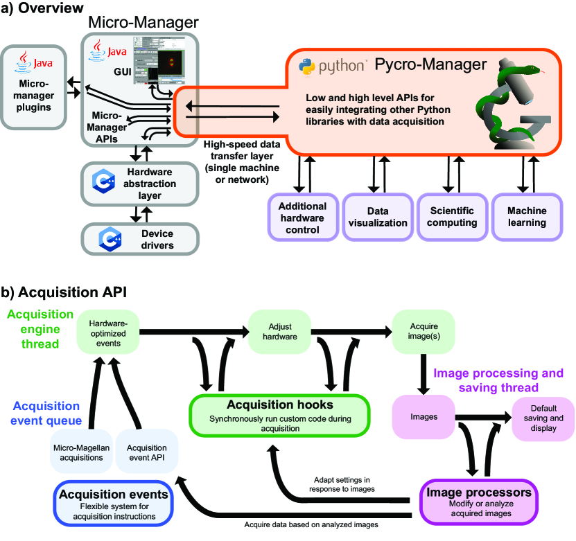
To address this need, we developed Pycro-Manager. The foundation of Pycro-Manager is a high-speed data transfer layer that dynamically translates between Java and Python (Fig. 1a), enabling users to call Java libraries as if they had been written in Python. This creates an easy access point through Python for all the existing capabilities of Manager, without requiring rewriting of any code or developers in the Java and Python communities to switch languages. The transfer layer can also be run over a network, enabling, for example, splitting out of control and processing to a different machine than the one connected to the microscope.
On top of this layer, Pycro-Manager implements an acquisition API with the flexibility to independently customize what is acquired (”acquisition events”), what happens while it is being acquired (”acquisition hooks”), and what happens to the data after it is acquired (”image processors”), without having to write all the boilerplate code that usually accompanies such customized experiments (Fig. 1b). This API can be used, for example, to synchronize external hardware with the acquisition process, modify acquired images on-the-fly before saving/visualization, or implement customized data saving/visualization. Combining these features can be used for even more powerful applications, such as data-adaptive microscopy.
Acquisition events describe what to acquire. For example, a -stack would be a series of acquisition events, each corresponding to a single image and containing the position of the focus drive. They can be created either from a GUI (such as Micro-Magellan [7]) or programmatically through the acquisition event API.
Acquisition hooks enable the synchronization of arbitrary code with acquisition operations. For example, they can be used to add in control of external devices, or apply auto-focusing operations before proceeding with acquisition.
Image processors give access to image data and metadata as soon it is acquired. They can be used to modify data before reentering the default saving/visualization pipeline, or to divert it to custom alternatives. They can also be used in conjunction with acquisition hooks to create feedback loops for updating hardware based on data, or with acquisition events for controlling future data acquisition.
In summary, Pycro-Manager provides a much needed interface between over a decade’s worth of open source microscopy development compatible with the cutting edge of scientific computing. It removes the need to write boilerplate code when developing new types of smart microscopes, freeing developers to rapidly test new ideas. Finally, it does this in a hardware-agnostic way to maximize the portability of code across different microscopes.
The source code for Pycro-Manager can be found at: https://github.com/micro-manager/pycro-manager, and the documentation is at: https://pycro-manager.readthedocs.io/en/latest/#
Acknowledgements
We thank Stéfan van der Walt for helpful discussion during development and and Kyle Marchuk for early beta testing.
References
- [1] Eric M Christiansen, Samuel J Yang, D Michael Ando, Lee L Rubin, Philip Nelson, Steven Finkbeiner, Eric M Christiansen, Samuel J Yang, D Michael Ando, Ashkan Javaherian, and Gaia Skibinski. In Silico Labeling: Predicting Fluorescent Labels in Unlabeled Images. Cell, pages 1–12, 2018.
- [2] Arthur Edelstein, Nenad Amodaj, Karl Hoover, Ron Vale, and Nico Stuurman. Computer control of microscopes using manager, 2010.
- [3] Arthur D Edelstein, Mark a Tsuchida, Nenad Amodaj, Henry Pinkard, Ronald D Vale, and Nico Stuurman. Advanced methods of microscope control using Micro-Manager software. Journal of Biological Methods, 1(2):10, 11 2014.
- [4] Syuan-Ming Guo, Li-Hao Yeh, Jenny Folkesson, Ivan E Ivanov, Anitha Priya Krishnan, Matthew G Keefe, David Shin, Bryant Chhun, Nathan Cho, Manuel Leonetti, Tomasz J Nowakowski, Shalin B Mehta, Chan Zuckerberg Biohub, and J F Contributed. Revealing architectural order with quantitative label-free imaging and deep learning. bioRxiv, pages 1–26, 2019.
- [5] Chawin Ounkomol, Sharmishtaa Seshamani, Mary M. Maleckar, Forrest Collman, and Gregory R. Johnson. Label-free prediction of three-dimensional fluorescence images from transmitted-light microscopy. Nature Methods, 15(11):917–920, 2018.
- [6] Henry Pinkard, Zachary Phillips, Arman Babakhani, Daniel A. Fletcher, and Laura Waller. Deep learning for single-shot autofocus microscopy. Optica, 6(6):794, 2019.
- [7] Henry Pinkard, Nico Stuurman, Kaitlin Corbin, Ronald Vale, and Matthew F Krummel. Micro-Magellan: open-source, sample-adaptive, acquisition software for optical microscopy. Nature Methods, 13(10):807–809, 9 2016.
- [8] Loïc A. Royer, William C. Lemon, Raghav K. Chhetri, Yinan Wan, Michael Coleman, Eugene W. Myers, and Philipp J. Keller. Adaptive light-sheet microscopy for long-term, high-resolution imaging in living organisms. Nature Biotechnology, 34(12):1267–1278, 2016.
- [9] Pauli Virtanen, Ralf Gommers, Travis E. Oliphant, Matt Haberland, Tyler Reddy, David Cournapeau, Evgeni Burovski, Pearu Peterson, Warren Weckesser, Jonathan Bright, Stéfan J. van der Walt, Matthew Brett, Joshua Wilson, K. Jarrod Millman, Nikolay Mayorov, Andrew R.J. Nelson, Eric Jones, Robert Kern, Eric Larson, C. J. Carey, İlhan Polat, Yu Feng, Eric W. Moore, Jake VanderPlas, Denis Laxalde, Josef Perktold, Robert Cimrman, Ian Henriksen, E. A. Quintero, Charles R. Harris, Anne M. Archibald, Antônio H. Ribeiro, Fabian Pedregosa, Paul van Mulbregt, Aditya Vijaykumar, Alessandro Pietro Bardelli, Alex Rothberg, Andreas Hilboll, Andreas Kloeckner, Anthony Scopatz, Antony Lee, Ariel Rokem, C. Nathan Woods, Chad Fulton, Charles Masson, Christian Häggström, Clark Fitzgerald, David A. Nicholson, David R. Hagen, Dmitrii V. Pasechnik, Emanuele Olivetti, Eric Martin, Eric Wieser, Fabrice Silva, Felix Lenders, Florian Wilhelm, G. Young, Gavin A. Price, Gert Ludwig Ingold, Gregory E. Allen, Gregory R. Lee, Hervé Audren, Irvin Probst, Jörg P. Dietrich, Jacob Silterra, James T. Webber, Janko Slavič, Joel Nothman, Johannes Buchner, Johannes Kulick, Johannes L. Schönberger, José Vinícius de Miranda Cardoso, Joscha Reimer, Joseph Harrington, Juan Luis Cano Rodríguez, Juan Nunez-Iglesias, Justin Kuczynski, Kevin Tritz, Martin Thoma, Matthew Newville, Matthias Kümmerer, Maximilian Bolingbroke, Michael Tartre, Mikhail Pak, Nathaniel J. Smith, Nikolai Nowaczyk, Nikolay Shebanov, Oleksandr Pavlyk, Per A. Brodtkorb, Perry Lee, Robert T. McGibbon, Roman Feldbauer, Sam Lewis, Sam Tygier, Scott Sievert, Sebastiano Vigna, Stefan Peterson, Surhud More, Tadeusz Pudlik, Takuya Oshima, Thomas J. Pingel, Thomas P. Robitaille, Thomas Spura, Thouis R. Jones, Tim Cera, Tim Leslie, Tiziano Zito, Tom Krauss, Utkarsh Upadhyay, Yaroslav O. Halchenko, and Yoshiki Vázquez-Baeza. SciPy 1.0: fundamental algorithms for scientific computing in Python. Nature Methods, 17(3):261–272, 2020.
- [10] Martin Weigert, Uwe Schmidt, Tobias Boothe, Andreas Müller, Alexandr Dibrov, Akanksha Jain, Benjamin Wilhelm, Deborah Schmidt, Coleman Broaddus, Siân Culley, Mauricio Rocha-Martins, Fabián Segovia-Miranda, Caren Norden, Ricardo Henriques, Marino Zerial, Michele Solimena, Jochen Rink, Pavel Tomancak, Loic Royer, Florian Jug, and Eugene W. Myers. Content-aware image restoration: pushing the limits of fluorescence microscopy. Nature Methods, 15(12):1090–1097, 2018.