Two-photon Doppler-free ultraviolet laser spectroscopy on sulphur atoms
Abstract
The 3PJ - 3PJ transition in the sulphur atom is investigated in a precision two-photon excitation scheme under Doppler-free and collision-free circumstances yielding an absolute accuracy of 0.0009 cm-1, using a narrowband pulsed laser. This verifies and improves the level separations between amply studied odd parity levels with even parity levels in S I. An improved value for the 3P2 - 3P1 ground state fine structure splitting is determined at (7) cm-1. A 34S - 32S atomic isotope shift was measured from combining time-of-flight mass spectrometry with laser spectroscopy.
I Introduction
The odd parity level energies for the neutral sulfur atom have been extensively studied through VUV absorption spectroscopy from the ground electronic configuration Kaufman (1982); Sarma and Joshi (1984); Joshi et al. (1987); Zhou et al. (2008). The connection with even parity excited states is studied through visible and infrared spectroscopy involving transition between excited states Frerichs (1933); Meissner et al. (1933); Jakobsson (1967); Baclawski and Musielok (2011). In addition, direct measurements of transitions between even parity states are studied through 2+1 resonance enhanced multiphoton ionization (REMPI) spectroscopy Venkitachalam and Rao (1991); Woutersen et al. (1997).
The level energies of the 1D2 and 1S0 states of the ground electronic configuration, were investigated via electric dipole-forbidden transitions, first measured by McConkey et al. McConkey et al. (1968), and revisited at higher accuracy by Eriksson Eriksson (1978). Based on the combination differences between the forbidden transitions, 3P1 - 1S0, 1D2 - 1S0 and 3P2 - 1D2 the level energies of the lowest five levels are determined at an uncertainty of 0.005 cm-1 Eriksson (1978). Later, Brown et al. measured the fine structure transition 3P1 - 3P0 using laser magnetic resonance yielding an accuracy better than 10-4 cm-1 Brown et al. (1994). The resulting level structure of the sulphur atom including a comprehensive compilation of lines and level energies is now well documented Martin et al. (1990); Kaufman and Martin (1993); Morton (2003).
In the present study, high-resolution spectra of 3PJ - 3PJ transitions of 32S are measured by using 2+1 REMPI employing a narrowband pulsed laser amplifier in a scheme with counter-propagating laser beams, thus allowing for Doppler-free spectroscopy at high resolution and high accuracy. The study is aimed at accurately bridging the large energy gap between the ground state and the manifold of excited states, which can be probed at high accuracy via infrared and visible spectroscopies. Via this means the measurement of a few transitions will allow for improving the accuracy of the entire level structure of the S atom. Moreover it will be shown that isotope shifts can be resolved in such Doppler-free precision experiment.
II Experiment
The experimental setup, schematically shown in Fig. 1, is similar to that used for the production and detection of vibrationally excited states in molecular hydrogen, also obtained from photolysis of H2S Niu et al. (2015); Trivikram et al. (2016, 2019). Two ultraviolet (UV) pulsed laser systems are used to produce sulphur atoms and perform the precise two-photon spectroscopy. Sulphur atoms in the 3PJ ground state triplet are formed by UV-photodissociation of H2S molecules, a well studied photolysis process Steadman and Baer (1988, 1989); Cook et al. (2001); Zhou et al. (2020). The first UV-laser pulse, inducing the dissociation, is obtained from a frequency-doubled pulsed dye laser (PDL) pumped by an injection-seeded pulsed Nd-YAG laser. Pulse energies of up to 4.5 mJ pulse are used for the photolysis. The wavelength of the dissociation laser is chosen at 291 nm following the original work of Steadman and Baer Steadman and Baer (1988, 1989).
The two-photon transition is measured by a traveling-wave pulsed-dye-amplifier (PDA) system amplifying the output of a narrowband cw-ring dye laser. The amplification is realized in three consecutive dye cells, pumped with the same Nd-YAG pump laser also used to pump the PDL Eikema et al. (1997). The output of the PDA at 616 - 621 nm is frequency-doubled in a KDP crystal to provide UV-pulses in the range 308 - 311 nm with ns pulse width. The frequency of the cw-seed light is calibrated against the standard of I2 saturated hyperfine lines combined with the transmission markers of a stabilized Fabry-Perot interferometer Xu et al. (2000). The chirp effect on the pulses, giving rise to an effective frequency offset between the pulsed output of the PDA and the cw-light is assessed via optical heterodyne measurements and analyzed via known techniques Melikechi et al. (1994); Eikema et al. (1997). The narrowband UV beam is then split and configured in a counter-propagating beam setup to induce the Doppler-free two-photon transitions. The angle mismatch of the counter-propagating beams is reduced based on Sagnac interference fringes Hannemann et al. (2007).
| Initial state | Excited state | Obs. (cm-1) |
|---|---|---|
| 3P1 | 64888.9317 (9) | |
| 3P2 | 3P0 | 64891.3536 (9) |
| 3P2 | 64892.5494 (9) | |
| 3P1 | 3P1 | 64492.8751 (9) |
| 3P2 | 64496.4917 (23) | |
| 3P0 | 3P0 | 64317.7561 (9) |
| 3P2 | 64318.9561 (9) |
The UV beams are focused into a spot of size few tens of m spatially overlapping a pulsed H2S beam, in a low-density region of a skimmed and collimated pulsed effusive gas expansion. To avoid ac-Stark disturbances from the dissociation laser, the PDA spectroscopy laser is optically delayed by 10 ns with respect to the photolysis laser, such that there is no temporal overlap. Sulphur atom signal is generated via 2+1 REMPI, whereby ions are extracted through a mass-resolving time-of-flight (TOF) tube, detecting S+ ions. Ion optics are triggered at a delay of ns from the spectroscopy laser, so that the laser-excitation takes place in zero DC field. The ion signal is amplified by a microchannel plate (MCP) with phosphor imaging screen with detection on a photomultiplier tube. Mass-selected spectra are recorded with a box-car integrator probing only a narrow channel of the TOF-trace. The large amounts of SH+ and H2S+ signal in nearby mass channels, as well as S+ background signals from various dissociation/ionization channels are limiting factors on the signal-to-noise-ratio of the S-atom spectra. In case of spectral recording of measuring spectra of 34S this is even more detrimental.
III Results and Interpretation
All of the seven two-photon allowed transitions between 3PJ and 3PJ were measured in the wavelength interval 308-311 nm. Figure 2 displays recordings of all observed lines under Doppler-broadened conditions. Note that the combination is forbidden by two-photon selection rules Dixit et al. (1988).
The spectra for the 3P2 - 3P2 and 3P2 - 3P1 lines, recorded under Doppler-free conditions, are shown in greater detail in Figs. 3 and 4. The width of the spectral lines, measured at the lowest power, is about 290 MHz (FWHM), only slightly larger than expected by assuming exact Fourier-transform limited laser pulses of Gaussian spectral profile. The power dependence (or ac-Stark effect) for the transition frequencies is studied by varying the PDA pulse energy as shown in the inset of the figures. Table 1 lists the transition frequencies, upon extrapolation to zero field, as measured for the seven transitions with the boxcar gate set to 32S.
| Contribution | Uncertainty ( cm-1) |
|---|---|
| Line profile (fitting) | 2 |
| Statistics | 3 |
| AC-Stark extrapolation | 5 |
| Frequency calibration | 3 |
| Cw-pulse offset (chirp) | 6 |
| Residual Doppler | |
| DC-Stark effect | |
| Total | 9 |
The sources of uncertainty are summarized in an error budget presented in Table 2. A statistical analysis of the determination of the line centres gives an uncertainty of cm-1, including averaging over multiple recordings. The ac-Stark effect is the dominant systematic effect in the present study. It causes a shift of line centres, accompanied by broadening, and due to the spatial variation of laser intensity over the laser focus, also results in an asymmetry of the line profile Trivikram et al. (2016). The asymmetry was addressed by fitting skewed Voigt profile fitting. Analysis of the line shape results in an additional contribution to the uncertainty of cm-1. The ac-Stark shift is further analyzed by performing measurements over a range of pulse energies of 20 - 100 J with extrapolation of the centre frequency to zero energy. This adds a contribution to the error budget of cm-1. Further contributions are associated with the frequency chirp in the PDA-system and the absolute frequency calibration against I2-hyperfine components, which were analyzed by established techniques Ubachs et al. (1997); Eikema et al. (1997) and result in a contribution of cm-1 for the frequency uncertainty. The absolute frequency calibration against I2 hyperfine components involves uncertainty in the reference frequencies Xu et al. (2000) and measurement of the FSR, amounting to cm-1. For the latter two contributions multiplication by four, for the frequency doubling and the two-photon process, is included. The experiment is essentially Doppler-free, although small shifts of the frequency centre may be associated with a non-isotropic velocity distribution of the S-atoms, similar to the case of H2 investigated Cheng et al. (2018). For this reason the counter-propagating laser beams were aligned in a Sagnac interferometer Hannemann et al. (2007) limiting this effect to below cm-1. Excitation was performed in zero field, hence the DC-Stark effect is negligible on the scale of the present accuracy. Taking the contributions in quadrature leads to a total uncertainty of 0.0009 cm-1 for the frequencies of the two-photon resonances for all observed transitions except for one. The uncertainty of 3P1 - 3P2 is estimated at 0.0023 cm-1 with larger uncertainty from statistics, a long measurement trace to be covered for reaching an I2 resonance, and problems encountered in ac-Stark extrapolation.
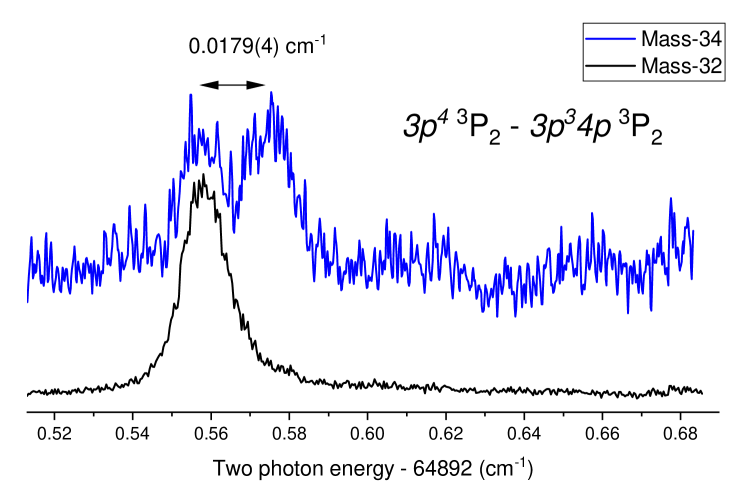
In a single case, for the strongest line 3P2 - 3P2, a study was made of the resonance line in 34S from the 5% naturally abundant isotope in the sample. The spectrum, shown in Fig. 5, and recorded with a boxcar gate probing mass-34, displays the much lower signal-to-noise ratio, caused by spurious signal on mass-34 of H2S+ ions. When only considering the statistical line fitting and relative calibration errors the isotope shift on the resonances amounts to 0.0179 (4) cm-1. The spectrum of Fig. 5 shows that the spectral contributions of 32S and 34S are well separated, thus verifying that the listed entries for the transition frequencies in Table 1 pertain to the main 32S isotope, and do not correspond to a mixture of isotopes.
The isotope shift for a transition can be separated into normal mass shift, specific mass shift and field shift contributions Carette et al. (2010). In view of the very small wave function amplitude of the outer electron within the nuclear charge radius, the field shift (or finite size) contribution is negligible for the differential isotope effect. The Bohr shift or normal mass shift () is calculated to be 0.0650 cm-1. From the experimental isotope shift obtained here, a specific mass shift of =-0.0471(4) cm-1 is extracted for the transition. The experimental value will be useful in validating ab initio calculations of electron correlations.
IV Discussion: Level energies
| Level | This work | Ref. Martin et al. (1990) | Difference |
|---|---|---|---|
| 3P2 | 0 | 0 | - |
| 3P1 | 396.0570 (11) | 396.055 (5) | 0.0020 |
| 3P0 | 573.5953 (9) | 573.640 (16) | -0.0447 |
| 3P1 | 64888.9319 (9) | 64888.964 (25) | -0.0321 |
| 3P0 | 64891.3525 (8) | 64891.386 (25) | -0.0335 |
| 3P2 | 64892.5503 (7) | 64892.582 (25) | -0.0317 |
| This work | Ref. Martin et al. (1990) | Ref. Brown et al. (1994) | |
| 3P0 - 3P1 | 177.5383 (14) | 177.585 (17) | 177.539253 (93) |
| Level | This work | Ref. Martin et al. (1990) |
|---|---|---|
| 3P2 | 0 | 0 |
| 3P1 | ||
| 3P0 | ||
| 1D2 | ||
| 1S0 |
The precise determination of transition frequencies can be cast into a least-squares analysis to determine level energies in both 3P states in the and configurations using the LOPT program Kramida (2011). First the internal consistency of the measurements can be tested by including only the presently obtained data set, as is done in Table 3. The LOPT analysis provides a consistent set of level energies at an accuracy in most cases below cm-1. More importantly the deduced ground state splitting between 3P1 and 3P0 levels is in full agreement (within ) with the very accurate LMR-measurement Brown et al. (1994), the most precise level splitting determined in the sulphur atom. This agreement provides proof that the uncertainties of the present study are not underestimated.
In Table 3 also a comparison is made with the results from VUV spectroscopy, which are at the basis of the comprehensive published line and level lists Kaufman (1982); Martin et al. (1990); Kaufman and Martin (1993). In these compilations a single spectral line in the VUV is included (a transition to the 3S1 level) for which an uncertainty as low as cm-1 is stated Kaufman (1982). We adopt this value as the general uncertainty for the level energies for the excited states, although the uncertainty for the overall level structure might be somewhat larger. The increased precision on the 3P ground state level energies in the compilations, so better than the quoted cm-1, in fact derive from the rather accurate measurement of the forbidden transitions by Eriksson Eriksson (1978). The 3P0 level was not accessed in the measurement of Ref. Eriksson (1978), hence its uncertainty relies on VUV-data. Viewed in this context the deviations between present results and the VUV-compilation Kaufman (1982), as listed in Table 3 both for ground state splittings and excitation energies are close to the expected uncertainties. This includes the consistent shift -0.032 cm-1 for all three levels in the 3PJ excited triplet. This finding is indicative for an overall systematic shift of all excited level in the data compilation by cm-1.
Finally a LOPT least-squares analysis can be performed to determine the level energies of the entire ground electronic configuration including 1D2 and 1S0 levels, based on the present study in combination with the high precision measurements of magnetic dipole transitions in Ref. Eriksson (1978); Brown et al. (1994). Table 4 lists the fitted level energy values with individual uncertainties relative to ground state and comparison with the corresponding values listed in the S-atom data compilation Martin et al. (1990). This results in an improved level structure for , in particular for the lowest fine structure splitting 3P2 - 3P1 which is determined at (7) cm-1, corresponding to a far-infrared wavelength of 25.24893 (4) m.
V Conclusion
In conclusion, seven transitions in the of 3PJ - 3PJ multiplet are measured by narrowband laser spectroscopy at an uncertainty of 0.0009 cm-1. For the first time a 34S - 32S isotope shift has been measured in atomic sulphur, from which a value for the specific mass-shift was derived, a measure for electron correlations in the atom. The accurate transition frequencies improve the level energies of the 3P ground electronic configuration by factor of two. The 3PJ excited state level energies are determined at an absolute accuracy of less than 0.001 cm-1. The present study provides an indication of an overall systematic shift for the excited level energies as listed in spectroscopic data compilations for the sulphur atom Kaufman (1982); Martin et al. (1990); Kaufman and Martin (1993). The precise measurement of even parity excited states may help optimizing the level energies of odd parity levels by future improved measurements between excited states in the infrared and visible regions, therewith using the 3PJ levels as anchor levels, in a similar fashion as applied to H2 Bailly et al. (2010).
Acknowledgement
WU acknowledges the European Research Council for an ERC Advanced grant (No: 670168).
References
- Kaufman (1982) V. Kaufman, Phys. Scripta 26, 439 (1982).
- Sarma and Joshi (1984) V. N. Sarma and Y. N. Joshi, Physica B+C 123, 349 (1984).
- Joshi et al. (1987) Y. N. Joshi, M. Mazzoni, A. Nencioni, W. H. Parkinson, and A. Cantu, J. Phys. B 20, 1203 (1987).
- Zhou et al. (2008) J. Zhou, B. Jones, X. Yang, W. M. Jackson, and C. Y. Ng, J. Chem. Phys. 128, 014305 (2008).
- Frerichs (1933) R. Frerichs, Z. Phys. 80, 150 (1933).
- Meissner et al. (1933) K. W. Meissner, O. Bartelt, and L. Eckstein, Z. Phys. 86, 54 (1933).
- Jakobsson (1967) L. R. Jakobsson, Ark. f. Fysik 34, 19 (1967).
- Baclawski and Musielok (2011) A. Baclawski and J. Musielok, J. Phys. B 44, 135002 (2011).
- Venkitachalam and Rao (1991) T. V. Venkitachalam and A. S. Rao, Appl. Phys. B 52, 102 (1991).
- Woutersen et al. (1997) S. Woutersen, J. B. Milan, W. J. Buma, and C. A. de Lange, J. Chem. Phys. 106, 6831 (1997).
- McConkey et al. (1968) J. W. McConkey, D. J. Burns, K. A. Moran, and J. A. Kernahan, Nature 217, 538 (1968).
- Eriksson (1978) K. B. S. Eriksson, Astroph. J. 222, 398 (1978).
- Brown et al. (1994) J. Brown, K. Evenson, and L. Zink, Astrophys. J. 431 (1994).
- Martin et al. (1990) W. C. Martin, R. Zalubas, and A. Musgrove, J. Phys. Chem. Ref. Data 19, 821 (1990).
- Kaufman and Martin (1993) V. Kaufman and W. C. Martin, J. Phys. Chem. Ref. Data 22, 279 (1993).
- Morton (2003) D. C. Morton, Astroph. J. Suppl. Series 149, 205 (2003).
- Niu et al. (2015) M. L. Niu, E. J. Salumbides, and W. Ubachs, J. Chem. Phys. 143, 081102 (2015).
- Trivikram et al. (2016) T. M. Trivikram, M. L. Niu, P. Wcislo, W. Ubachs, and E. J. Salumbides, Appl. Phys. B 122, 294 (2016).
- Trivikram et al. (2019) T. M. Trivikram, E. J. Salumbides, C. Jungen, and W. Ubachs, Mol. Phys. 117, 2961 (2019).
- Steadman and Baer (1988) J. Steadman and T. Baer, J. Chem. Phys. 89, 5507 (1988).
- Steadman and Baer (1989) J. Steadman and T. Baer, J. Chem. Phys. 91, 6113 (1989).
- Cook et al. (2001) P. A. Cook, S. R. Langford, R. N. Dixon, and M. N. R. Ashfold, J. Chem. Phys. 114, 1672 (2001).
- Zhou et al. (2020) J. Zhou, Y. Zhao, C. S. Hansen, J. Yang, Y. Chang, Y. Yu, G. Cheng, Z. Chen, Z. He, S. Yu, H. Ding, W. Zhang, G. Wu, D. Dai, C. M. Western, M. N. Ashfold, K. Yuan, and X. Yang, Nat. Comm. 11, 1547 (2020).
- Eikema et al. (1997) K. S. E. Eikema, W. Ubachs, W. Vassen, and W. Hogervorst, Phys. Rev. A 55, 1866 (1997).
- Xu et al. (2000) S. Xu, R. van Dierendonck, W. Hogervorst, and W. Ubachs, J. Mol. Spectr. 201, 256 (2000).
- Melikechi et al. (1994) N. Melikechi, S. Gangopadhyay, and E. E. Eyler, J. Opt. Soc. Am. A 11, 2402 (1994).
- Hannemann et al. (2007) S. Hannemann, E. J. Salumbides, and W. Ubachs, Opt. Lett. 32, 1381 (2007).
- Dixit et al. (1988) S. N. Dixit, D. A. Levin, and B. V. McKoy, Phys. Rev. A 37, 4220 (1988).
- Ubachs et al. (1997) W. Ubachs, K. S. E. Eikema, W. Hogervorst, and P. C. Cacciani, J. Opt. Soc. Am. B 14, 2469 (1997).
- Cheng et al. (2018) C.-F. Cheng, J. Hussels, M. Niu, H. L. Bethlem, K. S. E. Eikema, E. J. Salumbides, W. Ubachs, M. Beyer, N. Hölsch, J. A. Agner, F. Merkt, L.-G. Tao, S.-M. Hu, and C. Jungen, Phys. Rev. Lett. 121, 013001 (2018).
- Carette et al. (2010) T. Carette, C. Drag, O. Scharf, C. Blondel, C. Delsart, C. Froese Fischer, and M. Godefroid, Phys. Rev. A 81, 042522 (2010).
- Kramida (2011) A. E. Kramida, Comp. Phys. Comm. 182, 419 (2011).
- Bailly et al. (2010) D. Bailly, E. Salumbides, M. Vervloet, and W. Ubachs, Mol. Phys. 108, 827 (2010).