Multiscale simulation of the focused electron beam induced deposition process
Abstract
Focused electron beam induced deposition (FEBID) is a powerful technique for 3D-printing of complex nanodevices. However, for resolutions below 10 nm, it struggles to control size, morphology and composition of the structures, due to a lack of molecular-level understanding of the underlying irradiation-driven chemistry (IDC). Computational modelling is a tool to comprehend and further optimise FEBID-related technologies. Here we utilise a novel multiscale methodology which couples Monte Carlo simulations for radiation transport with irradiation-driven molecular dynamics for simulating IDC with atomistic resolution. Through an in depth analysis of W(CO)6 deposition on SiO2 and its subsequent irradiation with electrons, we provide a comprehensive description of the FEBID process and its intrinsic operation. Our analysis reveals that these simulations deliver unprecedented results in modelling the FEBID process, demonstrating an excellent agreement with available experimental data of the simulated nanomaterial composition, microstructure and growth rate as a function of the primary beam parameters.
Interaction of photon, neutron and charged particle beams with matter finds plenty of technological applications, particularly in materials science Robertson2012 ; Fowlkes2016 . Improvements in beam focusing and control are yielding cutting-edge methodologies for the fabrication of nanometre-size devices featuring unique electronic, magnetic, superconducting, mechanical and optical properties Sengupta2015 ; DeTeresa2016 ; Jesse2016 ; Fernandez-Pacheco2017 ; Winkler2017 ; Huth2018 . Among them, focused electron beam induced deposition (FEBID) is especially promising, as it enables reliable direct-write fabrication of complex, free-standing 3D nano-architectures Utke2008 ; Robertson2012 ; Huth2018 . Still, as the intended resolution falls below 10 nm, even FEBID struggles to yield the desired size, shape and chemical composition Utke2008 ; Thorman2015 ; Shawrav2016 , which primarily originates from the lack of molecular-level understanding of the irradiation-driven chemistry (IDC) underlying nanostructure formation and growth Utke2008 ; Huth2012 . Further progress requires to learn how to finely control IDC. This is achievable with the help of multiscale simulations DiazDeLaRubia2000 ; Sushko2016 ; Solovyov2017b , provided that the model is sufficiently accurate and detailed, but also computationally feasible to allow exploring the wide range of deposition parameters.
FEBID operates through successive cycles of organometallic precursor molecules replenishment on a substrate and irradiation by a tightly-focused electron beam, which induces the release of organic ligands and the growth of metal-enriched nanodeposits. It involves a complex interplay of phenomena, each of them requiring dedicated computational approaches: (a) deposition, diffusion and desorption of precursor molecules on the substrate; (b) multiple scattering of the primary electrons (PE) through the substrate, with a fraction of them being reflected (backscattered electrons, BSE) and the generation of additional secondary electrons (SE) by ionisation; (c) electron-induced dissociation of the deposited molecules; and (d) the subsequent chemical reactions, along with potential thermo-mechanical effects Mutunga2019 . While processes (b) and (c) typically happen on the femtosecond-picosecond timescale, (a) and (d) may require up to microseconds or even longer. Monte Carlo (MC) simulations have become an accurate tool for studying electron transport in condensed matter, and can also account for diffusion-reaction of molecules Smith2008 ; Plank2012 ; DaporBook , but without offering atomistic details. At the atomic/molecular level, ab initio methods permit the precise simulation of electronic transitions or chemical bond reorganisation Muthukumar2011 ; Stumpf2016 ; Rogers2018 , although their applicability is typically limited to the femtosecond–picosecond timescales and to relatively small molecular sizes. In between these approaches, classical molecular dynamics (MD) Solovyov2017b and particularly reactive MD Sushko2015 have proved to be very useful in the atomistic-scale analysis of molecular fragmentation and chemical reactions up to nanoseconds and microseconds Sushko2015 ; deVera2019frag . Still, a comprehensive and predictive multiscale simulation including all the FEBID-related processes has been, up to now, an elusive task.
A breakthrough into the atomistic description of FEBID was recently achieved Sushko2016 by means of the new method that permitted simulations of irradiation-driven MD (IDMD) with the use of the software package MBN Explorer Solovyov2012 . IDMD superimposes probabilities of various quantum processes (e.g., ionisation, dissociative electron attachment) occurring in large and complex irradiated systems, stochastically introducing chemically reactive sites in the course of affordable reactive MD simulations. In the present investigation we utilise a combination of the aforementioned MC and IDMD methodologies and perform the first inclusive simulation of radiation transport and effects in a complex system where all the FEBID-related processes (deposition, irradiation, replenishment) are accounted for. Here specifically, detailed space-energy distributions of electrons, obtained from MC DaporBook ; Dapor2017 ; Azzolini2018 at different irradiation conditions, were used as an input for IDMD simulations Sushko2016 ; Solovyov2017b on experimentally-relevant timescales, where a direct comparison could be performed.
The coupled MC-IDMD approach was employed, for the first time, to analyse IDC at the atomistic level of detail for W(CO)6 molecules deposited on hydroxylated SiO2. In particular, the dependence on the primary beam energy and current of the surface morphology, composition and growth rate of the created nanostructures was analysed and was shown to be in an excellent agreement with results of available experiments Porrati2009 . This new methodology provides the necessary molecular-level insights into the key processes behind FEBID for its further development. Furthermore, the approach being general and readily applicable to any combination of radiation type and material, opens unprecedented possibilities in the simulation of many other problems where IDC plays an essential role, including astrochemistry Tielens2013 ; Mason2014 , nuclear and plasma physics DiazDeLaRubia2000 , radiotherapy Solovyov2017 ; Surdutovich2019 or photoelectrochemistry Zhang2016 .
Results and discussion
Here we consider a multimolecular system, consisting of 1–2 layers of W(CO)6 molecules deposited on a 2020 nm2 hydroxylated SiO2 surface (in short, W(CO)6@SiO2), irradiated with PE beams of radius nm and energies 0.5 – 30 keV. This specific system is commonly used in FEBID and has been extensively studied experimentally Porrati2009 ; Fowlkes2010 ; Thorman2015 and theoretically Muthukumar2011 ; Sushko2016 ; deVera2019frag . However, it has still been impossible to reach an adequate understanding of the process, such that to provide full control of the emerging nanostructures.
The electron transport in the substrate is treated by means of the MC program SEED Dapor2017 ; Azzolini2018 , which uses accurate inelastic deVera2013 ; deVera2015 ; deVera2019 and elastic Jablonski2004 cross sections for the interaction of electrons with condensed-phase materials as input parameters. Its coupling to MBN Explorer Solovyov2012 is done by providing energy- and space-dependent electron distributions, which determine the space-dependent rates for dissociation of molecules at the substrate surface. The interaction of the precursor molecules both with the substrate and with PE, BSE and SE is described by the IDMD method Sushko2016 . See the section “Methods” for further details.
In the next subsections, all stages involved in the FEBID process of W(CO)6@SiO2 are individually studied and the parameters affecting the simulation of the whole process are determined. Once this is done, a detailed analysis of the nanostructure growth rate, composition and microstructure as a function of the PE beam energy and current is performed.
Precursor molecule interaction with the substrate
The first factor affecting the nanostructure growth process is the ability of the precursor molecules to migrate to the irradiated area. The surface diffusion coefficient depends on the strength of the binding of the molecule to the surface, and could be determined experimentally Utke2008 ; Fowlkes2010 . However, this is not an easy task for an arbitrary combination of precursor-substrate and temperature. Alternatively, molecular surface diffusion can be predicted by MD Sushko2016 if the parameters for molecule-substrate interaction are known. Here, we have simulated the diffusion of W(CO)6@SiO2 using the MBN Explorer software Solovyov2012 by means of the procedure described earlier Sushko2016 . The obtained value of the diffusion coefficient at room temperature turned out to be 7.71 m2/s, being close to the experimentally determined value of 6.4 m2/s Fowlkes2010 . See Supplementary Information S1 for further details.
Electron beam interaction with the substrate
The FEBID process is greatly influenced by the interaction between the PE beam and the substrate. PEs (of energies 0.5 – 30 keV in the present investigation) collide with precursor molecules, but also their multiple elastic and inelastic scattering in the substrate leads to the reflection of some of them (BSE), which re-emerge still keeping a significant fraction of their initial energy, as well as to the ionisation of the medium and the production of a large number of SE with energies mainly in the 1–100 eV range. PE, BSE and SE can interact with precursor molecules in very different ways, influencing the collision induced chemistry Thorman2015 , so it is essential to determine their yields and space and energy distributions.
MC simulations allow the analysis of the BSE and SE yields (total number of BSE and SE ejected per PE) as a function of the beam energy . The SE yield is available experimentally for SiO2 Glavatskikh2001 ; Yi2001 and is shown by symbols in Fig. 1(a) together with the present simulation results (line), which reproduce the main experimental features. The BSE yield is rather small, although comparable to the SE yield at large energies ( keV).
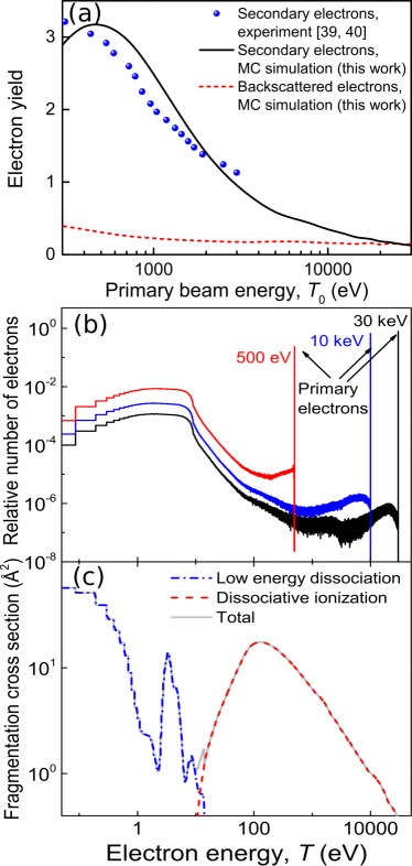
Figure 1(b) shows the relative number of electrons reaching the SiO2 surface with different energies. It can be seen that, for all PE energies , there is an intense SE peak at low energies, with its maximum at eV, while the number of BSE (those with larger energies closer to ) is in general small. Further benchmarks of energy distributions against experimental data appear in Supplementary Information S2.
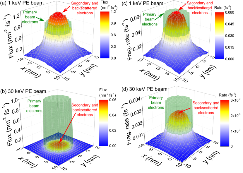
MC simulations also provide the space- and energy-dependent fluxes (electrons per unit area and unit time) of BSE and SE crossing the SiO2 surface at different positions. These are shown in Figs. 2(a) and (b) for uniform PE beams of 1 keV and 30 keV, respectively, and unit PE fluxes nm-2fs-1 within a circular area of radius nm. While the high energy 30 keV beam produces a small number of SE and BSE everywhere, the lower energy 1 keV beam produces a large number of SE and BSE, which spread outside the area covered by the PE beam and exceed the number of PE at the centre of the beam.
Electron-impact molecular fragmentation cross sections
Not only the number of electrons influences the properties of the structures emerging on the surface, but also the energy-dependent probability for W(CO)6 molecule fragmentation, given by the corresponding cross section , has an impact. This cross section includes dissociative ionisation (DI) for energies above the ionisation threshold ( eV Wnorowski2012a ) as well as dissociative electronic excitations and dissociative electron attachment Thorman2015 .
Measurement of for the molecular fragmentation channels on the substrate is rather complicated, since the influence of all PE, BSE and SE crossing the surface cannot be disentangled. Under these conditions, what is usually measured is an effective decomposition cross section due to a PE beam of energy , . Alternatively, gas-phase data may be used as a first approximation for the actual cross section . For W(CO)6 molecules, experimental information is available for DI Wnorowski2012a and lower energy dissociation channels Wnorowski2012 relative cross sections, but not the absolute values needed for our simulations. The absolute DI cross section can be calculated by means of the dielectric formalism deVera2019 . The corresponding result is shown in Fig. 1(c) by a dashed line. For energies below 14 eV, the experimental relative cross sections Wnorowski2012 can be scaled in order to get a decomposition cross section for 30 keV electrons incident in W(CO)6@SiO2 coinciding with the experimentally reported value Hoyle1994 (see Supplementary Information S3 for the details of the scaling procedure). The resulting low energy and total fragmentation cross sections appear in Fig. 1(c) as dash-dotted and solid lines, respectively. DI dominates above 12 eV, while a large fraction of SE will fragment precursor molecules through the lower energy dissociation channels.
Simulation of the FEBID process
The FEBID process relies on successive cycles of electron irradiation and precursor molecule replenishment Utke2008 ; Huth2018 . The irradiation phases are simulated by means of the IDMD method Sushko2016 by evaluating space-dependent bond dissociation rates for molecules on the substrate, which are calculated as explained in section “Methods”. In brief, these rates depend, in steady-state conditions, on both (i) the number and energies of the electrons crossing the SiO2 surface at each point per unit time and unit area (which in turn are determined by the PE beam energy and flux , see section “Electron beam interaction with the substrate”), and (ii) the energy-dependent molecular fragmentation cross section (determined in section “Electron-impact molecular fragmentation cross section”).
Figures 2(c) and (d) illustrate the space-dependent fragmentation rates induced by uniform 1 keV and 30 keV beams, respectively, of unit PE flux nm-2fs-1 within a circular area of radius nm. Although the number of BSE/SE electrons for 30 keV is small, their large cross section (in relation to PE) produces a significant fragmentation probability, but less than that due to PE at the beam area. However, for 1 keV, the fragmentation probability due to BSE/SE ( 80–90 % exclusively due to SE) is very large, and significantly extends beyond the PE beam area. These results clearly demonstrate the very different scenarios to be expected for beams of different energies and which will importantly influence the deposit properties, as well as the prominent role of low-energy SE on molecular fragmentation.
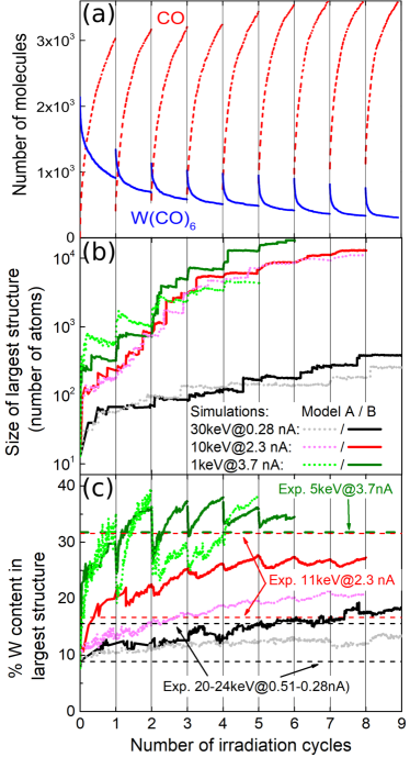
Each irradiation phase lasts for a time known as dwell time, which typical values in experiment (s) are still computationally demanding for MD. Instead, they are set here to 10 ns. Consequently, simulated PE fluxes (and hence PE beam currents ) must be scaled to match the same number of PE per unit area and per dwell time as in experiments Sushko2016 (see Supplementary Information S4.A). As for replenishment, its characteristic times are also typically very long (ms). In simulations, the CO molecules desorbed to the gas phase are simply removed during the replenishment stages and new W(CO)6 molecules are deposited. Figure 3(a) illustrates these successive irradiation-replenishment stages by depicting the number of W(CO)6 and free CO molecules during several of these cycles for a 30 keV PE beam of equivalent experimental current nA (in short, 30keV@5.9nA).
As the irradiation-replenishment cycles proceed, the process of nucleation of metal-enriched islands and its coalescence starts Sushko2016 . This is shown in Fig. 3(b), where the number of atoms in the largest island is shown for three simulation conditions close to reported in experiments Porrati2009 : 30keV@0.28nA, 10keV@2.3nA and 1keV@3.7nA. Results of two different models for the chemistry occurring within the growing nanostructure are presented Sushko2016 : in model A (dotted lines), dangling bonds of a given nanostructure can only react with unsaturated bonds belonging to a different molecule; in model B (solid lines), the restructuring of bonds within a growing nanostructure is also allowed (see Supplementary Information S4.C for further details). The jumps in the size of the largest island observed occasionally are due to the merging of independent nanoclusters that grow on the substrate.
As the islands grow, their average chemical composition also changes. The time evolution of the W-metal content of the largest island for the three aforementioned cases is depicted in Fig. 3(c) for the chemistry models A (dotted lines) and B (solid lines). The metal content grows fast during the first irradiation cycles, until it slowly starts to saturate for each set of beam parameters after 5 irradiation cycles. It is worth noting that our simulation results are consistent with experimental data Porrati2009 for the 20–24keV@0.28–0.51nA, 11keV@2.3nA and 5keV@3.7nA cases, represented by dashed horizontal lines in Fig. 3(c).
Experimental measurements were limited to particular values of energy and current due to the characteristics of the electron source Porrati2009 . Nonetheless, our simulation method allows for the exploration of much wider regions of electron beam parameters. To do so, we also considered the cases of 30keV@5.9nA, 0.5keV@0.25nA and 0.5keV@5.9nA, obtaining the deposit metal contents depicted by full symbols in Fig. 4(a), as a function of experimentally equivalent current . Error bars show the standard deviations obtained from three independent simulations for each case. Experimental results Porrati2009 are shown by open symbols. Numbers next to symbols represent the beam energies in keV. It is clearly seen that the results from simulations are within the range of experimental uncertainties, which indicates the predictive capabilities of the simulations.
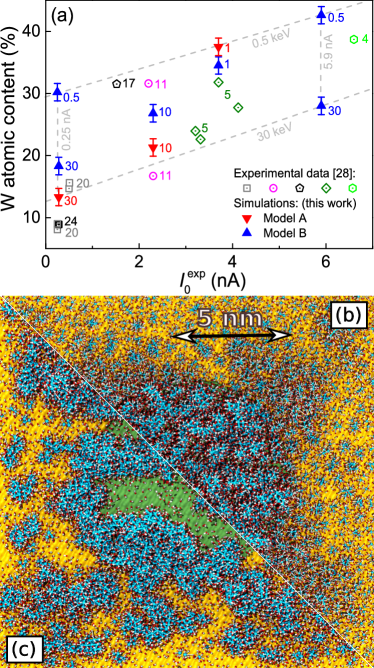
The cases analysed in this investigation provide a detailed “map” of the attainable metal contents in the deposits as a function of the beam parameters, which is a very valuable outcome for the optimisation of FEBID with W(CO)6@SiO2. This is marked in Fig. 4(a) by dashed lines corresponding to the limiting values of energy and current studied. These results clearly show that, within the analysed energy domain, a decrease in the beam energy and an increase in the current promote the faster growth of the deposit, as well as the augment in its metal content. Simulation results provide the grounds for clearly understanding such trends: an increment in the current means a larger number of PE per unit time, while a reduction in the energy produces an increase in the SE yield (Fig. 1(a)). These lead to both the greater size of the deposit and its larger metal content due to the increased probability for bond cleavage (Figs. 2(c)-(d)). It should be noted that a reduction of beam energy below 400 eV may diminish the metal content due to the lowering of the electron yields (Fig. 1).
Finally, Figs. 4(b) and (c) show top views of the simulated deposits for 1keV@3.7nA and 10keV@2.3nA, after 5 and 7 irradiation cycles, respectively (the number of atoms in the largest island is similar in these cases, ). The central green circular surface marks the area covered by the PE beam (of 5 nm radius). These figures help to understand how different energy-current regimes can lead to distinct deposit microstructures. While the higher energy beam of 10 keV produces a deposit almost entirely localised within the intended nanomanufacturing region (i.e., the PE beam area), the lower energy beam of 1 keV produces a more sparse and ramified deposit (at least during the early stage of the FEBID process), that significantly extends beyond the PE beam area, producing an undesired edge broadening of the structure. Although a detailed analysis of these effects deserves a more in-depth analysis (which is not possible within the limits of the present manuscript), it is worth to note that the SE yield goes from larger than 1 to lower than 1 in the 1–10 keV range (Fig. 1(a)), SE being the main responsible for the beam halo (Fig. 2). Such detailed predictions on the early stage of growth of metal deposits can be currently tested experimentally VanDorp2012a .
Conclusions
In this study we have demonstrated how to couple detailed space and energy distributions of electrons at the substrate surface (obtained from MC calculations Dapor2017 ; DaporBook ; Azzolini2018 ) with radiation-induced dynamics and chemical reactions simulations (by means of the IDMD technique Sushko2016 ; Solovyov2017b ) in order to describe radiation effects at the molecular level for experimentally relevant timescales. As a particular case study, and due to its relevance in nanotechnology, we have analysed the FEBID process for W(CO)6 precursor molecules on hydroxylated SiO2.
The presented results demonstrate how the novel MC-IDMD approach provides the necessary molecular insights into the key processes behind FEBID, which can be used for its further optimisation and development. Notably, the simulations (which rely on basic atomic and molecular data such as cross sections for electron scattering and molecular fragmentation) have demonstrated a great predictive power, yielding, for the first time, fabricated nanostructure compositions and morphologies in excellent agreement with available experimental data Porrati2009 . Particularly, the increase in both the growth rate and W-metal content of the deposits with the increase in PE beam current and with the decrease in its energy, have been shown to be related to the increase in the number of ejected low energy SE. The latter are also responsible for the different microsrtuctures and edge broadenings observed for beams of different energies. Many other aspects influencing FEBID and not addressed here (namely, other substrate-molecule combinations, different replenishment conditions Fowlkes2010 , the effects of contaminants or local heating by the PE beam Mutunga2019 , post-growth purification procedures…) can be analysed by utilising the protocols described in the present investigation.
Moreover, the new introduced methodology, which bridges the gap between other current approaches to describe radiation-induced effects spanning multiple space, time and energy scales, is general and readily applicable in many other important fields. It is worth noticing that mechanisms rather similar to the ones underlying FEBID (i.e., electron generation by different types of radiation, their transport and the chemistry induced on surfaces) are common to problems as diverse as the astrochemistry processes happening in interstellar ices due to cosmic radiation Tielens2013 ; Mason2014 , in the use of metallic nanoparticles as enhancers of modern radiotherapies Haume2016a ; Solovyov2017 or in photoelectrochemical devices Zhang2016 . This new MC-IDMD approach offers a valuable tool which might provide unprecedented insights in many relevant problems in physics, chemistry, materials science, biomedicine and related technologies, in which irradiation-driven chemistry and multiscale phenomena play an essential role.
Methods
Simulations were performed by means of the irradiation driven MD (IDMD) method Sushko2016 implemented in the MBN (Meso-Bio-Nano) Explorer software package Solovyov2012 ; Solovyov2017b . Within this framework, the space-dependent rate for bond cleavage in molecules on the substrate surface is given by:
| (1) | |||||
where a discrete set of values for the electron energies was assumed for simplicity, but without affecting the final results. are space- and energy-dependent fluxes of PE/SE/BSE (electrons per unit area and unit time) and is the energy-dependent molecular fragmentation cross section. The PE beam flux at the irradiated circular spot of radius is:
| (2) |
where corresponds to the PE beam current, to its area and is the elementary charge. Note that gives the space-dependent fluxes which are plotted in Figs. 2(a)-(b). For uniform PE beams, as used in this investigation, for every point with coordinates .
The electron distributions were simulated using the MC radiation transport code SEED (Secondary Electron Energy Deposition) Dapor2017 ; DaporBook ; Azzolini2018 . Molecular fragmentation and further chemical reactions were simulated by means of MBN Explorer Sushko2016 ; Solovyov2012 ; Solovyov2017b . Its dedicated user interface and multi-task toolkit, MBN Studio Sushko2019 , was employed for constructing the molecular system, performing the precursor molecule replenishment phases, as well as for analysing the IDMD simulation results.
Monte Carlo code SEED
The SEED code follows the classical trajectories of energetic electrons travelling inside a condensed phase material, by employing the usual Monte Carlo recipes for electron transport simulation DaporBook ; Dapor2017 ; Azzolini2018 . It is based on the calculation of (i) the differential inelastic scattering cross sections accurately obtained by using the dielectric formalism Dapor2017 ; deVera2013 ; deVera2015 , (ii) the electron-phonon quasi-elastic scattering cross-section computed by the use of the Fröhlich theory Frohlich1954 and (iii) the differential elastic scattering cross section performed by the relativistic partial wave expansion method (RPWEM) Jablonski2004 including the Ganachaud and Mokrani empirical correction for low electron energies ( 20–30 eV) Ganachaud1995 .
The empirical parameters for the Fröhlich and the Ganachaud-Mokrani theories are set in order to reproduce by simulation the experimentally known SE yield for SiO2 Glavatskikh2001 ; Yi2001 . See Supplementary Information S2 and Refs. DaporBook ; Dapor2017 ; Azzolini2018 for extended discussions on the SEED code and its validation.
MBN Explorer and Irradiation Driven Molecular Dynamics
MBN Explorer is a multi-purpose software package for advanced multiscale simulations of structure and dynamics of complex molecular systems Solovyov2012 ; Solovyov2017b , featuring a wide variety of computational algorithms for the simulation of atomistic and coarse-grained systems. It includes the advanced algorithms of reactive MD Sushko2015 and the unique IDMD Sushko2016 exploited in this investigation.
In the MD approach, the dynamics of a system is followed by numerically solving the coupled classical Langevin equations of motion of all its constituent atoms. The interaction forces are treated in this work by means of the CHARMM force field MacKerell1998 .
The IDMD algorithm implemented in MBN Explorer Sushko2016 superimposes random processes of molecular bond breakage due to irradiation during classical reactive MD. These processes are treated as local (involving the atoms participating in a chemical bond) energy deposition events occurring in the sub-femtosecond timescale, so they are considered to happen instantaneously between successive simulation time steps. They occur randomly, with a rate determined by the probabilities for quantum processes such as dissociative ionisation or dissociative electron attachment, Eq. (1). The fast relaxation of the excess energy after these interactions results in the cleavage of particular bonds and the formation of active species (radicals with unsaturated dangling bonds) which can undergo further chemical reactions.
The cleavage and formation of chemical bonds and the monitoring of the system’s dynamical topology, along with the redistribution of atomic partial charges, is managed by means of the reactive version of the CHARMM force field implemented in MBN Explorer Sushko2015 . Its parameterisation for the W(CO)6 molecule was described in an earlier study deVera2019frag . In this investigation we assume that every fragmentation event leads to the cleavage of a single W-C bond, while the much stronger C-O bonds will not react deVera2019frag . The energy deposited in the cleaved W-C bonds is chosen in accordance with average values obtained from mass spectrometry experiments Cooks1990 ; Wnorowski2012 and dedicated simulations of the molecule fragmentation deVera2019frag , see Supplementary Information S4.B.
The details of the IDMD methodology are explained in Sushko2016 , and all necessary details for its application to the system studied in this investigation are given in Supplementary Information S4.
Acknowledgements.
PdV gratefully acknowledges the Alexander von Humboldt Foundation/Stiftung and the Spanish Ministerio de Ciencia e Innovación for their financial support by means of, respectively, Humboldt (1197139) and Juan de la Cierva (FJCI-2017-32233) postdoctoral fellowships. MA is thankful to Prof. Nicola M. Pugno for managing her financial support. IAS acknowledges the Lundbeck Foundation and the Volkswagen Foundation (Lichtenberg Professorship) for their support. This work was also supported in part by the Deutsche Forschungsgemeinschaft (Projects no. 415716638 and GRK1885), the Spanish Ministerio de Ciencia e Innovación and the European Regional Development Fund (Project no. PGC2018-096788-B-I00), by the Fundación Séneca – Agencia de Ciencia y Tecnología de la Región de Murcia (Project No. 19907/GERM/15), by the Conselleria d’Educació, Investigació, Cultura i Esport de la Generalitat Valenciana (Project no. AICO/2019/070) and by the COST Action CA17126 “Towards understanding and modelling intense electronic excitation” (TUMIEE). The possibility to perform computer simulations at Goethe-HLR cluster of the Frankfurt Center for Scientific Computing is gratefully acknowledged.Additional information
Supplementary information is available in the online version of the manuscript.
References
- (1) Robertson, A. W. et al. Spatial control of defect creation in graphene at the nanoscale. Nature Communications 3, 1144 (2012).
- (2) Fowlkes, J. D. et al. Simulation-Guided 3D Nanomanufacturing via Focused Electron Beam Induced Deposition. ACS Nano 10, 6163–6172 (2016).
- (3) Sengupta, S. et al. Superconducting nanowires by electron-beam-induced deposition. Applied Physics Letters 106, 042601 (2015).
- (4) De Teresa, J. M. et al. Review of magnetic nanostructures grown by focused electron beam induced deposition (FEBID). Journal of Physics D: Applied Physics 49, 243003 (2016).
- (5) Jesse, S. et al. Directing Matter: Toward Atomic-Scale 3D Nanofabrication. ACS Nano 10, 5600–5618 (2016).
- (6) Fernández-Pacheco, A. et al. Three-dimensional nanomagnetism. Nature Communications 8, 15756 (2017).
- (7) Winkler, R. et al. Direct-Write 3D Nanoprinting of Plasmonic Structures. ACS Applied Materials and Interfaces 9, 8233–8240 (2017).
- (8) Huth, M., Porrati, F. & Dobrovolskiy, O. V. Focused electron beam induced deposition meets materials science. Microelectronic Engineering 185-186, 9–28 (2018).
- (9) Utke, I., Hoffmann, P. & Melngailis, J. Gas-assisted focused electron beam and ion beam processing and fabrication. Journal of Vacuum Science & Technology B: Microelectronics and Nanometer Structures 26, 1197–1276 (2008).
- (10) Thorman, R. M., Kumar, R., Fairbrother, D. H. & Ingólfsson, O. The role of low-energy electrons in focused electron beam induced deposition: Four case studies of representative precursors. Beilstein Journal of Nanotechnology 6, 1904–1926 (2015).
- (11) Shawrav, M. M. et al. Highly conductive and pure gold nanostructures grown by electron beam induced deposition. Scientific Reports 6, 34003 (2016).
- (12) Huth, M. et al. Focused electron beam induced deposition: A perspective. Beilstein Journal of Nanotechnology 3, 597–619 (2012).
- (13) Diaz De La Rubia, T. et al. Multiscale modelling of plastic flow localization in irradiated materials. Nature 406, 871–874 (2000).
- (14) Sushko, G. B., Solov’yov, I. A. & Solov’yov, A. V. Molecular dynamics for irradiation driven chemistry: application to the FEBID process. The European Physical Journal D 70, 217 (2016).
- (15) Solov’yov, I. A., Korol, A. V. & Solov’yov, A. V. Multiscale Modelling of Complex Molecular Structure and Dynamics with MBN Explorer (Springer International Publishing AG, Cham, Switzerland, 2017).
- (16) Mutunga, E. et al. Impact of Electron-Beam Heating during 3D Nanoprinting. ACS Nano 13, 5198–5213 (2019).
- (17) Smith, D. A., Fowlkes, J. D. & Rack, P. D. Understanding the kinetics and nanoscale morphology of electron-beam- induced deposition via a three-dimensional Monte Carlo simulation: the effects of the precursor molecule and the deposited material. Small 4, 1382–1389 (2008).
- (18) Plank, H., Smith, D. A., Haber, T., Rack, P. D. & Hofer, F. Fundamental proximity effects in focused electron beam induced deposition. ACS Nano 6, 286–294 (2012).
- (19) Dapor, M. Transport of Energetic Electrons in Solids. Computer Simulation with Applications to Materials Analysis and Characterization (Springer International Publishing AG, Cham, Switzerland, 2020), 3 edn.
- (20) Muthukumar, K., Opahle, I., Shen, J., Jeschke, H. O. & Valentí, R. Interaction of W(CO)6 with SiO2 surfaces: A density functional study. Physical Review B - Condensed Matter and Materials Physics 84, 205442 (2011).
- (21) Stumpf, V., Gokhberg, K. & Cederbaum, L. S. The role of metal ions in X-ray-induced photochemistry. Nature Chemistry 8, 237–241 (2016).
- (22) Rogers, J. P., Anstöter, C. S. & Verlet, J. R. Ultrafast dynamics of low-energy electron attachment via a non-valence correlation-bound state. Nature Chemistry 10, 341–346 (2018).
- (23) Sushko, G. B., Solov’yov, I. A., Verkhovtsev, A. V., Volkov, S. N. & Solov’yov, A. V. Studying chemical reactions in biological systems with MBN Explorer: implementation of molecular mechanics with dynamical topology. European Physical Journal D 70, 12 (2016).
- (24) de Vera, P., Verkhovtsev, A., Sushko, G. & Solov’yov, A. V. Reactive molecular dynamics simulations of organometallic compound W(CO)6 fragmentation. European Physical Journal D 73, 215 (2019).
- (25) Solov’yov, I. A., Yakubovich, A. V., Nikolaev, P. V., Volkovets, I. & Solov’yov, A. V. MesoBioNano Explorer–a universal program for multiscale computer simulations of complex molecular structure and dynamics. Journal of computational chemistry 33, 2412–39 (2012).
- (26) Dapor, M., Abril, I., de Vera, P. & Garcia-Molina, R. Energy deposition around swift proton tracks in polymethylmethacrylate: How much and how far. Physical Review B 96, 064113 (2017).
- (27) Azzolini, M. et al. Secondary electron emission and yield spectra of metals from Monte Carlo simulations and experiments. Journal of Physics Condensed Matter 31, 055901 (2018).
- (28) Porrati, F., Sachser, R. & Huth, M. The transient electrical conductivity of W-based electron-beam-induced deposits during growth, irradiation and exposure to air. Nanotechnology 20, 195301 (2009).
- (29) Tielens, A. G. G. M. The molecular universe. Reviews of Modern Physics 85, 1021–1081 (2013).
- (30) Mason, N. J., Nair, B., Jheeta, S. & Szymanska, E. Electron Induced Chemistry: A New Frontier in Astrochemistry. Faraday Discussions 168, 235 (2014).
- (31) Solov’yov, A. V. (ed.) Nanoscale Insights into Ion-Beam Cancer Therapy (Springer International Publishing AG, Cham, Switzerland, 2017).
- (32) Surdutovich, E. & Solov’yov, A. V. Multiscale modeling for cancer radiotherapies. Cancer Nanotechnology 10, 1–22 (2019).
- (33) Zhang, X. & Bieberle-Hütter, A. Modeling and Simulations in Photoelectrochemical Water Oxidation: From Single Level to Multiscale Modeling. ChemSusChem 9, 1223–1242 (2016).
- (34) Fowlkes, J. D. & Rack, P. D. Fundamental Electron-Precursor-Solid Deposition Simulations and Experiments. ACS Nano 4, 1619–1629 (2010).
- (35) de Vera, P., Garcia-Molina, R., Abril, I. & Solov’yov, A. V. Semiempirical Model for the Ion Impact Ionization of Complex Biological Media. Physical Review Letters 110, 148104 (2013).
- (36) de Vera, P., Garcia-Molina, R. & Abril, I. Angular and Energy Distributions of Electrons Produced in Arbitrary Biomaterials by Proton Impact. Physical Review Letters 114, 018101 (2015).
- (37) de Vera, P. & Garcia-Molina, R. Electron inelastic mean free paths in condensed matter down to a few electronvolts. Journal of Physical Chemistry C 123, 2075–2083 (2019).
- (38) Jablonski, A., Salvat, F. & Powell, C. J. Comparison of Electron Elastic-Scattering Cross Sections Calculated from Two Commonly Used Atomic Potentials. Journal of Physical and Chemical Reference Data 33, 409–451 (2004).
- (39) Glavatskikh, I. A., Kortov, V. S. & Fitting, H. J. Self-consistent electrical charging of insulating layers and metal-insulator-semiconductor structures. Journal of Applied Physics 89, 440–448 (2001).
- (40) Yi, W. et al. Study of the secondary-electron emission from thermally grown SiO2 films on Si. Thin Solid Films 397, 170–175 (2001).
- (41) Wnorowski, K., Stano, M., Barszczewska, W., Jówko, A. & Matejčík, S. Electron ionization of W(CO)6: Appearance energies. International Journal of Mass Spectrometry 314, 42–48 (2012).
- (42) Wnorowski, K. et al. Low-energy electron interactions with tungsten hexacarbonyl - W(CO)6. Rapid Communications in Mass Spectrometry 26, 2093–2098 (2012).
- (43) Hoyle, P. C., Cleaver, J. R. A. & Ahmed, H. Ultralow-energy focused electron beam induced deposition. Applied Physics Letters 64, 1448–1450 (1994).
- (44) Van Dorp, W. F. et al. Molecule-by-molecule writing using a focused electron beam. ACS Nano 6, 10076–10081 (2012).
- (45) Haume, K. et al. Gold nanoparticles for cancer radiotherapy: a review. Cancer Nanotechnology 7, 8 (2016).
- (46) Sushko, G. B., Solov’yov, I. A. & Solov’yov, A. V. Modeling MesoBioNano systems with MBN Studio made easy. Journal of Molecular Graphics and Modelling 88, 247–260 (2019).
- (47) Fröhlich, H. Electrons in Lattice Fields. Advances in Physics 3, 325–361 (1954).
- (48) Ganachaud, J. P. & Mokrani, A. Theoretical study of the secondary electron emission of insulating targets. Surface Science 334, 329–341 (1995).
- (49) MacKerell, A. et al. All-atom empirical potential for molecular modeling and dynamics studies of proteins. Journal of Physical Chemistry B 102, 3586–3616 (1998).
- (50) Cooks, R. G., Ast, T., Kralj, B., Kramer, V. & Žigon, D. Internal energy distributions deposited in doubly and singly charged tungsten hexacarbonyl ions generated by charge stripping, electron impact, and charge exchange. Journal of the American Society for Mass Spectrometry 1, 16–27 (1990).