MIDL 2020Medical Imaging with Deep Learning 2020
\jmlrvolume
\jmlryear
\jmlrworkshopMIDL 2020 – Short Paper
\midlauthor\NameDaniel Moyer \nametag1,2,3 \Emaildmoyer@csail.mit.edu
\addr1 CSAIL, Massachusetts Institute of Technology, Cambridge, MA, United States
\addr2 Information Sciences Institute, Marina del Rey, CA, United States
\addr3 Imaging Genetics Center, Keck School of Medicine, University of Southern California, Los Angeles, CA, United States and \NameGreg Ver Steeg \nametag2 \Emailgregv@isi.edu
\NamePaul M Thompson \nametag3 \Emailpthomp@usc.edu
Overview of Scanner Invariant Representations
Abstract
Pooled imaging data from multiple sources is subject to bias from each source. Studies that do not correct for these scanner/site biases at best lose statistical power, and at worst leave spurious correlations in their data. Estimation of the bias effects is non-trivial due to the paucity of data with correspondence across sites, so called ”traveling phantom” data, which is expensive to collect. Nevertheless, numerous solutions leveraging direct correspondence have been proposed. In contrast to this, Moyer et al. (2019) proposes an unsupervised solution using invariant representations, one which does not require correspondence and thus does not require paired images. By leveraging the data processing inequality, an invariant representation can then be used to create an image reconstruction that is uninformative of its original source, yet still faithful to the underlying structure. In the present abstract we provide an overview of this method.
keywords:
Harmonization, diffusion MRI, Invariant Representation1 Introduction and Summary
In magnetic resonance imaging (MRI), variations in observational conditions, protocol, and equipment induce site-wise and scanner-wise biases in the collected data [Chen et al.(2014)Chen, Liu, Calhoun, Arias-Vasquez, Zwiers, Gupta, Franke, and Turner, Fortin et al.(2017)Fortin, Parker, Tunc, Watanabe, Elliott, Ruparel, Roalf, Satterthwaite, Gur, Gur, et al., Jovicich et al.(2006)Jovicich, Czanner, Greve, Haley, van Der Kouwe, Gollub, Kennedy, Schmitt, Brown, MacFall, et al.]. Without correcting for these biases, multi-site studies will at best lose statistical power, and in some cases may arrive at erroneous conclusions. It is therefore imperative in multi-site studies to make these corrections; the process of doing so, removing or compensating for unwanted scanner/site-wise variations, is known as harmonization. In the present work we focus on harmonization for diffusion MRI (dMRI), a modality known to have scanner/site biases [Correia et al.(2009)Correia, Carpenter, and Williams, Giannelli et al.(2010)Giannelli, Cosottini, Michelassi, Lazzarotti, Belmonte, Bartolozzi, and Lazzeri, Pagani et al.(2010)Pagani, Hirsch, Pouwels, Horsfield, Perego, Gass, Roosendaal, Barkhof, Agosta, Rovaris, et al., Papinutto et al.(2013)Papinutto, Maule, and Jovicich, Vollmar et al.(2010), White et al.(2009), Zhan et al.(2010)Zhan, Leow, Jahanshad, Chiang, Barysheva, Lee, Toga, McMahon, de Zubicaray, Wright, et al., Zhan et al.(2013)Zhan, Mueller, Jahanshad, Jin, Lenglet, Yacoub, Sapiro, Ugurbil, Harel, Toga, et al., Zhan et al.(2012)] as well as several extra possible degrees of freedom with respect to protocol (e.g., angular resolution, -values, gradient waveform choice, etc.).
Previous work has largely focused on the summary statistic level (e.g. Fractional Anisotropy) [Fortin et al.(2017)Fortin, Parker, Tunc, Watanabe, Elliott, Ruparel, Roalf, Satterthwaite, Gur, Gur, et al., Zavaliangos-Petropulu et al.(2018)Zavaliangos-Petropulu, Nir, Thomopoulos, et al.] or on supervised cases where pairs of images from the same subject collected with different scanners are provided [Blumberg et al.(2018)Blumberg, Tanno, Kokkinos, and Alexander, Tanno et al.(2017)Tanno, Worrall, Ghosh, Kaden, Sotiropoulos, Criminisi, and Alexander]. These methods attempt to estimate the relative effects of each scanner/site, either in derived measures or in the original data domain.
Moyer et al. [Moyer et al.(2019)Moyer, Steeg, Tax, and Thompson] present an unsupervised method that instead learns a representation of the images that is uninformed of the scanner/site at which they were collected, yet also one that is otherwise maximally informative of the image. Reconstructions from this uninformed representation will then be uninformed of their original scanner/site context, a result which follows from data processing inequality.
The construction of invariant representations is, generally, non-trivial. A previous paper by Moyer et al. [Moyer et al.(2018)Moyer, Gao, Brekelmans, Galstyan, and Ver Steeg] show that this can be done by compressed conditional auto-encoder, where the learned encoding becomes uninformed of the conditional factor under compression. This leads to the following procedure:
-
1.
Construct an auto-encoder for image data, using compressive regularization (e.g. penalizing for data and encoding ), and condition the output on the site.
-
2.
Train the auto-encoder on images from each scanner/site independently.
-
3.
At test time, manipulate the conditional decoder to remap images through the learned invariant code to a single scanner/site context.
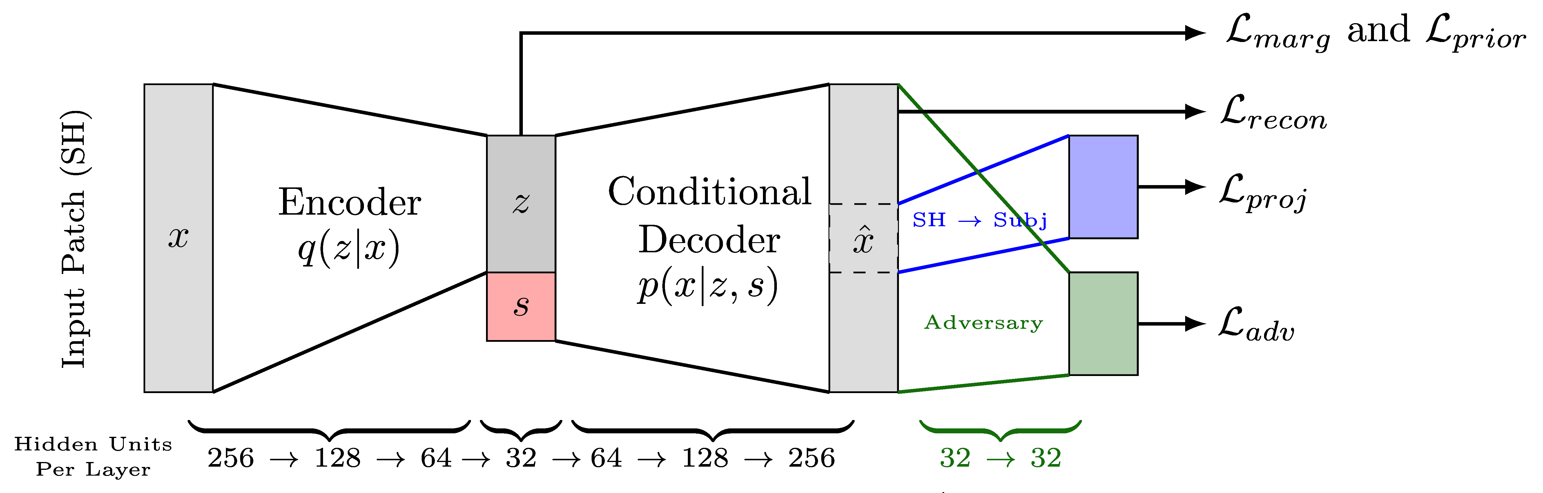
As described in [Moyer et al.(2018)Moyer, Gao, Brekelmans, Galstyan, and Ver Steeg], conditional architectures such as the one described here (Fig. 1) penalize learned site information . In reducing site information in this architecture reduces , the site information in the image reconstruction.
Due to hardware limitations, the method was applied patch-wise. The authors further included an adversary on the patch-wise output. To generalize representations across particular gradient directions, a spherical harmonics representation was used at the voxel level, which was projected back to subject specific directions when calculating reconstruction loss. The proposed auto-encoders may be learned either for the two-site case (“single-task”) or the multi-site case (“multi-task”).
2 Overview of the Empirical Evaluation
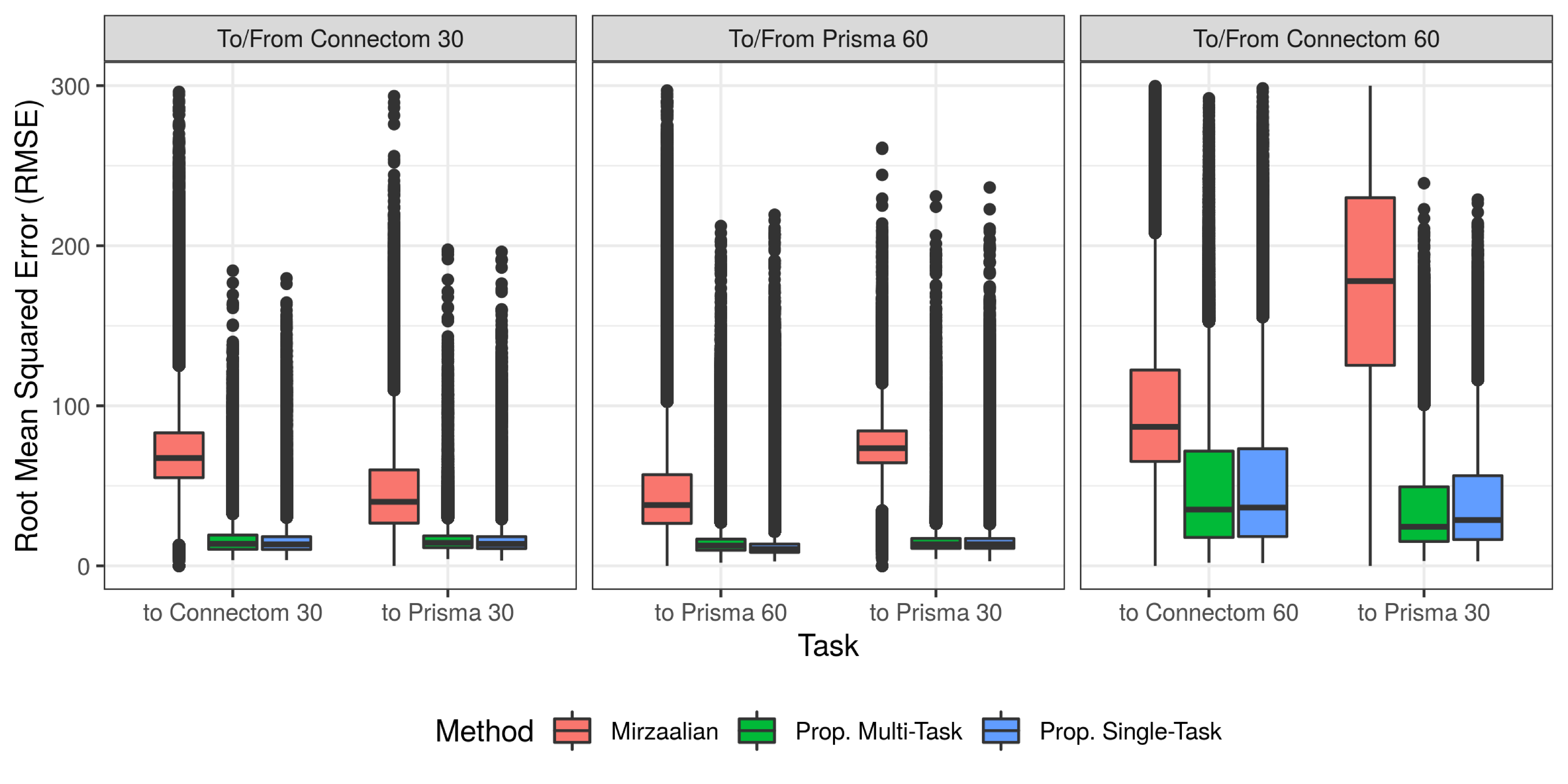
Moyer et al. [Moyer et al.(2019)Moyer, Steeg, Tax, and Thompson] present an evaluation of this method on the 2018 CDMRI Challenge Dataset [Ning et al.(2018)Ning, Bonet-Carne, Grussu, Sepehrband, Kaden, Veraart, Blumberg, Khoo, Palombo, Coll-Font, et al., Tax et al.(2019)Tax, Grussu, Kaden, Ning, Rudrapatna, Evans, St-Jean, Leemans, Koppers, Merhof, et al., Tax et al.(2018)Tax, Grussu, Kaden, Ning, Rudrapatna, Evans, St-Jean, Leemans, Puch, Rowe, et al.], which is composed of images from 15 subjects. As described in [Ning et al.(2018)Ning, Bonet-Carne, Grussu, Sepehrband, Kaden, Veraart, Blumberg, Khoo, Palombo, Coll-Font, et al.] data from each subject were collected on a 3 T GE Excite-HD “Connectom” and a 3 T Siemens Prisma scanner, each with two separate protocols. One protocol matches between the scanners at a low resolution (2.4 mm iso., with 30 grad. directions), and another which does not match at a high resolution (1.5 mm and 1.2mm iso., same shells 60 grad. directions). This creates 4 “sites” (denoted P30,P60,C30,C60). Scans are mapped to and from P30 for each other site.
The authors split this dataset into 9 training subjects, 1 validation subject, and 5 held out-test subjects. One baseline comparison was found in the literature and compared to, Mirzaalian et al [Mirzaalian et al.(2018)], which relies on a template based solution. For the proposed method, a post-hoc adversary was fit to the learned code, as a lower-bound proxy [Moyer et al.(2018)Moyer, Gao, Brekelmans, Galstyan, and Ver Steeg] for remaining mutual information ; a further ablation test for each network component was conducted. As show in Figure 2, the proposed methods significantly reduce predictive error, while also removing site information, as demonstrated by the post-hoc adversary.
| Post-hoc Adversarial Accuracy, Predicting from | ||||
| Oracle | Full Model | No | No or | |
| Proposed Single-task, C30 | 0.5 | 0.61 | 0.63 | 0.63 |
| Proposed Single-task, P60 | 0.5 | 0.5 | 0.51 | 0.54 |
| Proposed Single-task, C60 | 0.5 | 0.63 | 0.68 | 0.85 |
| Proposed Multi-task | 0.25 | 0.41 | 0.41 | 0.62 |
3 Discussion and Limitations
Moyer et al. [Moyer et al.(2019)Moyer, Steeg, Tax, and Thompson] presents an alternative method for harmonization, one using intermediate scanner invariant representations to remove scanner/site information. The proposed methods produces quality results, while removing site information from the intermediate encoding.
This summary was supported by NIH grants P41 EB015922, R01 MH116147, R56 AG058854, RF1 AG041915, and U54 EB020403, DARPA grant W911NF-16-1-0575, as well as the NSF Graduate Research Fellowship Program Grant Number DGE-1418060, and a GPU grant from NVidia.
References
- [Blumberg et al.(2018)Blumberg, Tanno, Kokkinos, and Alexander] Stefano B Blumberg, Ryutaro Tanno, Iasonas Kokkinos, and Daniel C Alexander. Deeper image quality transfer: Training low-memory neural networks for 3D images. In MICCAI, pages 118–125. Springer, 2018.
- [Chen et al.(2014)Chen, Liu, Calhoun, Arias-Vasquez, Zwiers, Gupta, Franke, and Turner] Jiayu Chen, Jingyu Liu, Vince D Calhoun, Alejandro Arias-Vasquez, Marcel P Zwiers, Cota Navin Gupta, Barbara Franke, and Jessica A Turner. Exploration of scanning effects in multi-site structural MRI studies. Journal of Neuroscience Methods, 230:37–50, 2014.
- [Correia et al.(2009)Correia, Carpenter, and Williams] Marta Morgado Correia, Thomas A Carpenter, and Guy B Williams. Looking for the optimal DTI acquisition scheme given a maximum scan time: are more b-values a waste of time? Magnetic Resonance Imaging, 27(2):163–175, 2009.
- [Fortin et al.(2017)Fortin, Parker, Tunc, Watanabe, Elliott, Ruparel, Roalf, Satterthwaite, Gur, Gur, et al.] Jean-Philippe Fortin, Drew Parker, Birkan Tunc, Takanori Watanabe, Mark A Elliott, Kosha Ruparel, David R Roalf, Theodore D Satterthwaite, Ruben C Gur, Raquel E Gur, et al. Harmonization of multi-site diffusion tensor imaging data. Neuroimage, 161:149–170, 2017.
- [Giannelli et al.(2010)Giannelli, Cosottini, Michelassi, Lazzarotti, Belmonte, Bartolozzi, and Lazzeri] Marco Giannelli, Mirco Cosottini, Maria Chiara Michelassi, Guido Lazzarotti, Gina Belmonte, Carlo Bartolozzi, and Mauro Lazzeri. Dependence of brain DTI maps of fractional anisotropy and mean diffusivity on the number of diffusion weighting directions. Journal of Applied Clinical Medical Physics, 11(1):176–190, 2010.
- [Jovicich et al.(2006)Jovicich, Czanner, Greve, Haley, van Der Kouwe, Gollub, Kennedy, Schmitt, Brown, MacFall, et al.] Jorge Jovicich, Silvester Czanner, Douglas Greve, Elizabeth Haley, Andre van Der Kouwe, Randy Gollub, David Kennedy, Franz Schmitt, Gregory Brown, James MacFall, et al. Reliability in multi-site structural MRI studies: effects of gradient non-linearity correction on phantom and human data. Neuroimage, 30(2):436–443, 2006.
- [Mirzaalian et al.(2018)] Hengameh Mirzaalian et al. Multi-site harmonization of diffusion MRI data in a registration framework. Brain Imaging and Behavior, 12(1):284–295, 2018.
- [Moyer et al.(2018)Moyer, Gao, Brekelmans, Galstyan, and Ver Steeg] Daniel Moyer, Shuyang Gao, Rob Brekelmans, Aram Galstyan, and Greg Ver Steeg. Invariant representations without adversarial training. In Advances in Neural Information Processing Systems 31, pages 9102–9111. Curran Associates, Inc., 2018.
- [Moyer et al.(2019)Moyer, Steeg, Tax, and Thompson] Daniel Moyer, Greg Ver Steeg, Chantal MW Tax, and Paul M Thompson. Scanner invariant representations for diffusion mri harmonization. arXiv preprint arXiv:1904.05375, 2019.
- [Ning et al.(2018)Ning, Bonet-Carne, Grussu, Sepehrband, Kaden, Veraart, Blumberg, Khoo, Palombo, Coll-Font, et al.] Lipeng Ning, Elisenda Bonet-Carne, Francesco Grussu, Farshid Sepehrband, Enrico Kaden, Jelle Veraart, Stefano B Blumberg, Can Son Khoo, Marco Palombo, Jaume Coll-Font, et al. Multi-shell diffusion MRI harmonisation and enhancement challenge (MUSHAC): progress and results. In International Conference on Medical Image Computing and Computer-Assisted Intervention, pages 217–224. Springer, 2018.
- [Pagani et al.(2010)Pagani, Hirsch, Pouwels, Horsfield, Perego, Gass, Roosendaal, Barkhof, Agosta, Rovaris, et al.] Elisabetta Pagani, Jochen G Hirsch, Petra JW Pouwels, Mark A Horsfield, Elisabetta Perego, Achim Gass, Stefan D Roosendaal, Frederik Barkhof, Federica Agosta, Marco Rovaris, et al. Intercenter differences in diffusion tensor MRI acquisition. Journal of Magnetic Resonance Imaging, 31(6):1458–1468, 2010.
- [Papinutto et al.(2013)Papinutto, Maule, and Jovicich] Nico Dario Papinutto, Francesca Maule, and Jorge Jovicich. Reproducibility and biases in high field brain diffusion MRI: An evaluation of acquisition and analysis variables. Magnetic Resonance Imaging, 31(6):827–839, 2013.
- [Tanno et al.(2017)Tanno, Worrall, Ghosh, Kaden, Sotiropoulos, Criminisi, and Alexander] Ryutaro Tanno, Daniel E Worrall, Aurobrata Ghosh, Enrico Kaden, Stamatios N Sotiropoulos, Antonio Criminisi, and Daniel C Alexander. Bayesian image quality transfer with CNNs: Exploring uncertainty in dMRI super-resolution. In MICCAI, pages 611–619. Springer, 2017.
- [Tax et al.(2018)Tax, Grussu, Kaden, Ning, Rudrapatna, Evans, St-Jean, Leemans, Puch, Rowe, et al.] Chantal MW Tax, Francesco Grussu, Enrico Kaden, Lipeng Ning, Umesh Rudrapatna, John Evans, Samuel St-Jean, Alexander Leemans, Santi Puch, Matt Rowe, et al. Cross-vendor and cross-protocol harmonisation of diffusion MRI data: a comparative study. In International Symposium on Magnetic Resonance in Medicine (Paris), volume 471, 2018.
- [Tax et al.(2019)Tax, Grussu, Kaden, Ning, Rudrapatna, Evans, St-Jean, Leemans, Koppers, Merhof, et al.] Chantal MW Tax, Francesco Grussu, Enrico Kaden, Lipeng Ning, Umesh Rudrapatna, John Evans, Samuel St-Jean, Alexander Leemans, Simon Koppers, Dorit Merhof, et al. Cross-scanner and cross-protocol diffusion mri data harmonisation: A benchmark database and evaluation of algorithms. Neuroimage, 2019.
- [Vollmar et al.(2010)] Christian Vollmar et al. Identical, but not the same: intra-site and inter-site reproducibility of fractional anisotropy measures on two 3.0 T scanners. Neuroimage, 51(4):1384–1394, 2010.
- [White et al.(2009)] Tonya White et al. Global white matter abnormalities in schizophrenia: a multisite diffusion tensor imaging study. Schizophrenia Bulletin, 37(1):222–232, 2009.
- [Zavaliangos-Petropulu et al.(2018)Zavaliangos-Petropulu, Nir, Thomopoulos, et al.] Artemis Zavaliangos-Petropulu, Talia M Nir, Sophia I Thomopoulos, et al. Diffusion MRI indices and their relation to cognitive impairment in brain aging: The updated multi-protocol approach in ADNI3. bioRxiv, page 476721, 2018.
- [Zhan et al.(2010)Zhan, Leow, Jahanshad, Chiang, Barysheva, Lee, Toga, McMahon, de Zubicaray, Wright, et al.] Liang Zhan, Alex D Leow, Neda Jahanshad, Ming-Chang Chiang, Marina Barysheva, Agatha D Lee, Arthur W Toga, Katie L McMahon, Greig I de Zubicaray, Margaret J Wright, et al. How does angular resolution affect diffusion imaging measures? Neuroimage, 49(2):1357–1371, 2010.
- [Zhan et al.(2013)Zhan, Mueller, Jahanshad, Jin, Lenglet, Yacoub, Sapiro, Ugurbil, Harel, Toga, et al.] Liang Zhan, Bryon A Mueller, Neda Jahanshad, Yan Jin, Christophe Lenglet, Essa Yacoub, Guillermo Sapiro, Kamil Ugurbil, Noam Harel, Arthur W Toga, et al. Magnetic resonance field strength effects on diffusion measures and brain connectivity networks. Brain Connectivity, 3(1):72–86, 2013.
- [Zhan et al.(2012)] Liang Zhan et al. How do spatial and angular resolution affect brain connectivity maps from diffusion MRI? In Biomedical Imaging (ISBI), 2012 9th IEEE International Symposium on, pages 1–4. IEEE, 2012.