MIDL 2020Medical Imaging with Deep Learning 2020
\jmlrvolume
\jmlryear
\jmlrworkshopMIDL 2020 – Short Paper
\midlauthor\NameBianca Lassen-Schmidt\nametag1 \Emailbianca.lassen@mevis.fraunhofer.de
\NameAlessa Hering\nametag1 \Emailalessa.hering@mevis.fraunhofer.de
\NameStefan Krass\nametag1 \Emailstefan.krass@mevis.fraunhofer.de
\NameHans Meine\nametag1 \Emailhans.meine@mevis.fraunhofer.de
\addr1 Fraunhofer MEVIS, Bremen, Germany
Automatic segmentation of the pulmonary lobes with a 3D u-net and optimized loss function
Abstract
Fully-automatic lung lobe segmentation is challenging due to anatomical variations, pathologies, and incomplete fissures. We trained a 3D u-net for pulmonary lobe segmentation on 49 mainly publically available datasets and introduced a weighted Dice loss function to emphasize the lobar boundaries. To validate the performance of the proposed method we compared the results to two other methods. The new loss function improved the mean distance to 1.46 mm (compared to 2.08 mm for simple loss function without weighting).
keywords:
pulmonary lobes, lung lobes, segmentation, deep learning, CNN, 3D U-net1 Introduction
The human lungs are subdived into five lobes with separated supply branches for both vessels and airways. Usually, a visceral pleura called the pulmonary fissure can be found between adjacent lobes. Accurate segmentation of the five pulmonary lobes is important for diagnosis, treatment planning and monitoring for lung diseases such as COPD and fibrosis. In the last two decades several automatic approaches were developed [Doel et al.(2015)Doel, Gavaghan, and Grau] each has different drawbacks and limitations. A few recently published approached are based on deep learning [Gerard and Reinhardt(2019), Wang et al.(2019)Wang, Chen, Zhao, Chi, Xie, Zhang, and Hua, Tang et al.(2019)Tang, Zhang, and Xie, Park et al.(2019)Park, Yun, Kim, Park, Cho, Park, Song, Lee, and Seo, Lee et al.(2019)Lee, Matin, Gleeson, and Grau, Imran et al.(2018)Imran, Hatamizadeh, Ananth, Ding, Terzopoulos, and Tajbakhsh, George et al.(2017)George, Harrison, Jin, Xu, and Mollura]. But none of them includes explicit knowledge from pulmonary fissures as weighting into the loss function. In this paper, we propose a fully automatic lobe segmentation method based on a 3D u-net with an optimized loss function focussing the lobar boundaries. We compare the new method with a proven fully automatic segmentation approach [Lassen et al.(2013)Lassen, van Rikxoort, Schmidt, Kerkstra, van Ginneken, and Kuhnigk] that won the LOLA11 challenge [LOLA11()] at publication time.
2 Method & Data
Pulmonary fissures are the most accurate feature for lobe segmentation but these are often incomplete and there can be accessory fissures in the lungs. We train a 3D u-net to rely on visible fissures and simultaneously learn that fissures are often incomplete.
Data: We use 70 lung CT scans, randomly subdivided into 49 training, 7 validation and 14 testing data sets. 50 datasets are taken from LIDC/IDRI [Armato et al.(2011)Armato, McLennan, Bidaut, McNitt-Gray, Meyer, Reeves, Zhao, Aberle, Henschke, Hoffman, Kazerooni, MacMahon, Van Beeke, Yankelevitz, Biancardi, Bland, Brown, Engelmann, Laderach, Max, Pais, Qing, Roberts, Smith, Starkey, Batrah, Caligiuri, Farooqi, Gladish, Jude, Munden, Petkovska, Quint, Schwartz, Sundaram, Dodd, Fenimore, Gur, Petrick, Freymann, Kirby, Hughes, Casteele, Gupte, Sallamm, Heath, Kuhn, Dharaiya, Burns, Fryd, Salganicoff, Anand, Shreter, Vastagh, and Croft] with public available reference segmentations by Tang et al. [Tang et al.(2019)Tang, Zhang, and Xie] and 20 datasets are from the University Medical Center Utrecht that are also used in [Lassen et al.(2013)Lassen, van Rikxoort, Schmidt, Kerkstra, van Ginneken, and Kuhnigk]. Reference segmentations for the 20 datasets are generated with an automatic segmentation and interactive correction.
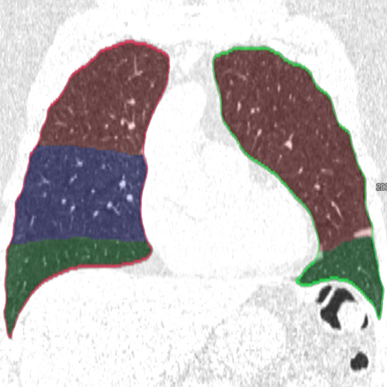
fig:method
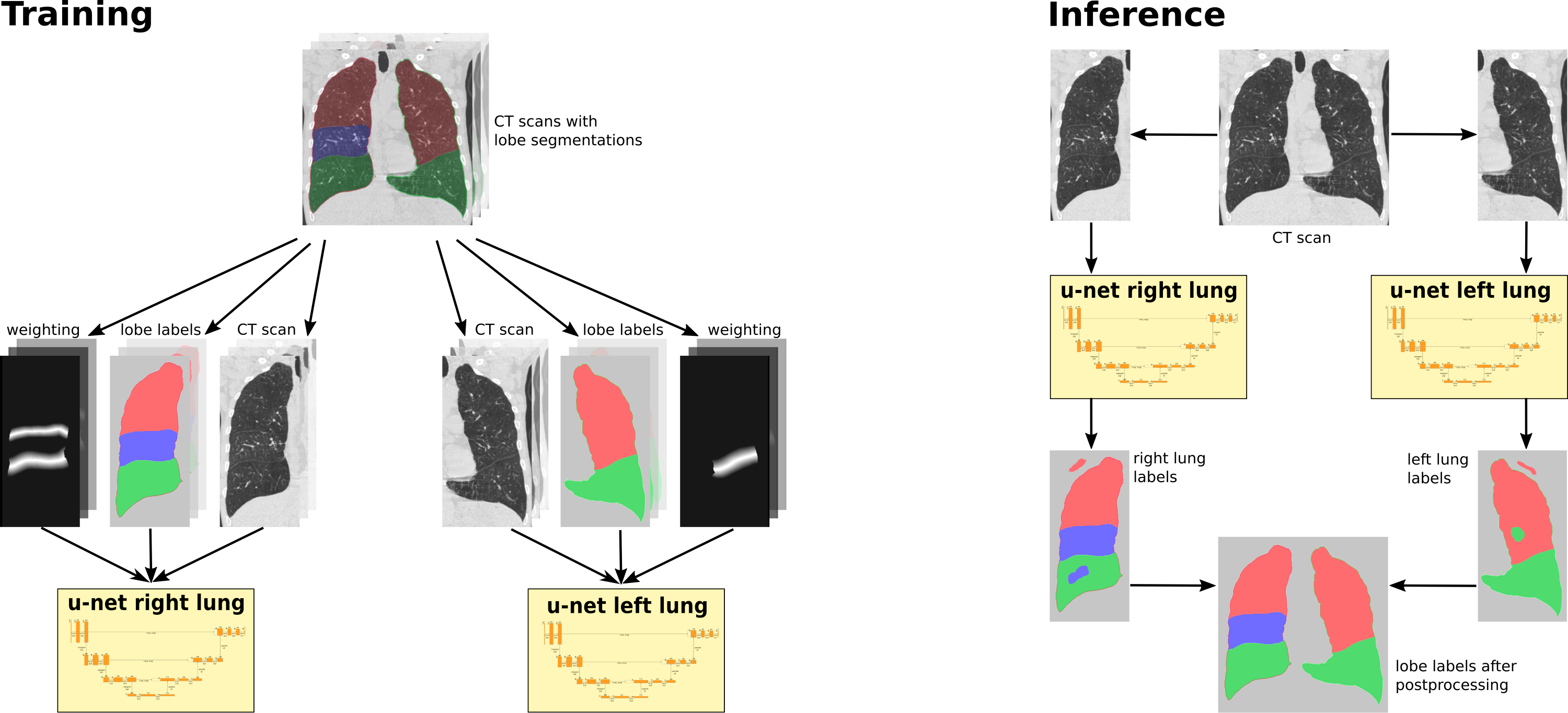
Training: We train two separate 3D u-nets (see Figure LABEL:fig:method), one for the left lung with 3 labels (background, upper lobe, lower lobe) and one for the right lung with 4 labels (background, upper lobe, middle lobe, lower lobe). We use 4 resolution levels, a filter size of for the convolutions, PReLu activation, batch normalization and dropout.
The most promising features for lung lobe segmentation can be found at the lobar boundaries. We want our networks to learn two facts: 1. pulmonary fissures are at the lobar boundaries, but 2. there might be no fissures at the lobar boundaries (incomplete fissures). Therefore we introduce a weighted Dice loss in the following manner: We calculate an Euclidean distance transformation (EDT) from the lobar boundaries of the reference segmentation. Next, we invert the distance dist within a radius of 10 mm by setting the values to 10 - dist and set the remaining part of the lungs to a value of 1. This results in a weighting image with a value of 10 at the lobar boundaries descending to value 1 within 10 mm radius. We use the EDT because the reference segmentation might not be exact on the lobar boundaries and we do not want to miss information from visible fissures. Thus, we use the following loss function:
As a preprocessing step the input images are resampled to 1.5 . The 49 training datasets are subdivided into patches of size voxels with 44 voxels padding on all six sides. We start the training with a learning rate of 0.005, a batchsize of 2 and stop after 70000 iterations.
Inference: For segmentation, we first resample a CT scan to 1.5 . Next, we apply our trained networks for the left and the right lung lobes and upsample the image back to the original resolution. We propose the following postprocessing to deal with remaining misclassifications regions (see Figure LABEL:fig:method): We perform a connected component analysis and keep the largest two (three) components in the left (right) lung. Finally, we fill the holes with a Voronoi division and use a given lung mask to delete all objects outside the lungs.
3 Evaluation & Results
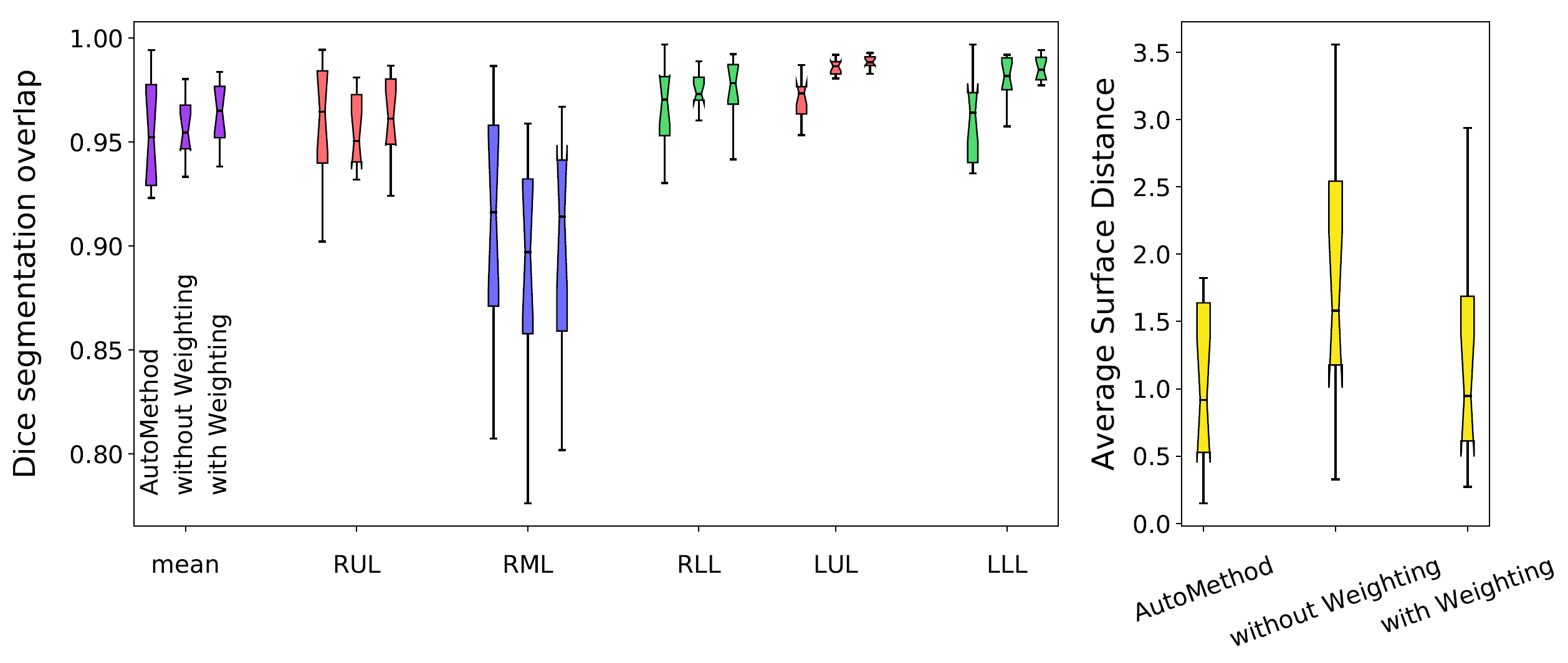
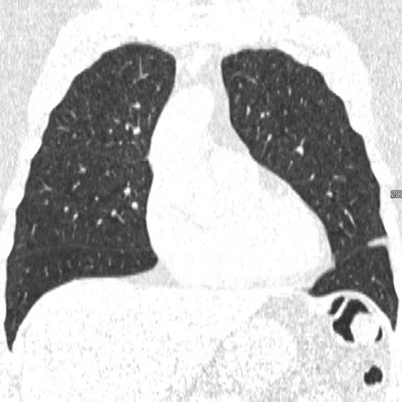
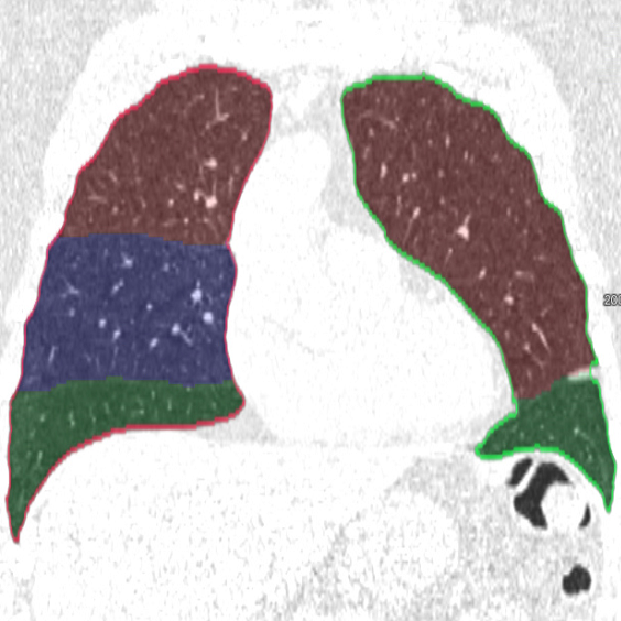
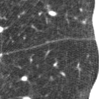
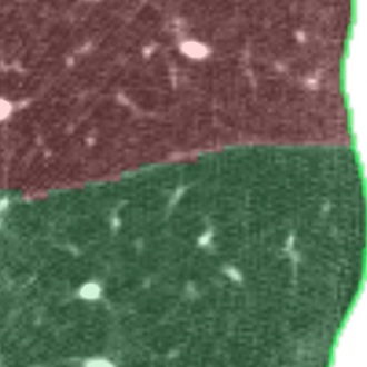
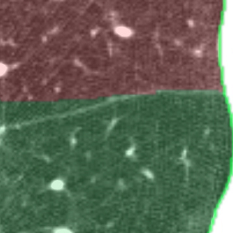
We applied the described segmentation pipeline to the 14 testing datasets which were not used for training and validation. Segmentation including postprocessing takes less than 6 seconds for a case. We compared our method to two other approaches: 1. a non-deep-learning-based automatic method [Lassen et al.(2013)Lassen, van Rikxoort, Schmidt, Kerkstra, van Ginneken, and Kuhnigk] 2. the same u-net as proposed but without weighting. The mean distance from the visible fissure improved to 1.46 mm (without weighting: 2.08 mm). See Figure 2 for plots and Figure 3 for screenshots.

4 Discussion
We trained a 3D u-net for a lung lobe segmentation task and showed that emphasizing the lobar boundaries in the loss function improved the segmentation results (see Figure 2 and 3). The segmentation quality is comparable to the method proposed in [Lassen et al.(2013)Lassen, van Rikxoort, Schmidt, Kerkstra, van Ginneken, and Kuhnigk] and even slightly better for the left lobes. This study was performed on a small amount of data. In future work, we plan to train with the same architecture on a much larger database including a wide range of pathologies and performing an extensive evaluation with participation in the LOLA11 [LOLA11()] challenge.
References
- [Armato et al.(2011)Armato, McLennan, Bidaut, McNitt-Gray, Meyer, Reeves, Zhao, Aberle, Henschke, Hoffman, Kazerooni, MacMahon, Van Beeke, Yankelevitz, Biancardi, Bland, Brown, Engelmann, Laderach, Max, Pais, Qing, Roberts, Smith, Starkey, Batrah, Caligiuri, Farooqi, Gladish, Jude, Munden, Petkovska, Quint, Schwartz, Sundaram, Dodd, Fenimore, Gur, Petrick, Freymann, Kirby, Hughes, Casteele, Gupte, Sallamm, Heath, Kuhn, Dharaiya, Burns, Fryd, Salganicoff, Anand, Shreter, Vastagh, and Croft] Samuel G Armato, Geoffrey McLennan, Luc Bidaut, Michael F McNitt-Gray, Charles R Meyer, Anthony P Reeves, Binsheng Zhao, Denise R Aberle, Claudia I Henschke, Eric A Hoffman, Ella A Kazerooni, Heber MacMahon, Edwin J R Van Beeke, David Yankelevitz, Alberto M Biancardi, Peyton H Bland, Matthew S Brown, Roger M Engelmann, Gary E Laderach, Daniel Max, Richard C Pais, David P Y Qing, Rachael Y Roberts, Amanda R Smith, Adam Starkey, Poonam Batrah, Philip Caligiuri, Ali Farooqi, Gregory W Gladish, C Matilda Jude, Reginald F Munden, Iva Petkovska, Leslie E Quint, Lawrence H Schwartz, Baskaran Sundaram, Lori E Dodd, Charles Fenimore, David Gur, Nicholas Petrick, John Freymann, Justin Kirby, Brian Hughes, Alessi Vande Casteele, Sangeeta Gupte, Maha Sallamm, Michael D Heath, Michael H Kuhn, Ekta Dharaiya, Richard Burns, David S Fryd, Marcos Salganicoff, Vikram Anand, Uri Shreter, Stephen Vastagh, and Barbara Y Croft. The lung image database consortium (LIDC) and image database resource initiative (IDRI): a completed reference database of lung nodules on CT scans. Medical Physics, 38(2):915–31, 2011. ISSN 0094-2405.
- [Doel et al.(2015)Doel, Gavaghan, and Grau] Tom Doel, David J. Gavaghan, and Vicente Grau. Review of automatic pulmonary lobe segmentation methods from CT. Computerized Medical Imaging and Graphics, 40:13–29, 2015. ISSN 18790771. 10.1016/j.compmedimag.2014.10.008.
- [George et al.(2017)George, Harrison, Jin, Xu, and Mollura] Kevin George, Adam P. Harrison, Dakai Jin, Ziyue Xu, and Daniel J. Mollura. Pathological pulmonary lobe segmentation from CT images using progressive holistically nested neural networks and random walker. In Lecture Notes in Computer Science (including subseries Lecture Notes in Artificial Intelligence and Lecture Notes in Bioinformatics), volume 10553 LNCS, pages 195–203, 2017. ISBN 9783319675572. 10.1007/978-3-319-67558-9_23.
- [Gerard and Reinhardt(2019)] Sarah E Gerard and Joseph M Reinhardt. Pulmonary lobe segmentation using a sequence of convolutional neural networks for marginal learning. In Proceedings - International Symposium on Biomedical Imaging, volume 2019-April, pages 1207–1211. IEEE, 2019. ISBN 9781538636411. 10.1109/ISBI.2019.8759212.
- [Imran et al.(2018)Imran, Hatamizadeh, Ananth, Ding, Terzopoulos, and Tajbakhsh] Abdullah Al Zubaer Imran, Ali Hatamizadeh, Shilpa P Ananth, Xiaowei Ding, Demetri Terzopoulos, and Nima Tajbakhsh. Automatic segmentation of pulmonary lobes using a progressive dense V-network. In Lecture Notes in Computer Science (including subseries Lecture Notes in Artificial Intelligence and Lecture Notes in Bioinformatics), volume 11045 LNCS, pages 282–290, 2018. ISBN 9783030008888. 10.1007/978-3-030-00889-5_32.
- [Lassen et al.(2013)Lassen, van Rikxoort, Schmidt, Kerkstra, van Ginneken, and Kuhnigk] Bianca Lassen, Eva M. van Rikxoort, Michael Schmidt, Sjoerd Kerkstra, Bram van Ginneken, and Jan Martin Kuhnigk. Automatic segmentation of the pulmonary lobes from chest CT scans based on Fissures, Vessels, and Bronchi. IEEE Transactions on Medical Imaging, 32(2):210–222, 2013. ISSN 02780062. 10.1109/TMI.2012.2219881.
- [Lee et al.(2019)Lee, Matin, Gleeson, and Grau] Hoileong Lee, Tahreema Matin, Fergus Gleeson, and Vicente Grau. Efficient 3D Fully Convolutional Networks for Pulmonary Lobe Segmentation in CT Images. arXiv:1909.07474 [eess.IV], pages 1–11, 2019.
- [LOLA11()] LOLA11. LObe and Lung Analysis 2011 (LOLA11). URL https://lola11.grand-challenge.org.
- [Park et al.(2019)Park, Yun, Kim, Park, Cho, Park, Song, Lee, and Seo] Jongha Park, Jihye Yun, Namkug Kim, Beomhee Park, Yongwon Cho, Hee Park, Mijeong Song, Minho Lee, and Joon Beom Seo. Fully automated lung lobe segmentation in volumetric chest ct with 3D u-net: Validation with intra- and extra-datasets. Journal of Digital Imaging, 05 2019. 10.1007/s10278-019-00223-1.
- [Tang et al.(2019)Tang, Zhang, and Xie] Hao Tang, Chupeng Zhang, and Xiaohui Xie. Automatic pulmonary lobe segmentation using deep learning. CoRR, abs/1903.09879, 2019.
- [Wang et al.(2019)Wang, Chen, Zhao, Chi, Xie, Zhang, and Hua] Wenjia Wang, Junxuan Chen, Jie Zhao, Ying Chi, Xuansong Xie, Li Zhang, and Xiansheng Hua. Automated segmentation of pulmonary lobes using coordination-guided deep neural networks. In Proceedings - International Symposium on Biomedical Imaging, volume 2019-April, pages 1353–1357, apr 2019. ISBN 9781538636411. 10.1109/ISBI.2019.8759492.