MIDLMedical Imaging with Deep Learning
\jmlrpages
\jmlryear2020
\jmlrworkshopShort Paper – MIDL 2020
\jmlrvolume
\midlauthor\NameNishanth Thumbavanam Arun\midljointauthortextContributed equally\nametag1 and \NameNathan Gaw\midlotherjointauthor\nametag2 and \NamePraveer Singh\midlotherjointauthor\nametag1 and \NameKen Chang\midlotherjointauthor\nametag1 and \NameKatharina Viktoria Hoebel\nametag1 and \NameJay Patel\nametag1 and \NameMishka Gidwani\nametag1 and \NameJayashree Kalpathy-Cramer\nametag1\Emailkalpathy@nmr.mgh.harvard.edu
\addr1 Athinoula A. Martinos Center for Biomedical Imaging, Department of Radiology, Massachusetts General Hospital, Boston, MA, USA
\addr2 Arizona State University-Mayo Clinic Center for Innovative Imaging, School of Computing, Informatics, and Decision Systems Engineering, Tempe, AZ, USA
Assessing the validity of saliency maps for abnormality localization in medical imaging
Abstract
Saliency maps have become a widely used method to assess which areas of the input image are most pertinent to the prediction of a trained neural network. However, in the context of medical imaging, there is no study to our knowledge that has examined the efficacy of these techniques and quantified them using overlap with ground truth bounding boxes. In this work, we explored the credibility of the various existing saliency map methods on the RSNA Pneumonia dataset. We found that GradCAM was the most sensitive to model parameter and label randomization, and was highly agnostic to model architecture.
keywords:
Saliency maps, localization, deep learning.1 Introduction
Saliency maps have become a popular approach for post-hoc interpretability of Convolutional Neural Networks (CNNs). [Adebayo et al.(2018)Adebayo, Gilmer, Muelly, Goodfellow, Hardt, and Kim] These maps are designed to highlight the salient components of the input images that are important to the model prediction. As a result, many deep learning medical imaging studies have used saliency maps to rationalize model prediction and provide localization. [Rajpurkar et al.(2017)Rajpurkar, Irvin, Zhu, Yang, Mehta, Duan, Ding, Bagul, Langlotz, Shpanskaya, et al., Bien et al.(2018)Bien, Rajpurkar, Ball, Irvin, Park, Jones, Bereket, Patel, Yeom, Shpanskaya, et al., Mitani et al.(2019)Mitani, Huang, Venugopalan, Corrado, Peng, Webster, Hammel, Liu, and Varadarajan] However, the validity of saliency maps has been called into question in a recent study showing that many popular saliency map approaches are not sensitive to model weight or label randomization for models evaluated on several datasets. [Adebayo et al.(2018)Adebayo, Gilmer, Muelly, Goodfellow, Hardt, and Kim] In this study, we extend this work by evaluating popular saliency map methods both quantitatively and qualitatively for classification models trained on the RSNA Pneumonia dataset. [Shih et al.(2019)Shih, Wu, Halabi, Kohli, Prevedello, Cook, Sharma, Amorosa, Arteaga, Galperin-Aizenberg, et al.] Specifically, we assess the performance of these methods in localizing abnormalities in medical imaging by quantifying overlap with ground truth bounding boxes. Furthermore, we assess the effect of model weight and label randomization on localization performance. Lastly, we empirically study repeatability of the saliency maps, both within the same model architecture and across different model architectures.
2 Methods and Results
2.1 Model and Data Randomization
The saliency methods examined in our experiments are Gradient Explanation [Simonyan et al.(2013)Simonyan, Vedaldi, and Zisserman], Smoothgrad Integrated Gradients (IG) [Sundararajan et al.(2017)Sundararajan, Taly, and Yan], GradCAM [Selvaraju et al.(2016)Selvaraju, Das, Vedantam, Cogswell, Parikh, and Batra], XRAI [Kapishnikov et al.(2019)Kapishnikov, Bolukbasi, Viégas, and Terry], and Smoothgrad [Smilkov et al.(2017)Smilkov, Thorat, Kim, Viégas, and Wattenberg]. Along with using Spearman rank correlation to compare maps before and after model weight and label randomization, we leverage the ground-truth bounding box coordinates
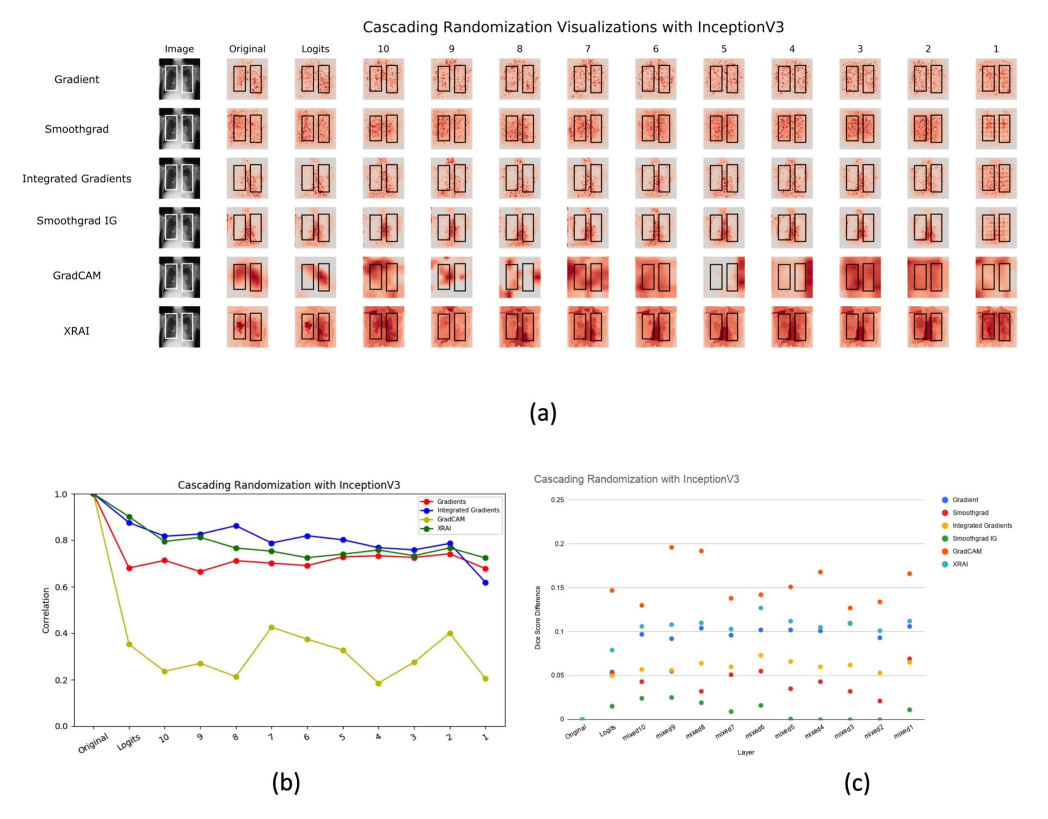
provided in the RSNA Pneumonia dataset to establish a quantitative baseline using the dice metric. To investigate the sensitivity of saliency methods under changes to model parameters, we employ cascading randomization. [Adebayo et al.(2018)Adebayo, Gilmer, Muelly, Goodfellow, Hardt, and Kim] We observed that among these saliency techniques, GradCAM degraded with model randomization to a large degree whereas the other methods did not (Fig 1). This is also verified in a label randomization experiment shown in Fig 2(c) wherein we randomly flipped the labels and retrained the model to observe the difference in the dice scores of the saliency maps. In both the tests, it can be observed that gradient explanation, Smoothgrad IG, and XRAI do not degrade significantly under randomization, suggesting an undesirable invariance to model parameters and labels.
2.2 Repeatability and Reproducibility
We also conducted repeatability tests on these saliency methods by comparing maps from a) models with the same architecture trained independently (intra-architecture repeatability)
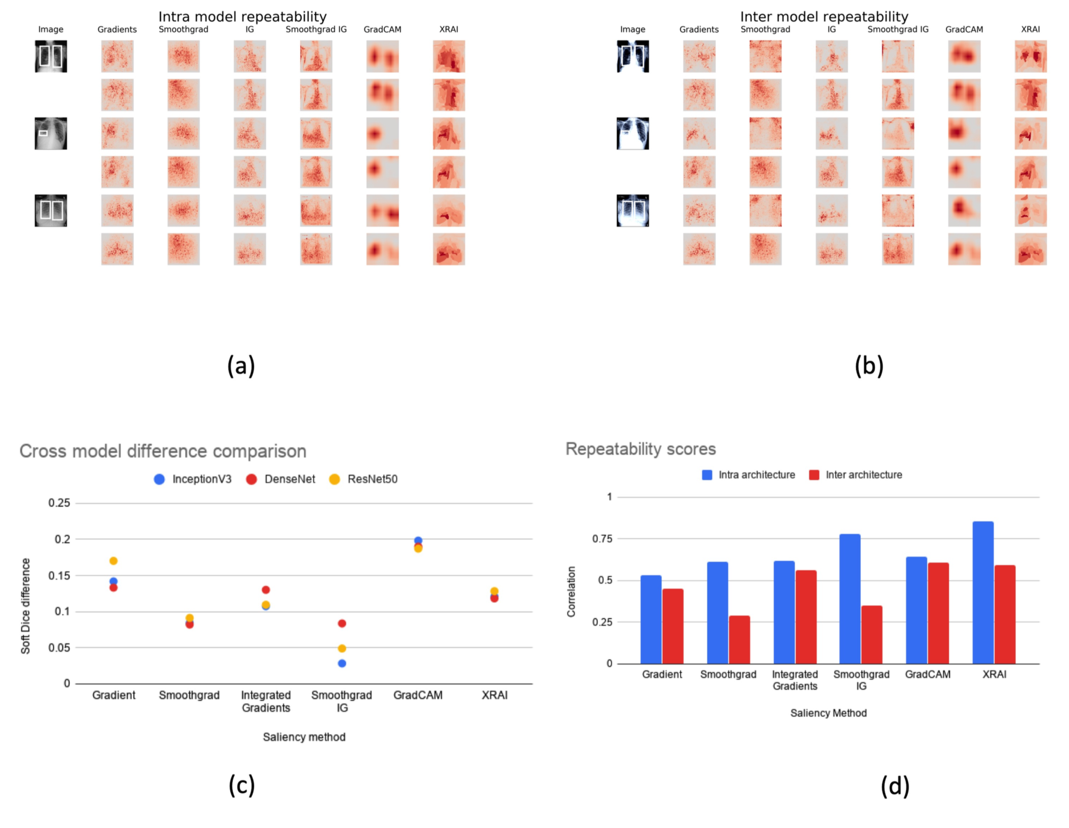
and b) models with different architectures (inter-architecture reproducibility). These experiments are designed to test if these saliency methods produce similar maps with a different set of weights and whether they are architecture agnostic. Fig 2(a) shows considerable differences across all the different maps from two independently trained InceptionV3 models. Furthermore, Fig 2(b) shows saliency maps differences between those produced from InceptionV3 (top row) versus those from DenseNet121 (bottom row). Fig 2(d) demonstrates that both the Smoothgrad and Smoothgrad IG yielded the most dissimilar maps across architectures while GradCAM yielded maps that were most similar.
3 Discussion and Conclusion
In this study, we evaluated the performance of several popular saliency methods on the RSNA Pneumonia Detection dataset in regards to their localization capabilities, robustness to model parameter and label randomization, as well as repeatability and reproducibility with model architectures. It was found that GradCAM showed superior sensitivity to model parameter and label randomization, and was highly agnostic to model architecture. In future studies, we will further examine the effect of different model architectures on saliency maps and validate our findings on a separate medical imaging dataset.
We would like to thank Julius Adebayo for providing us with the cascading randomization code used in his work. [Adebayo et al.(2018)Adebayo, Gilmer, Muelly, Goodfellow, Hardt, and Kim]
Research reported in this publication was supported by a training grant from the National Institute of Biomedical Imaging and Bioengineering (NIBIB) of the National Institutes of Health under award number 5T32EB1680 to K. Chang and J. B. Patel and by the National Cancer Institute (NCI) of the National Institutes of Health under Award Number F30CA239407 to K. Chang. The content is solely the responsibility of the authors and does not necessarily represent the official views of the National Institutes of Health.
This publication was supported from the Martinos Scholars fund to K. Hoebel. Its contents are solely the responsibility of the authors and do not necessarily represent the official views of the Martinos Scholars fund.
This study was supported by National Institutes of Health (NIH) grants U01 CA154601, U24 CA180927, and U24 CA180918 and National Science Foundation (NSF) grant NSF 1622542 to J. Kalpathy-Cramer.
This research was carried out in whole or in part at the Athinoula A. Martinos Center for Biomedical Imaging at the Massachusetts General Hospital, using resources provided by the Center for Functional Neuroimaging Technologies, P41EB015896, a P41 Biotechnology Resource Grant supported by the National Institute of Biomedical Imaging and Bioengineering (NIBIB), National Institutes of Health.
References
- [Adebayo et al.(2018)Adebayo, Gilmer, Muelly, Goodfellow, Hardt, and Kim] Julius Adebayo, Justin Gilmer, Michael Muelly, Ian Goodfellow, Moritz Hardt, and Been Kim. Sanity checks for saliency maps. In Advances in Neural Information Processing Systems, pages 9505–9515, 2018.
- [Bien et al.(2018)Bien, Rajpurkar, Ball, Irvin, Park, Jones, Bereket, Patel, Yeom, Shpanskaya, et al.] Nicholas Bien, Pranav Rajpurkar, Robyn L Ball, Jeremy Irvin, Allison Park, Erik Jones, Michael Bereket, Bhavik N Patel, Kristen W Yeom, Katie Shpanskaya, et al. Deep-learning-assisted diagnosis for knee magnetic resonance imaging: development and retrospective validation of mrnet. PLoS medicine, 15(11):e1002699, 2018.
- [Kapishnikov et al.(2019)Kapishnikov, Bolukbasi, Viégas, and Terry] Andrei Kapishnikov, Tolga Bolukbasi, Fernanda Viégas, and Michael Terry. Xrai: Better attributions through regions. In Proceedings of the IEEE International Conference on Computer Vision, pages 4948–4957, 2019.
- [Mitani et al.(2019)Mitani, Huang, Venugopalan, Corrado, Peng, Webster, Hammel, Liu, and Varadarajan] Akinori Mitani, Abigail Huang, Subhashini Venugopalan, Greg S Corrado, Lily Peng, Dale R Webster, Naama Hammel, Yun Liu, and Avinash V Varadarajan. Detection of anaemia from retinal fundus images via deep learning. Nature Biomedical Engineering, pages 1–10, 2019.
- [Rajpurkar et al.(2017)Rajpurkar, Irvin, Zhu, Yang, Mehta, Duan, Ding, Bagul, Langlotz, Shpanskaya, et al.] Pranav Rajpurkar, Jeremy Irvin, Kaylie Zhu, Brandon Yang, Hershel Mehta, Tony Duan, Daisy Ding, Aarti Bagul, Curtis Langlotz, Katie Shpanskaya, et al. Chexnet: Radiologist-level pneumonia detection on chest x-rays with deep learning. arXiv preprint arXiv:1711.05225, 2017.
- [Selvaraju et al.(2016)Selvaraju, Das, Vedantam, Cogswell, Parikh, and Batra] Ramprasaath R Selvaraju, Abhishek Das, Ramakrishna Vedantam, Michael Cogswell, Devi Parikh, and Dhruv Batra. Grad-cam: Why did you say that? arXiv preprint arXiv:1611.07450, 2016.
- [Shih et al.(2019)Shih, Wu, Halabi, Kohli, Prevedello, Cook, Sharma, Amorosa, Arteaga, Galperin-Aizenberg, et al.] George Shih, Carol C Wu, Safwan S Halabi, Marc D Kohli, Luciano M Prevedello, Tessa S Cook, Arjun Sharma, Judith K Amorosa, Veronica Arteaga, Maya Galperin-Aizenberg, et al. Augmenting the national institutes of health chest radiograph dataset with expert annotations of possible pneumonia. Radiology: Artificial Intelligence, 1(1):e180041, 2019.
- [Simonyan et al.(2013)Simonyan, Vedaldi, and Zisserman] Karen Simonyan, Andrea Vedaldi, and Andrew Zisserman. Deep inside convolutional networks: Visualising image classification models and saliency maps. arXiv preprint arXiv:1312.6034, 2013.
- [Smilkov et al.(2017)Smilkov, Thorat, Kim, Viégas, and Wattenberg] Daniel Smilkov, Nikhil Thorat, Been Kim, Fernanda Viégas, and Martin Wattenberg. Smoothgrad: removing noise by adding noise. arXiv preprint arXiv:1706.03825, 2017.
- [Sundararajan et al.(2017)Sundararajan, Taly, and Yan] Mukund Sundararajan, Ankur Taly, and Qiqi Yan. Axiomatic attribution for deep networks. In Proceedings of the 34th International Conference on Machine Learning-Volume 70, pages 3319–3328. JMLR. org, 2017.