MIDL 2020Medical Imaging with Deep Learning 2020
\jmlrvolume
\jmlryear
\jmlrworkshopMIDL 2020 – Short Paper
\midlauthor\NameLiset Vázquez Romaguera\nametag1 \Emailliset.vazquez@polymtl.ca
\NameRosalie Plantefeve\nametag2 \Emailrosalie@plantefeve.fr
\NameSamuel Kadoury\nametag1,2 \Emailsamuel.kadoury@polymtl.ca
\addr1 MedICAL Laboratory, Polytechnique Montreal, Montreal, Canada
\addr2 CHUM Research Center, Montreal, Canada
Spatiotemporal motion prediction in free-breathing liver scans via a recurrent multi-scale encoder decoder
Abstract
In this work we propose a multi-scale recurrent encoder-decoder architecture to predict the breathing induced organ deformation in future frames. The model was trained end-to-end from input images to predict a sequence of motion labels. Targets were created by quantizing the displacement fields obtained from deformable image registration. We report results using MRI free-breathing acquisitions from 12 volunteers. Experiments were aimed at investigating the proposed multi-scale design and the effect of increasing the number of predicted frames on the overall accuracy of the model. The proposed model was able to predict vessel positions in the next temporal image with a mean accuracy of mm showing increased performance in comparison with state-of-the-art approaches.
keywords:
motion prediction, liver, MRI, free-breathing, LSTM1 Introduction
According to the last American Cancer Society’s report (Ame, 2020), about 42,810 new cases of primary liver cancer will be diagnosed this year in the US. Radiation therapy is the first line of treatment for the majority of these cases. Its goal is to focus the radiation beams in the target and to avoid surrounding anatomy. However, respiratory motion is one of the major issues with large dosimetric impact Mechalakos et al. (2004). Image-guided radiation treatments can greatly benefit from future frame prediction models since the beam can be re-positioned compensating for motion. State-of-the-art approaches rely on statistical Samei et al. (2012); Preiswerk et al. (2014) and biomechanical modeling (Nguyen et al., 2008; Fuerst et al., 2014). Recently, deep learning-based solution have been reported for motion prediction in the medical context Wang et al. (2018); Teo et al. (2018); Krebs et al. (2019). In this work, we propose a recurrent multi-scale encoder-decoder framework to perform in-plane spatio-temporal motion prediction from sequential images.
2 Method
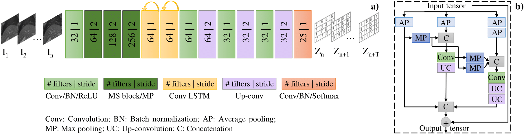
The proposed model aims at learning a representation that predicts the sequence of encoded motion over future time steps given an input image sequence of length . Consecutive pair of images were non-rigidly registered applying the B-spline transformation model and optimization based on normalized mutual information as implemented by NiftyReg software Modat et al. (2010). The resulting two-dimensional displacement fields were encoded using an auxiliary representation space = where and is the number of classes. is a mapping function to encode the displacement fields into labels. To that end, the ranges of values for each vectorial component, i.e. axes and , were quantized into bins. A codebook was built by assigning a class to each possible combination between the bins of each axis. As the probability distribution of the motion vectors obtained from deformable registration has an approximately-Gaussian shape, bins were selected near to the mean, standard deviation, minimum and maximum distribution values. Using this scheme, we effectively represent the motion observed in the dataset. The motion learning architecture is composed by a multi-scale (MS) encoder, recurrent units and a decoder, as illustrated in Figure 1 (a). The MS encoder extracts feature representations at multiple scales through the network: fully, medium and low resolution in order to fully exploit the image features (see Figure 1 (b)). The motion learning architecture also contains recurrent units and a fully convolutional spatial decoder. The spatio-temporal features extracted by the MS encoder are extrapolated in time by the convolutional Long Short-Term Memory (LSTM) units and further processed by the spatial decoder to recover the desired dimensions in the form of motion labels. We used a weighted cross entropy loss function as proposed by Zhang et al. (2016) to promote class rebalancing since the distribution is strongly biased toward classes representing the superior-inferior motion. Adam optimizer with an initial learning rate of was used. This learning rate was reduced by 2 after 10 epochs without improvements in the validation set accuracy.
3 Results and discussion
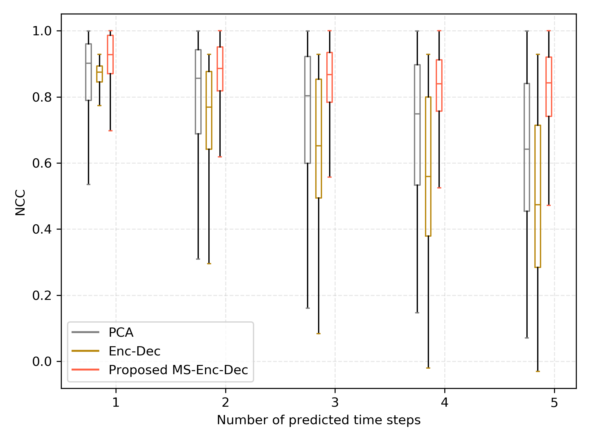
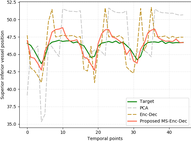
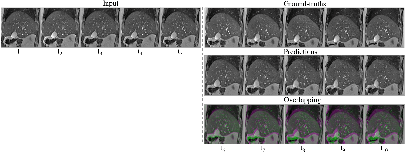
Experiments were conducted using sagittal slices covering 15 positions on the right liver lobe from 12 volunteers. Each anatomical position comprises 50 dynamics. These cine-phase scans were acquired on a Siemens Skyra 3T scanner using a 2D T2-weighted true FISP sequence. Pixel spacing, slice thickness and temporal resolution are equal to mm2, 3 mm and 320 ms, respectively. The dataset was divided in train, validation and test sets following a leave-one-out scheme on a subject level. Thus, the model was tested on all the slice positions belonging to an unseen subject. We compared the proposed network with statistical modeling Li et al. (2011) and with a similar architecture which uses the traditional feature extraction scheme (Conv-Pool stacking) (Luo et al., 2017). Two blood vessels were manually annotated in each image. We report results when varying the number of extrapolated times given 5 input images. Table LABEL:tab:vessel_tracking presents mean Euclidean distances between ground truth and predicted vessel positions. Figure 3 shows a comparison based on the Normalized Cross Correlation (NCC) metric. The proposed model outperforms the compared methods for the in-plane motion prediction task. Results show a lower performance when more time steps are predicted. It is natural that, based on the same information, the error increases when extrapolating more time points. Moreover, the model does not cope with out-of-plane motion which also influences the reported values. Figure 3 illustrates the vessel trajectory through the target and predicted temporal images. Our multi-scale encoder-decoder model showed the closest alignment with the target trajectory. Finally, Figure 4 shows an example of the output sequence obtained by deforming the last input image with the predicted deformations. While the model showed a competitive performance, some limitations should be considered. The first is related to the inherent error introduced by the quantization, which depends directly on the selected number of bins. Also, since the predicted displacement fields are recovered from motion labels, potential misclassification may lead to unrealistic and ambiguous motion. This aspect should be investigated in a future study.
tab:vessel_tracking Model t=1 t=2 t=3 t=4 t=5 (320 ms) (640 ms) (960 ms) (1280 ms) (1600 ms) PCA 2.25 3.46 2.49 3.76 3.45 4.20 3.96 4.42 4.39 4.01 Enc-Dec 2.71 3.21 3.41 3.40 3.98 4.17 4.41 3.65 4.87 4.11 Proposed 2.07 2.95 2.24 3.16 2.91 3.52 3.11 3.42 3.81 3.63
This research was undertaken thanks, in part, to funding from the Canada First Research Excellence Fund through the TransMedTech Institute.
References
- Ame (2020) Facts and Figures 2020. American Cancer Society, Atlanta, Ga., 2020.
- Fuerst et al. (2014) Bernhard Fuerst, Tommaso Mansi, Francois Carnis, Martin Sälzle, Jingdan Zhang, Jerome Declerck, Thomas Boettger, John Bayouth, Nassir Navab, and Ali Kamen. Patient-specific biomechanical model for the prediction of lung motion from 4-d ct images. IEEE transactions on medical imaging, 34(2):599–607, 2014.
- Krebs et al. (2019) Julian Krebs, Tommaso Mansi, Nicholas Ayache, and Hervé Delingette. Probabilistic motion modeling from medical image sequences: Application to cardiac cine-mri. arXiv preprint arXiv:1907.13524, 2019.
- Li et al. (2011) Ruijiang Li, John H Lewis, Xun Jia, Tianyu Zhao, Weifeng Liu, Sara Wuenschel, James Lamb, Deshan Yang, Daniel A Low, and Steve B Jiang. On a pca-based lung motion model. Physics in Medicine & Biology, 56(18):6009, 2011.
- Luo et al. (2017) Zelun Luo, Boya Peng, De-An Huang, Alexandre Alahi, and Li Fei-Fei. Unsupervised learning of long-term motion dynamics for videos. In Proceedings of the IEEE Conference on Computer Vision and Pattern Recognition, pages 2203–2212, 2017.
- Mechalakos et al. (2004) James Mechalakos, Ellen Yorke, Gikas S Mageras, Agung Hertanto, Andrew Jackson, Ceferino Obcemea, Kenneth Rosenzweig, and C Clifton Ling. Dosimetric effect of respiratory motion in external beam radiotherapy of the lung. Radiotherapy and Oncology, 71(2):191–200, 2004.
- Modat et al. (2010) Marc Modat, Gerard R Ridgway, Zeike A Taylor, Manja Lehmann, Josephine Barnes, David J Hawkes, Nick C Fox, and Sébastien Ourselin. Fast free-form deformation using graphics processing units. Computer methods and programs in biomedicine, 98(3):278–284, 2010.
- Nguyen et al. (2008) Thao-Nguyen Nguyen, Joanne L Moseley, Laura A Dawson, David A Jaffray, and Kristy K Brock. Adapting population liver motion models for individualized online image-guided therapy. In 2008 30th Annual International Conference of the IEEE Engineering in Medicine and Biology Society, pages 3945–3948. IEEE, 2008.
- Preiswerk et al. (2014) Frank Preiswerk, Valeria De Luca, Patrik Arnold, Zarko Celicanin, Lorena Petrusca, Christine Tanner, Oliver Bieri, Rares Salomir, and Philippe C Cattin. Model-guided respiratory organ motion prediction of the liver from 2d ultrasound. Medical image analysis, 18(5):740–751, 2014.
- Samei et al. (2012) Golnoosh Samei, Christine Tanner, and Gabor Székely. Predicting liver motion using exemplar models. In International MICCAI Workshop on Computational and Clinical Challenges in Abdominal Imaging, pages 147–157. Springer, 2012.
- Teo et al. (2018) Troy P Teo, Syed Bilal Ahmed, Philip Kawalec, Nadia Alayoubi, Neil Bruce, Ethan Lyn, and Stephen Pistorius. Feasibility of predicting tumor motion using online data acquired during treatment and a generalized neural network optimized with offline patient tumor trajectories. Medical physics, 45(2):830–845, 2018.
- Wang et al. (2018) Ran Wang, Xiaokun Liang, Xuanyu Zhu, and Yaoqin Xie. A feasibility of respiration prediction based on deep bi-lstm for real-time tumor tracking. IEEE Access, 6:51262–51268, 2018.
- Zhang et al. (2016) Richard Zhang, Phillip Isola, and Alexei A Efros. Colorful image colorization. In European conference on computer vision, pages 649–666. Springer, 2016.