First passage time study of DNA strand displacement
Abstract
DNA strand displacement, where a single-stranded nucleic acid invades a DNA duplex, is pervasive in genomic processes and DNA engineering applications. The kinetics of strand displacement have been studied in bulk; however, the kinetics of the underlying strand exchange were obfuscated by a slow bimolecular association step. Here, we use a novel single-molecule Fluorescence Resonance Energy Transfer (smFRET) approach termed the “fission” assay to obtain the full distribution of first passage times of unimolecular strand displacement. At a frame time of , the first passage time distribution for a 14-nt displacement domain exhibited a nearly monotonic decay with little delay. Among the eight different sequences we tested, the mean displacement time was on average and varied by up to a factor of 13. The measured displacement kinetics also varied between complementary invaders and between RNA and DNA invaders of the same base sequence except for TU substitution. However, displacement times were largely insensitive to the monovalent salt concentration in the range of . Using a one-dimensional random walk model, we infer that the single-step displacement time is in the range of depending on the base identity. The framework presented here is broadly applicable to the kinetic analysis of multistep processes investigated at the single-molecule level.
I Statement of Significance
DNA strand displacement occurs when a single nucleic acid strand invades and replaces another nearly identical strand in a duplex. This process is ubiquitous in biology and is fundamental to the field of DNA nanotechnology. Previous kinetic studies of strand displacement either used DNA strands much longer than those found in practical applications or were obscured by a rate-limiting bimolecular step known as toehold formation. In this study, we introduce a new, single-molecule scheme that enables direct measurement of the strand displacement first passage time. Our observed kinetics demonstrate highly non-trivial sequence dependence as well as surprising differences between RNA and DNA invaders.
II Introduction
Nucleic acids’ ability to form hydrogen bonds between complementary Watson-Crick bases allows for a rich set of complicated, multi-step kinetic behaviors such as duplex hybridizationOuldridge et al. (2013) and dehybridizationSanstead and Tokmakoff (2018), Holliday junction structural dynamicsMcKinney et al. (2003); Bugreev et al. (2006), and strand invasionWright et al. (2018). In particular, strand displacement, which is the exchange of bases between two competing nucleic acid strands of identical sequence, occurs in homologous recombinationChen et al. (2008); Savir and Tlusty (2010), DNA replicationMi et al. (2020) and RNA transcriptionKIreeva et al. (2018), as well as CRISPR/CasSingh et al. (2016) and the related Cascade complexIvančić-Baće et al. (2012). In addition to fundamental genomic processes, DNA nanotechnology exploits strand displacement to create nanoscale gadgetsAndersen et al. (2009); Thubagere et al. (2017); Chang et al. (2019) and computational circuitsZhang and Seelig (2011); Cherry and Qian (2018); Wang et al. (2020a); Simmel et al. (2019). Strand displacement also aids in the development of quantitative assays for detection of nucleic acidWang et al. (2018); Li et al. (2019); Sapkota et al. (2019) and enzymatic activityCui et al. (2019); Lee et al. (2019) with improved probe specificityFigg et al. (2020); Tang et al. (2020); Garcia et al. (2020).
For practical applications, strand displacement is implemented with the “invader” strand and a partial duplex composed of the “incumbent” strand and the “substrate” strandSimmel et al. (2019)(Fig. 1). The partial duplex has two distinct domains: 1) the single-stranded overhang called the toehold which is critical to the speed and efficiency of the reactionZhang and Winfree (2009) and 2) the duplex region called the displacement domain. Toehold-mediated strand displacement is initiated when the invader strand anneals to the toehold in a bimolecular reaction. Once a stable toehold interaction is formed, the incumbent can be displaced by the dangling strand of the invader through spontaneous opening of a base pair between substrate and incumbent and closing of a base pair between substrate and invader. This unimolecular strand displacement is also called branch migrationRadding et al. (1977); Green and Tibbetts (1981); Srinivas et al. (2013).

Great attention has been paid to the kinetics of toehold-mediated strand displacement Zhang and Winfree (2009); Qian and Winfree (2011); Srinivas et al. (2013); Machinek et al. (2014); Broadwater and Kim (2016). However, these kinetics have been measured mostly in bulk, where the reaction kinetics are limited by bimolecular toehold association. Therefore, the measured kinetics do not shed light on the unimolecular branch migration. Other studies using long () DNA estimated the branch migration time per base pair step to be Radding et al. (1977); Green and Tibbetts (1981), but strand displacement in those studies took place in a D-loop geometry, which is different from the geometry of current interest where dangling strands are unrestricted during branch migration. Also, scale extends far beyond the length scales of interest for DNA nanotechnology. To understand whether and how sequence can be used to control displacement kinetics, we require experimental studies on unimolecular displacement of short oligos that can be modeled at the single base level.
In this study, we introduce a DNA “fission” assay to study toehold-mediated DNA strand displacement kinetics. The fission assay employs single-molecule Fluorescence Resonance Energy Transfer (smFRET) in order to directly measure the first passage displacement timeChou and D’Orsogna (2014); Polizzi et al. (2016) for the unimolecular reaction that occurs between toehold formation and incumbent dissociation. Using a wide-field total internal reflection fluorescence microscope, we measured the displacement kinetics for a 14-nucleotide displacement domain of eight different sequences. The mean displacement time varied by more than 10-fold between the slowest and fastest sequence and was on average , and the histograms of displacement times obtained at resolution showed a monotonous decay with little to no lag. We found that the displacement kinetics depend on the base sequence and the nucleic acid type (DNA vs. RNA) of the invader, but not on monovalent salt concentration. We analyzed the first passage time histograms of strand displacement using a symmetric random walk model to extract single base pair step times. The best fit to the histograms was obtained with , , , and for A,C,T, and G respectively. Our study reports the displacement rates of short DNA oligos and reveals biophysical mechanisms that govern DNA strand displacement kinetics.
III Materials and Methods
III.1 Sample Preparation
Custom DNA oligomers were purchased from Integrated DNA Technologies. The 26-nt substrate was internally labeled near the end distal to the toehold with a Cy3 fluorophore. The 24-nt invader molecule was labeled with a BioTEG linker at the end proximal to the toehold for surface immobilization. The 14-nt incumbent sequences were labeled with a Cy5 fluorophore at the end distal to the toehold. All oligos were HPLC purified by the manufacturer. The specific sequences are in Tables S1S3 in the Supporting Material. Partial duplexes were constructed by combining substrate and incumbent at a 1:10 ratio ( substrate, incumbent) in buffer at 7 pH containing NaCl and Tris. The excess of incumbent strands was meant to minimize the number of single-stranded substrates in solution; unpaired substrates can compete with the partial duplexes for binding with the surface-bound invaders, while lone incumbent strands do not bind to the invaders and will not fluoresce on their own. The mixture was heated to and slowly cooled for to to ensure the partial duplex was fully annealed.
III.2 Experimental Setup
Molecules were observed with an objective-type total internal reflection fluorescence microscope assembled on a commercial microscope body (IX81; Olympus). Fluorophores were excited by a laser (BWN-532-50E, B&W Tek). Images were binned and captured with an EMCCD (DU-897ECS0-#BV; Andor Technology), and images were recorded at 228 fps with exposure time using Micro-Manager softwareEdelstein et al. (2014). This high frame rate was achieved by cropping the image height to 64 super pixels. Experiments were performed on flow cells constructed as previously described in Le and KimLe and Kim (2014) while flow volume and flow rate () was controlled by a syringe pump (NE-1000; New Era Pump System).
The surface was passivated with polyethylene glycol (PEG) to minimize nonspecific bindingLe and Kim (2014). After neutravidin coating, the biotin-containing invader molecules were immobilized by flowing in at a concentration of . Next, of partial duplexes were pumped into the flow cell at in an oxygen-scavenging imaging bufferAitken et al. (2008), which contained 6-hydroxy-2,5,7,8-tetramethylchroman-2-carboxylic acid (Trolox), protocatechuic acid, protocatechuate 3,4-dioxygenase, and Tris-HCl (pH 7).
An appearance of high FRET signal marked formation of the toehold. A low FRET signal appeared as strand displacement concluded. The FRET signal time series was recorded and analyzed using in-house MATLAB software. The lifetime of the high FRET state was observed for many molecules to collect a distribution of displacement times.
III.3 Statistics of displacement times
Here, we provide an analytical expression we used to fit the histograms of displacement times. We model strand displacement as a one-dimensional random walk:
| (1) |
In this model, each state is denoted by , the number of displaced bases, and the measured displacement time corresponds to the first passage time from the reflecting state on the left boundary to the absorbing state on the right boundary. The forward rate and reverse rate from state are denoted as and , respectively. Since state is the absorbing state, . The time dependence of the system is governed by the master equation
| (2) |
where is the transition matrix operator, and the ket vector represents the system state. The probability amplitude to be in the absorbing state at some time after starting in is then given byHartich and Godec (2019)
| (3) |
The experimentally accessible datapoints in single-molecule experiments are the number of displacement events detected during a short time interval or bin time . These numbers form the so-called dwell-time or survival-time histogram. For sufficiently large number of total events , in the -th bin is related to the probability amplitudes according to
| (4) |
This can be expanded using the left and right eigenvectors of , and that satisfy , and their corresponding eigenvalue :
| (5) |
In the limit of , Eq. 5 yields the first passage time density
| (6) |
We used Eq. 5 to fit the measured histograms of displacement times with a fixed that corresponds to the frame time of 4.4 ms. In the representation of , is an asymmetric tridiagonal matrix:
| (7) |
whose left and right eigenvectors and eigenvalues can be obtained using MATLAB.
We also present here the expression we use to analyze the mean first passage time Kim (1958). can be computed using Eq. 6 as
| (8) |
A more useful expression can be obtained in terms of an invertible submatrix of , which we term :
| (9) |
Using the normalization of probability amplitude
| (10) |
we can express in terms of the inverse of
| (11) |
In the matrix presentation, the inverse matrix is related to matrix cofactors by
| (12) |
| (13) |
This sum of cofactors can be equated to the determinant of matrix which replaces the first row of with 1’s. Hence, the mean first passage time is given by the ratio of two matrix determinantsBroadwater and Kim (2016):
| (14) |
where represent the matrix elements of . Eq. 14 can also be expressed in terms of the bias factor asBroadwater and Kim (2016)
| (15) |

IV Results
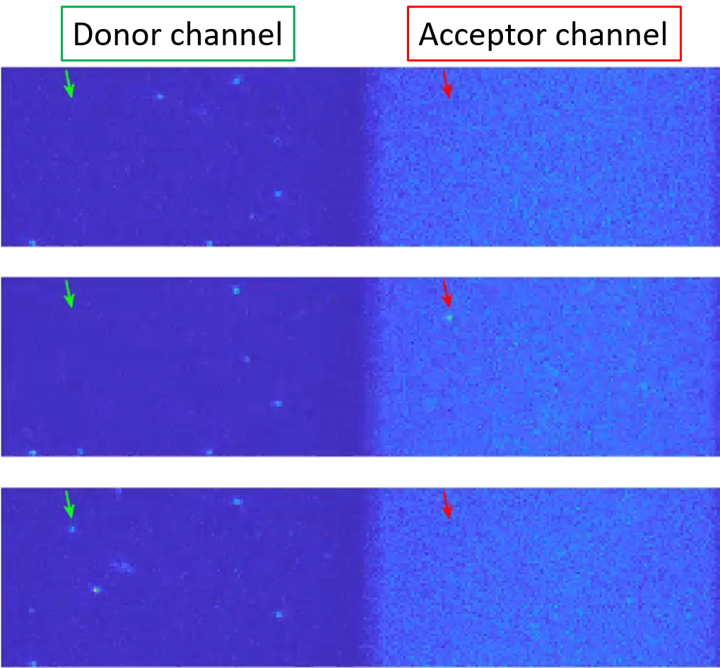
To focus on the unimolecular kinetics of strand displacement, we took a surface-based single-molecule FRET approach (Fig. 2). In this approach, the invader is immobilized on the glass surface of a flow chamber, and the partial duplex between the donor (Cy3)-labeled substrate and the acceptor (Cy5)-labeled incumbent are perfused into the chamber. The toehold length (10-bp) is chosen so that toehold formation is practically irreversible throughout the experiment. Upon toehold formation, a diffraction-limited spot emerges out of the diffusive background in the Cy5 channel. Upon incumbent dissociation, the spot changes fluorescence emission from the Cy5 channel to the Cy3 channel. We termed this experimental scheme “fission” because the duplex labeled with the FRET pair is split as a result of strand displacement.
Partial duplexes were constructed by annealing Cy3-labeled substrate molecules and Cy5-labeled incumbent molecules. Invader molecules were biotinylated near the end containing the toehold sequence and immobilized onto the surface (see Fig. 2(A)). As shown in Fig. 2(B) and SFig. S1, high FRET signals started to appear in the field of view after partial duplexes were flowed into the chamber. The average time at which spots appeared became shorter at a higher concentration of partial duplexes, and the transition of FRET from high to low only occurred in the presence of the matching displacement domain. Without the matching displacement domain, the high-FRET spots remained until they photobleached, which confirms that dissociation of the 10-bp toehold is much slower than the typical minute-long observation period. The red signal jumped to a high level in one or two frames, which suggests that toehold formation is much faster than our time resolution, and can therefore be considered instantaneous for analysis purposes. This high-level red signal lasted for variable periods of time from trace to trace, but the eventual transition back to low-FRET always occurred in one or two frames. Simultaneously with the disappearance of the signal from the Cy5 channel, a new signal appeared in the Cy3 channel, consistent with the fission scheme (Fig. 2(A)). Based on these observations, the first arrival of a high-FRET spot was attributed to toehold formation, and the transition from high- to low-FRET was attributed to completion of strand displacement. Hence, the dwell time in the high-FRET state (Fig. 2(B)) represents the displacement time.

By performing the fission assay multiple times, we could record hundreds of strand displacement events for one particular displacement system and build a histogram of displacement times. To investigate the sequence dependence of strand displacement kinetics, we tested 8 unique strand displacement systems, each with a different sequence in the displacement domain. We obtained these histograms at the finest bin width of ms, two of which are shown in Fig. 2(C). Note that displacement events faster than the exposure time do not produce a clear signal in the acceptor channel, and therefore, the first bin of the histogram starts from ms. For comparison of the histogram across all eight different sequences, we also present the histograms as a normalized heat map in Fig. 3. The salient feature of these histograms is that they decay monotonically with little or no delay. Six out of eight sequences show decay from the first bin; only two sequences show more events in the second than in the first bin. Nonetheless, we find a significant difference in the characteristic decay time among the tested sequences (black dots, Fig. 3). The fastest mean displacement time is 8 ms, while the slowest is 107 ms. The average over all sequences is .
To ensure that the observed difference between different sequences is not due to the uncertainties of the histograms, we need to establish the baseline uncertainties in the empirical histograms. As explained above, each histogram is obtained by combining displacement events taken from multiple runs of the fission assay in one day using the same reagents and flow cell. Hence, each histogram possesses statistical uncertainty due to the finite number of events and empirical uncertainty due to fluctuations in the experimental conditions. To estimate these uncertainties, we randomly sampled of the events collected on the same day and re-evaluated the mean displacement time (Supp. Fig. S2). The spread of the mean values is nonuniform among different sequences. For example, Sequences 4 and 6 have a similar total mean, but show different uncertainties. Nonetheless, the uncertainty in the mean for each sequence is much narrower than the variation among different sequences. We also documented the variability of the histogram means obtained at different times over a 4-year span by two users (Supp. Fig. S3). This empirical variability is much higher than the statistical variability due to reasons not completely clear. For transparency, we present these individual mean values in Fig. 4.

In addition to the base pair sequence of the displacement domain, the base sequence of the invader can also affect the displacement kinetics. As shown in Fig. 4(A), the same displacement domain can be invaded using a toehold extended from either the -end or the -end of the displacement domain. We refer to these complementary invasions as top and bottom invasions. The mean displacements times of top and bottom invasions are clearly different for all three displacement domains we tested. No particular invasion side was consistently faster: for Sequence 1, bottom invasion is faster, but for Sequence 2, top invasion is. Interestingly, when RNA with an identical sequence except for TU substitution was used as an invader in place of DNA, the faster side was switched (Fig. 5). All of these results suggest that the displacement kinetics are not completely determined by the base pair sequence or the thermodynamic stability of the displacement domain, but rather that the measured displacement kinetics are sensitive down to the chemical makeup of invading bases.

Lastly, we investigated the salt dependence of displacement kinetics. Monovalent salt can screen the negative charges on the phosphate backbone and alter the thermodynamics and kinetics of base pairingHuguet et al. (2010). However, its effect on the kinetics of branch migration is less clear because branch migration involves the base pairing dynamics of two competing strands. As shown in Fig. 6, the mean displacement time shows little change from 250 mM to 1 M [NaCl].

V Discussion
Using the fission assay, we measured the unimolecular branch migration kinetics in toehold-mediated DNA strand displacement. Using wide-field TIRFM and subregion readout of an EMCCD camera, we were able to record tens of strand displacement events at 4.4 ms frame rate. Our fission assay begins in a dark field-of-view with unlabeled invader strands immobilized on the surface, and monitors displacement events through the appearance and disappearance of FRET signal on the surface. The experimental design permits us to use high excitation intensity to detect fast displacement events at high signal-to-noise; strong excitation of fluorescent molecules begins only at the start of branch migration. Hence, the undesirable effect of photobleaching is eliminated.
Our fission assay produces data that could not be obtained to date. It separates out the bimolecular toehold formation step from the rest so that the apparent displacement time truly reflects a unimolecular process. In the language of stochastic processes, the displacement time represents the first passage time: the time taken for the branch point to start from the first position and reach the last for the first time. The fission scheme allows access to the full distribution of individual displacement times, which is more informative than just the average values. Below, we use the first passage time analysis to extract single-step migration rates from the measured histograms and discuss potential microscopic mechanisms that may control these rates.

.
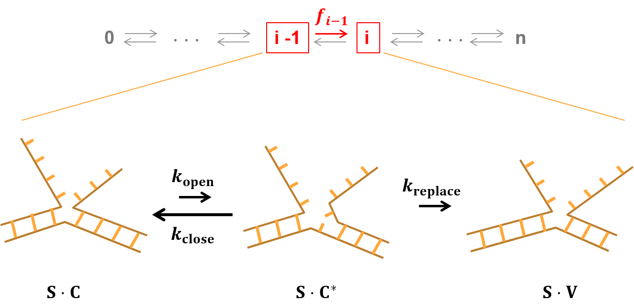
The most elementary model to describe DNA strand displacement is a one-dimensional random walk among states defined by the number of displaced base pairs (Eq. 1). Displacement is initiated after the invader hybridizes to the toehold and continues until the incumbent loses all base pairs with the substrate to the invader. Any intermediate state during this process can be envisioned as two dangling strands branching off from the duplex stem (Fig. 7). At the junction or the branch point, an incumbent (invader) base can spontaneously break away from the substrate base, and the most adjacent invader (incumbent) base can base-pair with the substrate base. As a result, the branch point can move by one base in either direction. The branch point, however, cannot recede into the toehold region because the incumbent is shorter than the substrate. Therefore, branch migration can be modeled as a one-dimensional random walk with single base steps from a reflecting boundary on one end (state 0) to an absorbing boundary on the other (state n).
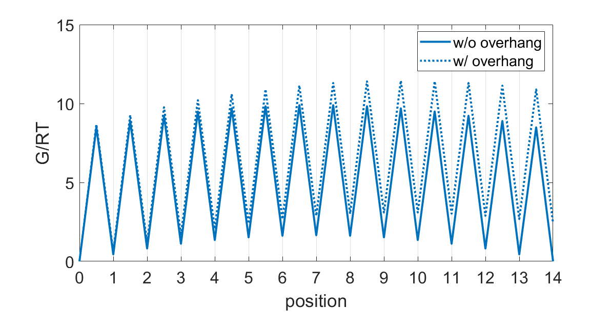
It is straightforward to derive the first passage time statistics from a Markov chain like Eq. 1. The simplest model is a uniform random walk where all transition rates are equal (). Such a model can be represented by a free energy landscape shown in Fig. 8 with troughs separated by equal height barriers. Based on Eq. 15, the mean first passage time () is given by
| (16) |
Using Eq. 16, , and the measured mean first passage time of , we can estimate the single-step migration time () to be . This estimate is also consistent with the measured histogram of displacement times. If single-step migration occurs more slowly than the time resolution, the histogram of displacement times must exhibit a strong delay or lag in early times (SFig. S4). However, our measured histograms at 4.4 ms bin width show little or no lag, which points to a single-step migration time much shorter than 4.4 ms.

However, this estimated time of per step is likely to be longer than the true value because displacement events faster than the exposure time are not included in our measurement. To extract the single-step migration rates in a more accurate, unbiased way despite this missing fraction of events, we fit the analytical solution Eq. 5 to all eight histograms with four shared parameters representing rates for A,G,C, and T. This global fitting procedure looks for the best set of rates that describe all eight histograms in the least-squares sense, excluding the missing first bin. It also implies a nonuniform symmetric random walk (Fig. 8) where the single-step migration rate depends only on the identity of the base to be displaced. Therefore, each step has the same forward and reverse rates (). The extracted step times for A,C, and T base are , , and , respectively. The step time for the G base did not converge, but the goodness of fit increased with faster values. Thus, we estimate the step time for G to be . As predicted, these times obtained by fitting histograms in their entirety are all faster than obtained from the mean values that omit fast events.
Our estimated single-step migration rates () appear to be much slower than the rate of base pair fraying or base flipping ()Andreatta et al. (2006); Banavali (2013); Zgarbová et al. (2014); Lindahl et al. (2017). Similarly, a previous study by Srivinas et al.Srinivas et al. (2013) also inferred the single-step migration rate to be much slower than the fraying rate. This discrepancy suggests that a single base pair opening event does not always lead to single-step branch migration. As shown in Fig. 7, a base pair between the substrate and the incumbent can transiently open and close with rate constants of and , respectively. While the substrate base is transiently unbound (), the invading base can base pair with the substrate base and replace the incumbent base at a rate of . We can safely assume that is much faster than based on the known base pair stabilityFrank-Kamenetskii and Prakash (2014). Coarse-grained molecular dynamics simulationsSrinivas et al. (2013) show that the branch migration intermediate frequently adopts a coaxially unstacked state where a transiently open incumbent base would be closer to the substrate base than the invading base (, Fig. 7). Therefore, we reason that is also much faster than . Given , will appear at the rate of
| (17) |
Hence, the single-step migration rate is expected to be much slower than the single base-pair opening rate.
We stress that a symmetric random walk is an oversimplification of strand displacement. As shown in SFig. S5, the symmetric random walk model significantly underrepresents the range of observed displacement times: the fastest observed histogram and the slowest observed histogram are outside the range represented by the fitted curves. Therefore, the observed sequence dependence calls for a more complicated model. We list below several microscopic mechanisms which indicate strand displacement is more properly described as an asymmetric random walk ().
First, displacement of the first base pair is energetically less favorable than the rest because it creates steric exclusion between dangling basesSrinivas et al. (2013); Šulc et al. (2015). Srinivas et al.Srinivas et al. (2013) measured the thermodynamic penalty for the steric exclusion to be at , which corresponds to -fold slower than all other rates (). According to Eq. 15, a bias in the first step () alters the mean first passage time to
| (18) |
With a strong reverse bias () in the first step, the single-step time () is estimated to be , faster than our previous estimate of based on a completely symmetric random walk. per step also falls well within the range () inferred by Srinivas et al.Srinivas et al. (2013). Second, the stability of a base pair is highly influenced by its nearest neighboring base pair, which would render the base pair opening rate direction-dependent. For example, let us consider an A base in two adjacent branch migration intermediates G∨AC and GA∨C, where ∨ refers to the branch point. In G∨AC, A is stacked more closely on C, whereas in GA∨C, A is stacked more closely on G. Therefore, the rate of A flipping out would be different between forward and reverse transitions. Third, the incumbent and the invader base at the branch point carry dangling strands of variable lengths depending on the state. These dangling strands will inevitably affect the diffusion rates of the bases at the branch point. To demonstrate this idea, we performed the fission assay with an invader extended by 5 nucleotides at the -end. As shown in Fig. 9, the displacement kinetics become significantly slower even with the same displacement domain. This result is consistent with the idea that a base with a longer dangling strand invades more slowly. As strand displacement progresses, the dangling part of the invader becomes shorter, and the dangling part of the incumbent becomes longer. Hence, the forward rate should become faster (), and the reverse rate slower (). These dangling-strand dependent rates produce asymmetric barriers in the free energy landscape, causing the basins to follow a concave curve (Fig. 8, SFig. S6). Previous oxDNA simulations also predicted a concave free energy landscapeSrinivas et al. (2013); Šulc et al. (2015). In the asymmetric random walk model, single-step rates are not only base-dependent but also position-dependent. Determining these rates would require measurements at a much larger scale, which is beyond the scope of this study.

We assumed that the number of steps is equal to the number of base pairs for modeling purposes. This could raise concern that this number may not accurately reflect the number of actual branch migration steps taken because the incumbent can spontaneously dissociate near the end of migration. In our previous work Broadwater and Kim (2016), we estimated spontaneous dissociation of a 2-bp incumbent to be which would be comparable to the branch migration step rate we measured in this study. This means that the last few steps can occur either via branch migration or spontaneous dissociation, and the rate would be dominated by the faster of the two. Regardless, our proposed asymmetric branch migration model, where forward rates become faster, and reverse rates become slower, would effectively account for such an effect.
We made an interesting observation that RNA invasion and DNA invasion occur at very different rates even with the same invader sequence (except for T to U substitution). For the one sequence we tested, RNA invaded faster than DNA (Fig. 5) from one side but more slowly from the other side. Several factors may contribute to this finding. Structurally, an RNA-DNA hybrid duplex adopts an A-form helixGyi et al. (1998); Conn et al. (1999); de Oliveira Martins et al. (2019), while a DNA-DNA duplex adopts a B-form helix. The thermodynamic stability difference between RNA-DNA and DNA-DNA duplexes depends on the sequenceSugimoto et al. (1995), with purine (AG)-rich substrate favoring RNA-DNA hyrbrid duplexesHuppert (2008). Directional differences in stacking between DNA and RNA are known to persist even in the single stranded formIsaksson et al. (2004) and could contribute to this inversion of side dependence. In a similar vein, a recent study shows that coaxial stacking between an RNA-DNA hybrid duplex and a DNA-DNA homoduplex is stronger when the interhelical junction contains a RNA end than when it contains a RNA end Cofsky et al. (2020). This effect may partially contribute to the faster top invasion by RNA shown in Fig 5. However, another RNA sequence we tested exhibited faster bottom invasion than top invasion, suggesting that the base sequence is a stronger determinant of the displacement rate than the invasion polarity. RNA invasion of a DNA duplex in particular is a fundamental feature of the CRISPR-Cas systemMulepati et al. (2014); Singh et al. (2016); Hong and Šulc (2019). R-loop formation appears to be the rate limiting step for DNA cleavageJeon et al. (2018); Gong et al. (2018) and is highly sequence dependentSzczelkun et al. (2014); Jeon et al. (2018); Zeng et al. (2018), but proceeds much more slowly ( second) than the spontaneous displacement rate we measured in this study. It will be thus interesting to investigate whether the sequence dependence is preserved between spontaneous and enzyme-mediated displacement reactions in the future.
The lack of salt dependence of the measured displacement kinetics was at first surprising to us because salt has a substantial effect on base pairing thermodynamicsSantaLucia Jr and Hicks (2004). Experimental measurements of salt-dependent opening and closing rates of a single base pair are scarce, but we can still infer their salt dependence from molecular dynamics studyWang et al. (2020b) and hybridization and dissociation measurements of short oligosCisse et al. (2012); Dupuis et al. (2013). These studies show that monovalent cations stabilize base pairing mainly by increasing the rate of base pair closing instead of decreasing the rate of base pair opening. Despite the strong salt dependence of base pair closing ( and ), our proposed three-state model for branch migration (Fig. 7 and Eq. 17) predicts that salt dependences of and will cancel each other out and render step migration rates, and , largely salt-independent.
Even in the case where ’s and ’s all carry a weak salt-dependence through , we can show that the overall salt dependence of the mean displacement time remains weak. Based on an experimental studyDupuis et al. (2013), we assume a simple power law dependence of on () so that all ’s and ’s change by the same factor upon changing . According to Eq. 14, the mean first passage time is equal to the ratio of two matrix determinants
| (19) |
Since , and , . Hence, the overall displacement of the base pair domain follows the weak salt dependence of . Either way, we are able to rationalize the weak salt dependence of the mean displacement time (Fig. 6).
We hope that our results will be beneficial to the field of DNA nanotechnology. Our work has provided sequence-specific branch migration step times that could be used to rationally design sequences with desired kinetics. For example, our results could aid in the design of complex interaction networks between competing reactions with specifically tuned kinetics. Further, our fission assay opens the door to understanding branch migration kinetics in more reaction conditions than we studied here (e.g. buffers, pH, and temperature).
In this study, we assumed that strand displacement proceeds through one-dimensional branch migration, but it is possible that other mechanisms are at play. The invader might invade through the end distal to the toehold when terminal base pairs fray or through internal base pairs that spontaneously open up. Although internal invasion is highly unlikely for the short displacement domain we used here, it would be more probable for longer displacement domains. We also cannot rule out direct swapping between segments of invader and incumbentParamanathan et al. (2014), invasion through triplex formationLee et al. (2012); Chen et al. (2017), or concurrent dissociation of a weakly bound incumbent strandMachinek et al. (2014); Broadwater and Kim (2016). All these processes can occur in parallel, which makes it difficult to predict the strand displacement rate for any given sequence. In this regard, a future study on a much larger set of displacement domain sequences would help us to attain more accurate phenomenological models for explaining the sequence dependence of strand displacement kinetics.
VI Conclusion
We developed a novel smFRET assay that we call fission in order to study the timing of the unimolecular reaction that occurs during toehold-mediated strand displacement. Our fission assay separates the timescales between the slower toehold formation step and the faster displacement step and enabled us to tally displacement first passage times distributions for 11 separate invasion schemes. We found non-trivial sequence dependence in the distributions, while the mean first passage times varied by an order of magnitude. Further, we highlighted significant differences between the “side” of invasion which suggest the kinetics are not completely determined by base-pair sequence alone. Curiously, we showed that DNA and RNA invaders can behave drastically differently despite having identical sequences (apart from a TU substitution). Finally, we demonstrated that displacement times were relatively unchanged over a wide range of salt concentrations. Motivated by these results, we developed a one-dimensional random walk model to estimate single-base displacement times. This model is widely relevant to multistep processes, and we anticipate our analysis to be highly important to an array of biological reactions.
VII Author Contributions
DWB and HDK designed research; DWB and AWC performed experiments; DWB, AWC, and HDK contributed analytical tools; DWB and AWC analyzed data; and DWB, AWC, and HDK wrote the manuscript.
VIII Acknowledgements
The authors thank the current and past members of the Kim laboratory for critical discussions during the research project and helpful comments on the manuscript. This work was supported by National Institutes of Health (R01GM112882) and National Science Foundation (1517507).
References
- Ouldridge et al. (2013) T. E. Ouldridge, P. Šulc, F. Romano, J. P. Doye, and A. A. Louis, Nucleic acids research 41, 8886 (2013).
- Sanstead and Tokmakoff (2018) P. J. Sanstead and A. Tokmakoff, The Journal of Physical Chemistry B 122, 3088 (2018).
- McKinney et al. (2003) S. A. McKinney, A.-C. Déclais, D. M. Lilley, and T. Ha, Nature Structural and Molecular Biology 10, 93 (2003).
- Bugreev et al. (2006) D. V. Bugreev, O. M. Mazina, and A. V. Mazin, Nature 442, 590 (2006).
- Wright et al. (2018) W. D. Wright, S. S. Shah, and W.-D. Heyer, Journal of Biological Chemistry , jbc (2018).
- Chen et al. (2008) Z. Chen, H. Yang, and N. P. Pavletich, Nature 453, 489 (2008).
- Savir and Tlusty (2010) Y. Savir and T. Tlusty, Molecular cell 40, 388 (2010).
- Mi et al. (2020) C. Mi, S. Zhang, W. Huang, M. Dai, Z. Chai, W. Yang, S. Deng, L. Ao, and H. Zhang, Biochimie (2020).
- KIreeva et al. (2018) M. KIreeva, C. Trang, G. Matevosyan, J. Turek-Herman, V. Chasov, L. Lubkowska, and M. Kashlev, Nucleic acids research (2018).
- Singh et al. (2016) D. Singh, S. H. Sternberg, J. Fei, J. A. Doudna, and T. Ha, Nature communications 7, 12778 (2016).
- Ivančić-Baće et al. (2012) I. Ivančić-Baće, J. A. Howard, and E. L. Bolt, Journal of molecular biology 422, 607 (2012).
- Andersen et al. (2009) E. S. Andersen, M. Dong, M. M. Nielsen, K. Jahn, R. Subramani, W. Mamdouh, M. M. Golas, B. Sander, H. Stark, C. L. Oliveira, et al., Nature 459, 73 (2009).
- Thubagere et al. (2017) A. J. Thubagere, W. Li, R. F. Johnson, Z. Chen, S. Doroudi, Y. L. Lee, G. Izatt, S. Wittman, N. Srinivas, D. Woods, et al., Science 357, eaan6558 (2017).
- Chang et al. (2019) Y. Chang, Z. Wu, Q. Sun, Y. Zhuo, Y. Chai, and R. Yuan, Analytical chemistry 91, 8123 (2019).
- Zhang and Seelig (2011) D. Y. Zhang and G. Seelig, Nature chemistry 3, 103 (2011).
- Cherry and Qian (2018) K. M. Cherry and L. Qian, Nature , 1 (2018).
- Wang et al. (2020a) F. Wang, H. Lv, Q. Li, J. Li, X. Zhang, J. Shi, L. Wang, and C. Fan, Nature Communications 11, 1 (2020a).
- Simmel et al. (2019) F. C. Simmel, B. Yurke, and H. R. Singh, Chemical reviews 119, 6326 (2019).
- Wang et al. (2018) K. Wang, M.-Q. He, F.-H. Zhai, J. Wang, R.-H. He, and Y.-L. Yu, Biosensors and Bioelectronics 105, 159 (2018).
- Li et al. (2019) Y. Li, H. Chen, Y. Dai, T. Chen, Y. Cao, and J. Zhang, Analytica chimica acta 1064, 25 (2019).
- Sapkota et al. (2019) K. Sapkota, A. Kaur, A. Megalathan, C. Donkoh-Moore, and S. Dhakal, Sensors 19, 3495 (2019).
- Cui et al. (2019) Y.-X. Cui, X.-N. Feng, Y.-X. Wang, H.-Y. Pan, H. Pan, and D.-M. Kong, Chemical Science 10, 2290 (2019).
- Lee et al. (2019) C. Y. Lee, H. Kim, H. Y. Kim, K. S. Park, and H. G. Park, Analyst 144, 3364 (2019).
- Figg et al. (2020) C. A. Figg, P. H. Winegar, O. G. Hayes, and C. A. Mirkin, Journal of the American Chemical Society (2020).
- Tang et al. (2020) W. Tang, W. Zhong, Y. Tan, G. A. Wang, F. Li, and Y. Liu, Topics in Current Chemistry 378, 1 (2020).
- Garcia et al. (2020) P. D. Garcia, R. W. Leach, G. M. Wadsworth, K. Choudhary, H. Li, S. Aviran, H. D. Kim, and V. A. Zakian, Nature Communications 11, 1 (2020).
- Zhang and Winfree (2009) D. Y. Zhang and E. Winfree, Journal of the American Chemical Society 131, 17303 (2009).
- Radding et al. (1977) C. M. Radding, K. L. Beattie, W. K. Holloman, and R. C. Wiegand, Journal of molecular biology 116, 825 (1977).
- Green and Tibbetts (1981) C. Green and C. Tibbetts, Nucleic Acids Research 9, 1905 (1981).
- Srinivas et al. (2013) N. Srinivas, T. E. Ouldridge, P. Šulc, J. M. Schaeffer, B. Yurke, A. A. Louis, J. P. Doye, and E. Winfree, Nucleic acids research 41, 10641 (2013).
- Qian and Winfree (2011) L. Qian and E. Winfree, Science 332, 1196 (2011).
- Machinek et al. (2014) R. R. Machinek, T. E. Ouldridge, N. E. Haley, J. Bath, and A. J. Turberfield, Nature communications 5, 1 (2014).
- Broadwater and Kim (2016) D. B. Broadwater and H. D. Kim, Biophysical journal 110, 1476 (2016).
- Chou and D’Orsogna (2014) T. Chou and M. R. D’Orsogna, in First-passage phenomena and their applications (World Scientific, 2014) pp. 306–345.
- Polizzi et al. (2016) N. F. Polizzi, M. J. Therien, and D. N. Beratan, Israel journal of chemistry 56, 816 (2016).
- Edelstein et al. (2014) A. D. Edelstein, M. A. Tsuchida, N. Amodaj, H. Pinkard, R. D. Vale, and N. Stuurman, Journal of biological methods 1 (2014).
- Le and Kim (2014) T. T. Le and H. D. Kim, Journal of visualized experiments: JoVE (2014).
- Aitken et al. (2008) C. E. Aitken, R. A. Marshall, and J. D. Puglisi, Biophysical journal 94, 1826 (2008).
- Hartich and Godec (2019) D. Hartich and A. Godec, Journal of Statistical Mechanics: Theory and Experiment 2019, 024002 (2019).
- Kim (1958) S. K. Kim, The Journal of Chemical Physics 28, 1057 (1958).
- Huguet et al. (2010) J. M. Huguet, C. V. Bizarro, N. Forns, S. B. Smith, C. Bustamante, and F. Ritort, Proceedings of the National Academy of Sciences 107, 15431 (2010).
- Andreatta et al. (2006) D. Andreatta, S. Sen, J. L. Pérez Lustres, S. A. Kovalenko, N. P. Ernsting, C. J. Murphy, R. S. Coleman, and M. A. Berg, Journal of the American Chemical Society 128, 6885 (2006).
- Banavali (2013) N. K. Banavali, Journal of the American Chemical Society 135, 8274 (2013).
- Zgarbová et al. (2014) M. Zgarbová, M. Otyepka, J. Sponer, F. Lankaš, and P. Jurečka, Journal of chemical theory and computation 10, 3177 (2014).
- Lindahl et al. (2017) V. Lindahl, A. Villa, and B. Hess, PLoS computational biology 13, e1005463 (2017).
- Frank-Kamenetskii and Prakash (2014) M. D. Frank-Kamenetskii and S. Prakash, Physics of life reviews 11, 153 (2014).
- Šulc et al. (2015) P. Šulc, T. E. Ouldridge, F. Romano, J. P. Doye, and A. A. Louis, Biophysical journal 108, 1238 (2015).
- Gyi et al. (1998) J. I. Gyi, A. N. Lane, G. L. Conn, and T. Brown, Biochemistry 37, 73 (1998).
- Conn et al. (1999) G. L. Conn, T. Brown, and G. A. Leonard, Nucleic acids research 27, 555 (1999).
- de Oliveira Martins et al. (2019) E. de Oliveira Martins, V. B. Barbosa, and G. Weber, Chemical Physics Letters 715, 14 (2019).
- Sugimoto et al. (1995) N. Sugimoto, S.-i. Nakano, M. Katoh, A. Matsumura, H. Nakamuta, T. Ohmichi, M. Yoneyama, and M. Sasaki, Biochemistry 34, 11211 (1995).
- Huppert (2008) J. L. Huppert, Molecular BioSystems 4, 686 (2008).
- Isaksson et al. (2004) J. Isaksson, S. Acharya, J. Barman, P. Cheruku, and J. Chattopadhyaya, Biochemistry 43, 15996 (2004).
- Cofsky et al. (2020) J. C. Cofsky, D. Karandur, C. J. Huang, I. P. Witte, J. Kuriyan, and J. A. Doudna, Elife 9, e55143 (2020).
- Mulepati et al. (2014) S. Mulepati, A. Héroux, and S. Bailey, Science , 1256996 (2014).
- Hong and Šulc (2019) F. Hong and P. Šulc, Journal of Structural Biology 207, 241 (2019).
- Jeon et al. (2018) Y. Jeon, Y. H. Choi, Y. Jang, J. Yu, J. Goo, G. Lee, Y. K. Jeong, S. H. Lee, I.-S. Kim, J.-S. Kim, et al., Nature communications 9, 1 (2018).
- Gong et al. (2018) S. Gong, H. H. Yu, K. A. Johnson, and D. W. Taylor, Cell reports 22, 359 (2018).
- Szczelkun et al. (2014) M. D. Szczelkun, M. S. Tikhomirova, T. Sinkunas, G. Gasiunas, T. Karvelis, P. Pschera, V. Siksnys, and R. Seidel, Proceedings of the National Academy of Sciences 111, 9798 (2014).
- Zeng et al. (2018) Y. Zeng, Y. Cui, Y. Zhang, Y. Zhang, M. Liang, H. Chen, J. Lan, G. Song, and J. Lou, Nucleic acids research 46, 350 (2018).
- SantaLucia Jr and Hicks (2004) J. SantaLucia Jr and D. Hicks, Annu. Rev. Biophys. Biomol. Struct. 33, 415 (2004).
- Wang et al. (2020b) Y. Wang, T. Liu, T. Yu, Z.-J. Tan, and W. Zhang, RNA 26, 470 (2020b).
- Cisse et al. (2012) I. I. Cisse, H. Kim, and T. Ha, Nature Structural and Molecular Biology 19, 623 (2012).
- Dupuis et al. (2013) N. F. Dupuis, E. D. Holmstrom, and D. J. Nesbitt, Biophysical journal 105, 756 (2013).
- Paramanathan et al. (2014) T. Paramanathan, D. Reeves, L. J. Friedman, J. Kondev, and J. Gelles, Nature communications 5, 1 (2014).
- Lee et al. (2012) I.-B. Lee, S.-C. Hong, N.-K. Lee, and A. Johner, Biophysical journal 103, 2492 (2012).
- Chen et al. (2017) J. Chen, Q. Tang, S. Guo, C. Lu, S. Le, and J. Yan, Nucleic acids research 45, 10032 (2017).
Supporting Material
Oligonucleotide sequences used in this study
| 5′-/BioTEG/ACCTGGTGTTGTCGGAATTTTAAT-3′ |
| 5′-ATTAAAATTCCGACAACACCAGGT/BioTEG/-3′ |
| 5′-/BioTEG/GGTTATTGGGTTGGGTGTGTGAAA-3′ |
| 5′-TTTCACACACCCAACCCAATAACC/BioTEG/-3′ |
| 5′-/BioTEG/CAATCAAATAAAACTCTCTCTAAA-3′ |
| 5′-TTTAGAGAGAGTTTTATTTGATTG/BioTEG/-3′ |
| 5′-TTTCCTCCTAAAAGACCACCACCT/BioTEG/-3′ |
| 5′-CACACGTACGAACAAACACCAGGT/BioTEG/-3′ |
| 5′-/BioTEG/ACCUGGUGUUGUCGGAAUUUUAAU-3′ |
| 5′-AUUAAAAUUCCGACAACACCAGGU/BioTEG/-3′ |
| 5′-/BioTEG/ACCTGGTGTTGTCGGAATTTTAATTTTTT-3′ |
| 5′-GTCGGAATTTTAAT/Cy5/-3′ |
| 5′-/Cy5/CACACGTACGAACA-3′ |
| 5′-TTGGGTGTGTGAAA/Cy5/-3′ |
| 5′-/Cy5/TTTCACACACCCAA-3′ |
| 5′-AAACTCTCTCTAAA/Cy5/-3′ |
| 5′-CTTTTAGGAGGAAA/Cy5/-3′ |
| 5′-/Cy5/GACGACTATAGTTC-3′ |
| 5′-TGTTCGTACGTGTG/Cy5/-3′ |
| 5′-CA/Cy3/ATTAAAATTCCGACAACACCAGGT-3′ |
| 5′-ACCTGGTGTTGTCGGAATTTTAAT/Cy3/AC-3′ |
| 5′-CA/Cy3/TTTCACACACCCAACCCAATAACC-3′ |
| 5′-GGTTATTGGGTTGGGTGTGTGAAA/Cy3/AC-3′ |
| 5′-CA/Cy3/TTTAGAGAGAGTTTTATTTGATTG-3′ |
| 5′-CAATCAAATAAAACTCTCTCTAAA/Cy3/AC-3′ |
| 5′-AGGTGGTGGTCTTTTAGGAGGAAA/Cy3/AC-3′ |
| 5′-ACCTGGTGTTTGTTCGTACGTGTG/Cy3/AC-3′ |


Asymmetric random walk model
We attempt to expand the random walk model to explain the 3.3-fold slower mean strand displacement time with the longer invader. This result suggests that the forward transition barrier would decrease with position as the overhang length of the invader becomes shorter. Likewise, the reverse transition barrier should increase with position. Although the exact position dependence of the barrier height is not known, we assume a simple analytical form (Hill equation or exponential function) for forward and reverse barrier heights (SEqs. S1 and S2).
| (S1) |
| (S2) |
Here, represents the position along the energy landscape (), the numerator comes from a previous estimate Srinivas et al. (2013), and the factor in the denominator determines the concavity of the landscape. For example, by substituting , we obtain a concave energy landscape as shown in SFig. S6, whose midpoint (position 7) of the landscape is about higher than the end states (position 0 or 14). This concavity is similar to that seen in the landscape constructed from simulations Srinivas et al. (2013). If the invader is lengthened by 5 extra bases, all forward barriers become higher:
| (S3) |
whereas the reverse barriers are unchanged (SEq. S3). Hence, the energy landscape becomes tilted (dotted line, SFig. S6), and the corresponding mean first passage time is predicted to be slower by 3.3-fold based on Eq. 14 of the main text. This crude exercise shows that our observed overhang effect is semi-quantitatively consistent with a simple asymmetric random walk model.
