Tunnel Magnetoresistance in Self-Assemblies of Exchange Coupled Core/Shell Nanoparticles
Abstract
We report the precise control of tunneling magnetoresistance (TMR) in devices of self-assembled core/shell Fe3O4/Co1-xZnxFe2O4 nanoparticles (). Adjusting the magnetic anisotropy through the content of Co2+ in the shell, provides an accurate tool to control the switching field between the bistable states of the TMR. In this way, different combinations of soft/hard and hard/soft core/shell configurations can be envisaged for optimizing devices with the required magnetotransport response.
I Introduction
The possibility to manipulate the electrical resistive state of magnetic/non-magnetic multilayers by an external magnetic field (giant magnetoresistance, GMR) was demonstrated already 30 years ago.Baibich et al. (1988); Binasch et al. (1989) The strong coupling between the electron spin and charge degrees of freedom and the development of the tools for their manipulation, triggered the growth of a new field called spintronics.Zutic et al. (2004); Bader and Parkin (2010) The fabrication of magnetic tunnel junctions (MTJ) constitutes one of the most important advances in this field since then.Julliere (1975); Moodera et al. (1995) A MTJ is composed of two layers of ferromagnetic conductors separated by an insulating tunneling barrier, typically of 1 nm. The different density of states at the Fermi level, N(EF), of the spin up/down subbands of the ferromagnetic metals imply a spin-dependent tunneling probability. Therefore, the electrical resistance of the device switches between high/low resistance states as the magnetic field changes the relative orientation of the magnetizations of the two magnetic layers (tunneling magnetoresistance, TMR). The MTJ devices present high versatility and a great degree of functionalization, allowing to combine electrodes and barriers of different nature, where large tunneling magnetoresistance, up to hundreds of percents at room temperature, was obtained Yuasa et al. (2004); Djayaprawira et al. (2005). However their fabrication is a challenge, involves advanced thin film deposition techniques and complex microfabrication procedures. Tunneling magnetoresistance has also been studied in simpler nanostructures as granular or disordered single films López-Quintela et al. (2003); Hwang et al. (1996), where the grain boundaries act as tunnel junction barriers. However, the characteristic of the barrier cannot be controlled in these nanostructures and lower TMR values are obtained.
On the other hand the spectacular advances of the chemical synthetic methods produced over the last few years, offer an affordable route for the synthesis of complex nanostructures, with a precise control of their chemical composition, shape and size. Murray et al. (2000) These can be assembled in crystal-like structures over large areas, in which the organic capping layer or a non-magnetic shell protecting the particles acts as a tunneling barrier that controls the electronic transport. Sun et al. (2000); Zeng et al. (2006); Black et al. (2000); Wang et al. (2009); Dugay et al. (2011); Mohan Kant et al. (2008) Spin-dependent electrical transport and large magnetoresistance was also observed in devices formed by assembling of conducting magnetic nanoparticles (MNPs)Black et al. (2000); Taub et al. (2009); Tran et al. (2008) or binary nanoparticles superlattice Chen et al. (2013); Dong et al. (2010); Jiang et al. (2017).
However, an important challenge that must be addressed in this field is the design of strategies to tune the switching field of the TMR devices, which is entirely determined by the anisotropy of the magnetic material.López-Quintela et al. (2003); Hwang et al. (1996); Kumar et al. (2013); Zhang et al. (2010); Helman and Abeles (1976); El-Hilo et al. (1998) Therefore, a good handling over the coercivity of the magnetic nanoparticles would allow the control over the TMR of the assemblies, in a similar approach as that used in multilayers. Hu and Suzuki (2002)
In this regard, an exciting possibility is the fabrication of devices based on self-assemblies of exchange coupled core/shell MNPs with tailored magnetic properties.López Ortega et al. (2015) The coercive field in these systems can be finely modified through the interface magnetic couplingLavorato et al. (2017); Salazar-Alvarez et al. (2007); Skumryev et al. (2003); Sarkar et al. (2012); Winkler et al. (2012); Lavorato et al. (2016a), the core size and shell thickness,Liu et al. (2015); Lottini, E. and López-Ortega, A. and Bertoni, G. and Turner, S. and Meledina, M. and Tendeloo, G. Van and de Julián Fernández, C. and Sangregorio, C. (2016); Lavorato et al. (2014) or the magnetic anisotropy of the components.Sytnyk et al. (2013); Kumar et al. (2013); Fabris et al. (2019); Lavorato et al. (2015) Devices of this type should provide a way to manipulate at will the characteristic switching field of TMR by controlling the magnetic coupling across the core/shell interface. In this way, core-shell nanoparticles combine the properties of multilayered based tunnel junctions and granular or disordered thin films, offering very high versatility with a simple fabrication process.
Here we report the precise control of the TMR in self-assemblies of half metallic ferrimagnetic Fe3O4 nanoparticles encapsulated in ferrimagnetic electrical insulator Co1-xZnxFe2O4 (). Progressive replacement of Co2+ by Zn2+ in the shell reduces the magnetic anisotropy and shifts the maximum of the TMR of the self-assembled device in a perfect correlation with the magnetic response. These results demonstrate the feasibility of tunning the TMR switching field in self-assembled devices formed by magnetic core/shell nanoparticles.
II Experimental Procedure
Fe3O4/Co1-xZnxFe2O4 core/shell nanoparticles were fabricated by seed mediated high temperature decomposition of metal-acetylacetonates in benzyl ether assisted by oleic acid and oleylamine, based on the method described in Refs. Sun and Zeng (2002); Sun et al. (2004); Fabris et al. (2019). Initially monodisperse Fe3O4 seeds were obtained by mixing 12 mmol of Fe(III) acetylacetonate (Fe(acac)3) with 24 mmol of 1,2-octanediol, 210 mmol benzyl ether, 8 mmol oleic acid, and 40 mmol oleylamine into a three neck flask, under N2 flow. The mixture was slowly heated up to the reflux temperature (295 ∘C) and held for a total time of 120 minutes. Then the solution containing the nanoparticles was separated in five portions in order to overgrow the spinel ferrite shell. At this point 0.6 mmol of Co(II) acetylacetonate (Co(acac)2) and Zn(II) acetylacetonate (Zn(acac)2) were added to the mixture, according to a nominal molar ratio Co1-xZnxFe2O4, together with Fe(acac)3 (1.2 mmol), 1,2-octanediol (3 mmol), oleic acid (3 mmol), oleylamine (3 mmol) and benzyl ether (210 mmol), and the heating procedure was repeated. Five samples with Zn nominal concentration x=0.00, 0.25, 0.50, 0.75 and 1.00 were synthesized. The samples were washed by adding ethanol and centrifuged, followed by the addition of acetone, and magnetically separated. Finally the MNPs were dispersed in hexane. The nanoparticle composition were determined with an Inductively Coupled Optical Emission Spectrometer (ICP-OES) brand Agilent model 5110. To perform the measurements the samples were processed with a Berghoff microwave digester model SW4 in an acid mixture with .
The self-assembly of the core/shell MNPS was done at the liquid-air interface following the procedure reported in Refs. Dong et al. (2010); Chen et al. (2013); Jiang et al. (2017). In the assembly process schematized in the upper panel of Figure 1, a drop of 10 L of solution with 5 mg/mL of nanoparticles in hexane is drop-casted onto the surface of triethylene glycol in a Teflon container, which was then covered by a glass slide. In order to transfer the assemblies to a substrate, the teflon vessel of 1.5x1.5x1.0 cm3 was designed with a 30∘ inclined base-plane where the substrate was located previously and was completed cover by the triethylene glycol. A self-assembled structure is formed after complete evaporation of hexane (between 10-15 min). After that the triethylene glycol was removed very slowly using a syringe in order to gently deposit the assembled film on the substrate. All the samples received a thermal treatment in a vacuum atmosphere (10-3 Torr) in order to reduce the organic coating of the particles and to promote a closer contact between them. The decomposition temperature of the organic nanoparticle coating was determined from thermogravimetric analysis. The self-organized nanoparticles were heated from room temperature up to 400 ∘C at heating rate of 15∘C/min, kept at 400 ∘C by 30 min and then cooled to room temperature at 15 ∘C/min.
Structural characterization of core/shell powder samples was performed by conducting X-ray diffraction (XRD) experiments on a PANAlytical XPert diffractometer with Cu K radiation using a glass sample holder (step size 0.026o, range 15o-90o). Transmission electron microscopy (TEM) images and electron diffraction patterns of powder samples and self assembled nanoparticles were taken in a Philips CM200 transmission electron microscope equipped with a Ultra-Twin lens operating at 200 kV and a resolution of 0.19 nm. In order to perform the structural characterization of the self-assemblies of core/shell nanoparticles, they were transferred from the triethylene glycol surface to commercial silicon nitride TEM grids followed by thermal annealing. Atomic force microscopy (AFM) measurements were done in a Veeco (now Bruker) Dimension 3100 SPM in tapping mode using a standard tip. The 2 m scans were done using a scan frequency of 1Hz and after waiting 30 minutes for thermal stabilization and noise reduction. No modification of the surface was observed after the measurements.
The magnetic properties were studied using a commercial superconducting quantum interference device magnetometer (SQUID, MPMS Quantum Design). To perform the measurements the self-assembled nanoparticles were transferred them from the triethylene glycol surface to glass substrate (4 mm 6 mm) followed by the thermal annealing. The magnetoresistive devices were fabricated by thermal evaporation of the Au/Cr electrodes on glass substrates. The Au/Cr patterns of 7 m channel length and 6 mm channel width, were fabricated by photolithography as shown in the middle panel of Figure 1. Then, the self-assembled of core/shell nanoparticles floating on the triethylene glycol surface were transferred to the prepatterned glass substrates, and the obtained films were thermally annealed. The magnetotransport measurements were performed using a Keithley 4200 source-measure unit in a two probe configuration, with a maximum applied field of 12 kOe.
III Results and discussion
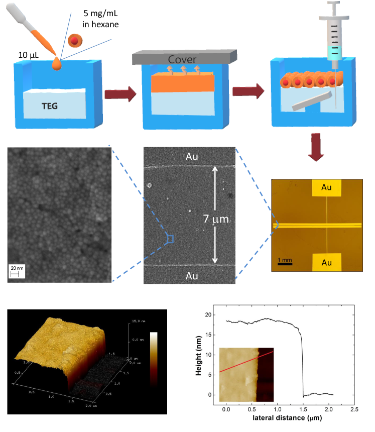
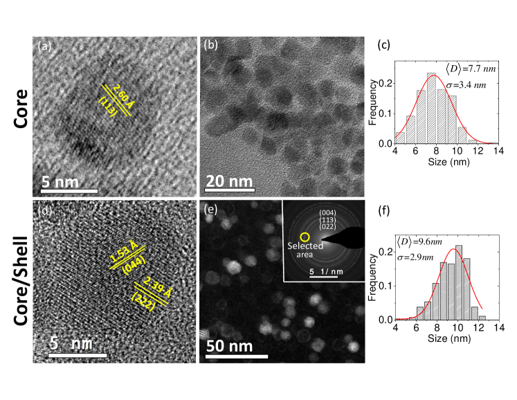
Figure 2 compares the morphology and size, measured by TEM, of Fe3O4 nanoparticles seeds with a representative core/shell system, Fe3O4/CoFe2O4, both samples were subjected to the same thermal annealing at 400 ∘C in vacuum atmosphere. From the size histograms, fitted with a Gaussian function, the mean particle sizes () were calculated, resulting 7.7 nm and 9.6 nm, for core and core/shell systems, respectively. From high resolution TEM (HRTEM) image it is noticed that the core is monocrystalline and the shell growth epitaxial over the core for most of the nanoparticles. Moreover different crystalline orientations for core and shell can be observed for most of the nanoparticles as noticed in Figure 2(d) where the (044) and (222) crystalline planes of spinel phase are signaled for core and shell, respectively. The core/shell structure is confirmed by dark field, as shown in Figure 2(e) where the TEM image was recorded with a small objective aperture positioned on the (113) brighter electron diffraction ring of the spinel phase. In this way the bright contrast in the reconstructed image corresponds to the spinel grains with the selected crystallographic orientation as schematically drawing in Figure S1(d-e) (see Suplemental Material). From the HRTEM and dark field TEM images, and from the comparison with the core size, the thickness of the Co1-xZnxFe2O4 shell was estimated as 1 nm. The composition of the nanoparticles was analyzed by inductively coupled plasma optical emission spectrometry (ICP-OES). From this analytical technique the concentration of the transition metal ions for all the samples were obtained and reported in Table 1. From these data, and assuming a Fe3O4 core of 7.7 nm of diameter, we calculated the shell stoichiometry which shows a systematic evolution consistent with the nominal concentration.
| Samples | Total concentration | Calculated | ||
|---|---|---|---|---|
| %Co | %Zn | %Fe | shell stoichiometry | |
| 9.40 | 0.00 | 90.60 | Co0.61Fe2.39O4 | |
| 7.71 | 2.36 | 89.93 | Co0.45Zn0.15Fe2.40O4 | |
| 6.01 | 4.80 | 89.19 | Co0.39Zn0.35Fe2.26O4 | |
| 2.68 | 5.78 | 91.54 | Co0.18Zn0.44Fe2.38O4 | |
| 0.00 | 9.94 | 90.06 | Zn0.80Fe2.20O4 | |
| Sample | ||||
|---|---|---|---|---|
| nm | nm | K | 106erg/cm3 | |
| Fe3O4 | 6.9 | 2.6 | 17 | 0.32 |
| 9.6 | 2.9 | 191 | 1.54 | |
| 9.9 | 3.8 | 171 | 1.25 | |
| 9.5 | 2.9 | 135 | 1.12 | |
| 9.4 | 2.7 | 115 | 0.99 | |
| 9.2 | 3.3 | 8 | 0.07 |
The self assemblies of Fe3O4-core/Co1-xZnxFe2O4-shell nanoparticles were obtained by the liquid-air interface process Dong et al. (2010); Chen et al. (2013); Jiang et al. (2017) as explained in the Experimental Procedure Section. In order to perform the different measurements, the self-assembly and the subsequent thermal treatment was reproduced using a commercial silicon nitride support grid for TEM characterization, and a glass substrate patterned with two Au electrodes separated by 7 m (as shown in the SEM image of Figure 1) for the magnetotransport studies. The topography of the annealed assemblies was analyzed by atomic force microscopy. Images acquired at different regions of the films reveal a large homogeneity with uniform and smooth surface, as observed in the bottom panel of Figure 1. From the AFM height profile cross section at the film boundary, an average film thickness of 20 nm was measured, which corresponds to two layers of nanoparticles.
Homogeneity and narrow size distribution are essential conditions to reach large area of self-organization; for core/shell nanoparticles, as observed from Figure 3 and Figure S1(a) (see Supplemental Material Sup ), assemblies of several microns are obtained. From Figure S1(f) it is also noticed that the self-organized nanoparticles are separated by a gap of 1 nm. As the thermogravimetric analysis indicates that approximately 7% of residual mass remains in the systems after the thermal treatment at 400 ∘C in vacuum atmosphere, and infrared spectroscopy measurements do not detect organic molecules (see Figure S3 in Supplemental Material Sup ), we conclude that the gap between the nanoparticles is formed by amorphous carbon. The nanoparticles size distributions measured from the TEM micrographs, are shown in the middle panels of Figure 3. From the fitting of the histograms with a Gaussian function, the mean nanoparticles size was calculated and summarized in Table 2, which vary between 9.2-9.9 nm for all the systems. We also notice that the nanoparticle size and also the superstructure of the self-assembly is preserved at higher annealing temperature, however at 600 ∘C the nanoparticles start to coalesce (see Figure S2 Supplemental Material Sup ). From the HRTEM images (shown in Figure 2 for 0 and in Figure 3 for 0.75 and 1) it is observed that the core/shell microstructure is preserved after the annealing at 400 ∘C, where different interplanar distances and crystallographic planes orientation for the inner and outer part of the particle can be measured. As mentioned before, this morphology is confirmed by dark field images, as shown in Figure 3 for x=0.00, 0.25 and 0.50. Although the core/shell microstructure is preserved, we can not discard some degree of interdiffusion at the interface as reported for similar nanoparticles systems López-Ortega et al. (2012); Krycka et al. (2013); however, as we are going to discuss later, the magnetoresistance measurements confirms the half-metallic nature of the Fe3O4 core.
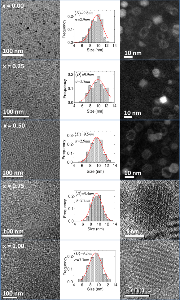
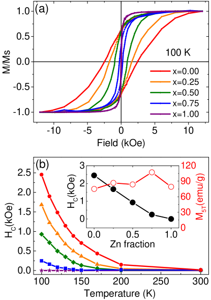
Figure 4(a) and Figure S4 (see Supplemental Material Sup ) show the magnetization hysteresis loops of the annealed self-assembled nanoparticles, measured with the magnetic field applied in the plane of the substrate. The hysteresis loops show a single magnetization reversal for all compositions, with the coercive field () decreasing approximately linearly with the Zn concentration , as shown in Figure 4(b). Notice that the magnetization hysteresis loop reported for binary nanocrystal superlattice CoFe2O4Fe3O4 results from the superposition of the individual component and an annealing treatment is an essential step to obtain an exchange interaction between the half-metallic Fe3O4 and the magnetic insulator CoFe2O4 Chen et al. (2013). On the contrary, in the core/shell morphology, both phases are strongly coupled at the interface and homogeneous loop is obtained even in the as-synthesized system as shown in Figure S5. The magnetization inversion process of Fe3O4/Co1-xZnxFe2O4 core/shell nanoparticles can be analyzed from the theoretical approach developed for bimagnetic soft/hard exchange coupled nanostructures.Skomski and Coey (1993); Kneller and Hawig (1991); Zhao and Wang (2006); Zhao et al. (2005); Leineweber and Kronmüller (1997) From these studies a critical size for the soft magnetic component, , was found bellow which both phases are rigidly coupled by interface exchange interaction and reverse their magnetization in a coherent mode at the nucleation field, . This critical size is approximately twice the magnetic Bloch wall width of the hard phase , where is the exchange stiffness. In this regime, a single square hysteresis loop is obtained, with ; where , and are the magnetization, film thickness (or volume fraction), and magnetic anisotropy of the core () and shell (), respectively.Fullerton et al. (1999); López Ortega et al. (2015); Zhao et al. (2007) Instead, if the size of the soft magnetic phase is larger than exchange spring behavior is found, the magnetization reversal is nonuniform and lower coercivities are obtained. Fullerton et al. (1999); Zhao et al. (2005); Zhao and Wang (2006) For CoFe2O4 the values reported for span in the range of 13-20 nm,Coey (2010); López Ortega et al. (2015) larger than the diameter of the Fe3O4 soft core used in this work. Therefore, rigid exchange coupling of the magnetizations of the core/shell phases is expected. This conclusion is supported by the study of Fe3O4/CoFe2O4 soft/hard bilayers, where a critical thickness of 8 nm was found for the crossover from rigid coupling to exchange spring behavior as a function of the Fe3O4 thicknessLavorato et al. (2016b). Therefore, the decrease of with can be accounted by the diminution of the magnetic anisotropy of the shell from 4 106ergcm3 for , to 2 105ergcm3 for Bullita et al. (2014) in agreement with the expression for in the rigid exchange coupled regime.
The systematic dependence of the magnetic anisotropy with the shell composition is also reflected in the zero-field-cooled (ZFC) and field-cooled (FC) magnetization curves shown in Figure S6 of Supplemental Material Sup . The temperature where the change from blocked to the superparamagnetic regime is observed decreases progressively with increasing . From the maximum of the energy barrier distribution calculated as the mean blocking temperature, , and the effective magnetic anisotropy constant, , can be obtained for particles of total volume , as reported in Table 2.Dormann et al. (1996) For comparison, Table S1 (see Supplemental Material Sup ) reports the parameters that characterize the magnetic properties of dispersed nanoparticles before the annealing process. Both systems, the dispersed nanoparticles and the annealed assemblies, present qualitative and quantitative similar behavior, with an enhancement of the effective magnetic anisotropy when the concentration of Co in the shell increases, which points out that the magnetic behavior is governed by the hard/soft rigid coupling magnetization inversion process, as analyzed previously. However, the annealed assemblies present an approximately 20% larger , and , probably due to an increase of the dipolar interaction and the improvement of the crystallinity in the annealed assemblies.
The magnetic measurements demonstrate that the effective magnetic anisotropy of the system can be controlled by adjusting the shell composition, without appreciably modifying the morphology and the overall magnetic saturation, as observed from the inset of Figure 4(b). Given that the anisotropy of the system is to a great extent responsible of the switching field of the TMR, devices made of self-assembled core/shell nanoparticles provide an ideal system for studying spin-dependent transport between magnetic nanoparticles. Although both materials at the core/shell structure are strongly exchange coupled and behave as a unique magnetic entity with an average magnetic anisotropy, the conductivity of each phase is different. While the Fe3O4 core is half-metallic, the Co1-xZnxFe2O4 shell is a semiconductor. Therefore, in order to observe TMR properties it is crucial to have Fe3O4 phase to provide the spin polarized transport, whereas the role of the shell is to modulate the switching field by tuning the magnetic anisotropy, while acting as a tunnel barrier.
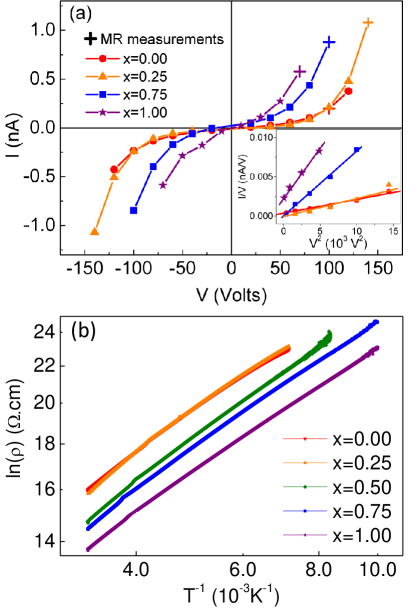
The electronic transport in the annealed self-assembled devices was studied from the current-voltage () measurements and from the temperature dependence of the resistivity , as reported in Figure 5. From this figure it can be affirmed that the electron conduction in the devices is given by two independent mechanisms: thermally activated hopping, which is revealed from the temperature dependence of ; and the tunneling conduction manifested by the non-Ohmic behavior in the I-V curve with the characteristic dependence at low temperature, as discussed next. From the Simmons’s model, which considers inelastic tunneling across an insulating barrier, the curves can be quantitatively linked to the physical parameters of the system, i.e. the tunnel barrier height () and width (), the effective contact area, etc. Simmons (1963a). This model also considers linear dependence approximation of the barrier potential profile with and . For voltage smaller than the potential barrier, the Simmons’s model can be approximated with the well known polynomial relationship:Simmons (1963b)
| (1) |
where is the equilibrium conductance and , where is the shape factor of the barrier.Vilan (2007) From the plot shown in the inset of Figure 5(a), the shape factor of the barrier is clearly increasing with . Given that the barrier width is approximately constant for all samples, this reflects a progressive decrease of the tunneling barrier height as the Zn content increases. Considering the geometry of the assemblies devices, i.e. the area measured by SEM and the film thickness obtained by AFM microscopy, we have plotted in Figure 5(b), which suggests that a thermally activated transport mechanism is also involved in the conduction of the devices. The temperature dependence follows the relation , where was found as the best fitted parameter for all the systems. This value is close to the dependence found in the Efros’s variable range hopping model , Efros and Shklovskii (1975) and it is consistent with the behavior measured in other nanoparticles arrays. Black et al. (2000); Dong et al. (2010); Song et al. (2013); Mohan Kant et al. (2008); Zhang et al. (2010). Moreover, in the core/shell assemblies is, at least, two orders of magnitude smaller than the reported for pellets of Co1-xZnxFe2O4 powder ferrite (for example at room temperature CoFe2O1.107 and Co0.4Zn0.6Fe2O1.108 Rani et al. (2013); Gul et al. (2007); Reddy et al. (1999)). This result shows that the tunnel conduction strongly increases the conductivity of the core/shell assemblies compared to the values measured in the semiconductor shell material. Notice that the larger the tunneling current, the lower the resistivity of the devices. Therefore, the material that has the lower tunneling barrier will be the one that is most influenced by this contribution. Consistently, from Figure 5 we have measured that the assemblies of nanoparticles with a ZnFe2O4 shell present the lowest energy barrier, and a systematic increase with decreasing is observed.
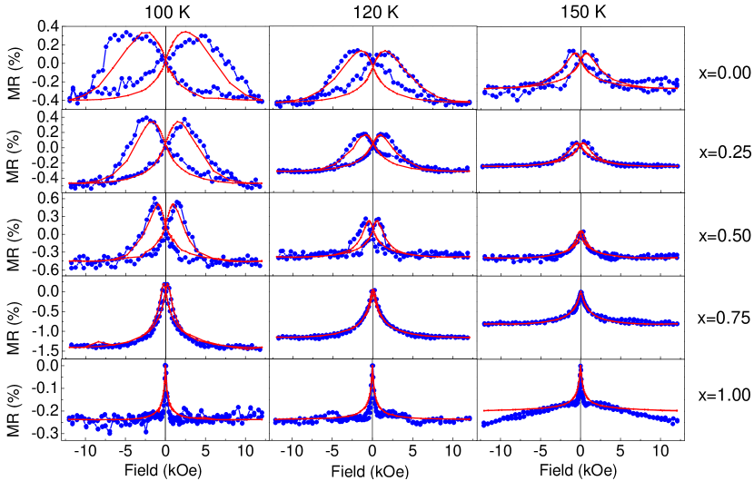
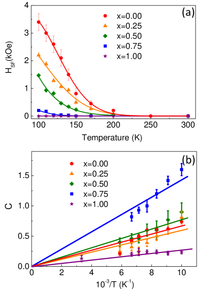
The main findings of this work are summarized in Figure 6 and 7. Magnetoresistance curves measured at different temperatures for the five devices studied in this work are shown in Figure 6 and Figure S7 of Supplemental Material Sup . At a given temperature, the switching field of the magnetoresistance curve monotonously decreases when the Zn concentration of the shell increases. Moreover, as shown in Figure 7, each sample shows a smooth decrease of the switching field with temperature, consistently with the temperature evolution of the coercive field. Also, a saturation behavior of TMR at high magnetic field is observed when decreases, in good correspondence with the M(H) curves, see Figure 4.
For spin-polarized intergrain tunneling the TMR is related to the macroscopic magnetization.López-Quintela et al. (2003); Hwang et al. (1996); Helman and Abeles (1976) For a ferromagnetic-insulator granular system, the electron tunneling across the insulating barrier was calculated including an additional exchange energy arising from the interaction between the tunneling electron spin and the non-parallel magnetic moment of the neighboring grains. Helman and Abeles (1976); Barzilai et al. (1981). Assuming that the exchange energy can be expressed in terms of the spin correlation function of two ferromagnetic neighboring grain, the magnetoresistance can be expressed as:
| (2) |
where accounts for the magnetic correlations when the electron tunnels through the insulating barrier and . Notice that within this model the magnetoresistance does not depend on the total resistivity of the sample. The fittings to this equation using as the single adjusted parameter for each temperature, are shown in Figure 6. The good agreement between both measurements from equation 2 confirms the spin polarized tunnel transport in the present devices, where the parameter gives the proportionality between two independent experiments, and . Moreover, in agreement with equation 2, the fittings show that varies approximately linearly with over the measured temperature range (see Figure 7(b)). However, although equation 2 adjusts the field and temperature dependence of TMR with the magnetization, due to the complex nature of the present system, it is hard to determine the dependence between the C parameter and the shell stoichiometry. Even though all the systems have the same Fe3O4 core, the Co1-xZnxFe2O4 shell was synthesized in a second step which lead to a shell thickness dispersion along the Zn composition, moreover from the synthesis and later thermal treatment the nanostructures could also present dispersion in the nanoparticle coating. These experimental factors could affect the magnetic correlations when the electron tunnels through the insulating magnetic barrier and also the surface Fe3O4 spin polarization. Nevertheless, although it is hard to estimate the evolution of the magnetic correlation with the shell stoichiometry, the magnitude of the interaction can be estimated. Assuming the spin polarization of magnetite, () Hu and Suzuki (2002) a coupling constant of meV ( K) is obtained, comparable to the calculations for the intergrain tunneling transport of manganese perovskites Balcells et al. (1998).
The TMR amplitude, which is in the range depending on the composition, is similar to other reported values for self assembled nanoparticles, Dugay et al. (2011); Dong et al. (2010); Zhang et al. (2010) however, it is much smaller than the calculated from the Julliere’s model: on the basis of the spin polarization values of magnetite. This reduction may be due to the fact that the tunneling probability decreases exponentially with the barrier width. According to Refs. Moodera et al. (1997); Joo et al. (2014) for TMR multilayers, the optimal barrier thickness is in the nm range; however, the tunnel current between Fe3O4 cores in the self-assembled structure must pass through the insulator barrier of 3 nm width, which is composed by the cobalt ferrite shell and the amorphous carbon nanoparticles coating. On the other hand, although it is known that the TMR diminishes with increasing bias voltage,Coey (2010) the high resistance of the magnetic nanoparticles devices determines the experimental parameters, and high voltage bias 100 V had to be applied to perform the transport measurements at low temperature. These factors make evident the importance of optimizing the different stages of the fabrication process in order to increase the conductivity to produce large amplitude and low switching field TMR devices based on core/shell magnetic nanoparticles. However, irrespective of the absolute value of TMR, this study demonstrates that the switching field of TMR can be tailored at will in self-assemblies of exchange coupled core-shell nanoparticles, synthesized by an affordable chemical route.
IV Conclusion
In summary, we have fabricated self-assemblies of core/shell nanoparticles with controlled TMR. Particularly, we have shown that the magnetic properties can be finely tuned by changing the shell composition which provides a tool to adjust the TMR switching field. We shows whereas that the Fe3O4 core provides the spin polarized transport, the shell acts as tunnel barrier in the self assembly and also modulates the switching field by tuning the magnetic anisotropy through the interface exchange coupling. This approach shows the feasibility to use assemblies of exchange coupled magnetic nanoparticles in TMR devices, where different combinations of soft/hard and hard/soft core/shell configurations can be envisaged. In this way, combinations of materials can be carefully designed to move across the rigid coupling to exchange bias regime, to design devices with tailored magnetotransport response, which gives a promising base for the design of core/shell nanoparticles based devices for fundamental studies or for spintronic applications.
Acknowledgements.
The authors thank the staff of the INVAP SE Chemistry Laboratory for the ICP-OES spectrometry measurements. The authors also acknowledges financial support of Argentinian governmental agency ANPCyT (Project No.PICT-2016-0288 and PICT-2015-0883) and UNCuyo (Project No. 06/C527 and 06/C528). The authors gratefully acknowledge the EU-commission financial support under the: H2020-MSCA-RISE-2016 SPICOLOST PROJECT No 734187. F. R. acknowledges financial support of the Ministerio de Economía y Competitividad of Spain (Project No. MAT2016-80762-R), and Xunta de Galicia (Centro Singular de Investigación de Galicia accreditation 2016-2019) and the European Union (European Regional Development Fund ERDF).References
- Baibich et al. (1988) M. N. Baibich, J. M. Broto, A. Fert, F. Nguyen Van Dau, F. Petroff, P. Etienne, G. Creuzet, A. Friederich, and J. Chazelas, “Giant magnetoresistance of (001)Fe/(001)Cr magnetic superlattices,” Phys. Rev. Lett. 61, 2472–2475 (1988).
- Binasch et al. (1989) G. Binasch, P. Grünberg, F. Saurenbach, and W. Zinn, “Enhanced magnetoresistance in layered magnetic structures with antiferromagnetic interlayer exchange,” Phys. Rev. B 39, 4828–4830 (1989).
- Zutic et al. (2004) I. Zutic, F. Jaroslav, and S. Das Sarma, “Spintronics: Fundamentals and applications,” Rev. Mod. Phys. 76, 323–410 (2004).
- Bader and Parkin (2010) S.D. Bader and S.S.P. Parkin, “Spintronics,” Annual Review of Condensed Matter Physics 1, 71–88 (2010).
- Julliere (1975) M. Julliere, “Tunneling between ferromagnetic films,” Physics Letters A 54, 225 – 226 (1975).
- Moodera et al. (1995) J. S. Moodera, L. R. Kinder, T. M. Wong, and R. Meservey, “Large magnetoresistance at room temperature in ferromagnetic thin film tunnel junctions,” Phys. Rev. Lett. 74, 3273–3276 (1995).
- Yuasa et al. (2004) S. Yuasa, T. Nagahama, A. Fukushima, Y. Suzuki, and K. Ando, “Giant room-temperature magnetoresistance in single-crystal Fe/MgO/Fe magnetic tunnel junctions,” Nature Materials 3, 868 (2004).
- Djayaprawira et al. (2005) D. D. Djayaprawira, K. Tsunekawa, M. Nagai, H. Maehara, S. Yamagata, N. Watanabe, S. Yuasa, Y. Suzuki, and K. Ando, “230% room-temperature magnetoresistance in CoFeB/MgO/CoFeB magnetic tunnel junctions,” Applied Physics Letters 86, 092502 (2005).
- López-Quintela et al. (2003) M. A. López-Quintela, L. E. Hueso, J. Rivas, and F. Rivadulla, “Intergranular magnetoresistance in nanomanganites,” Nanotechnology 14, 212 (2003).
- Hwang et al. (1996) H. Y. Hwang, S-W. Cheong, N. P. Ong, and B. Batlogg, “Spin-polarized intergrain tunneling in La2/3Sr1/3MnO3,” Physical Review Letters 77, 2041 (1996).
- Murray et al. (2000) C.B. Murray, C.R. Kagan, and M.G. Bawendi, “Synthesis and characterization of monodisperse nanocrystals and close-packed nanocrystal assemblies,” Annual Review of Materials Science 30, 545–610 (2000).
- Sun et al. (2000) S. Sun, C. B. Murray, D. Weller, L. Folks, and A. Moser, “Monodisperse FePt Nanoparticles and Ferromagnetic FePt Nanocrystal Superlattices,” Science 287, 1989–92 (2000).
- Zeng et al. (2006) H. Zeng, C. T. Black, R. L. Sandstrom, P. M. Rice, C. B. Murray, and Shouheng Sun, “Magnetotransport of magnetite nanoparticle arrays,” Phys. Rev. B 73, 020402 (2006).
- Black et al. (2000) C. T. Black, C. B. Murray, R. L. Sandstrom, and S. Sun, “Spin-dependent tunneling in self-assembled cobalt-nanocrystal superlattices,” 290, 1131–1134 (2000).
- Wang et al. (2009) S. Wang, F. J. Yue, D. Wua, F. M. Zhang, W. Zhong, and Y. W. Du, “Enhanced magnetoresistance in self-assembled monolayer of oleic acid molecules on Fe3O4 nanoparticles,” Applied Physics Letters 94, 012507 (2009).
- Dugay et al. (2011) J. Dugay, R. P. Tan, A. Meffre, T. Blon, L. M. Lacroix, J. Carrey, P. F. Fazzini, S. Lachaize, B. Chaudret, and M. Respaud, “Room-temperature tunnel magnetoresistance in self-assembled chemically synthesized metallic iron nanoparticles,” Nano Letters 11, 5128–5134 (2011).
- Mohan Kant et al. (2008) K. Mohan Kant, K. Sethupathi, and M. S. Ramachandra Rao, “Role of oxide barrier in intergranular tunnel junctions: An enhanced magnetoresistance in SiO2 and ZnO coated Fe3O4 nanoparticle compacts,” Journal of Applied Physics 103, 07F318 (2008).
- Taub et al. (2009) N. Taub, A. Tsukernik, and G. Markovich, “Interparticle spin-polarized tunneling in arrays of magnetite nanocrystals,” Journal of Magnetism and Magnetic Materials 321, 1933 (2009).
- Tran et al. (2008) T. B. Tran, I. S. Beloborodov, Jingshi Hu, X. M. Lin, T. F. Rosenbaum, and H. M. Jaeger, “Sequential tunneling and inelastic cotunneling in nanoparticle arrays,” Phys. Rev. B 78, 075437 (2008).
- Chen et al. (2013) J. Chen, X. Ye, J. O. Soong, J. M. Kikkawa, C. R. Kagan, and C. B. Murray, “Bistable Magnetoresistance Switching Binary Nanocrystal Superlattices by Self-Assembly and Thermal Annealing,” ACS Nano 7, 1478–1486 (2013).
- Dong et al. (2010) A. Dong, J. Chen, J. M. Vora, P. M.and Kikkawa, and C.B. Murray, “Binary Nanocrystal Superlattice Membranes Self-Assembled at the Liquid-Air Interface,” Nature 466, 474–477 (2010).
- Jiang et al. (2017) C. Jiang, C. W. Leung, and P. W.T. Pong, “Self-assembled thin films of Fe3O4-Ag composite nanoparticles for spintronic applications,” Applied Surface Science 419, 692 – 696 (2017).
- Kumar et al. (2013) P. A. Kumar, S. Ray, S. Chakraverty, and D. D. Sarma, “Engineered spin-valve type magnetoresistance in Fe3O4-CoFe2O4 coreshell nanoparticles,” Appl. Phys. Lett. 103, 102406 (2013).
- Zhang et al. (2010) Y. Zhang, H. Xing, N. Poudyal, V. Nandwana, C. B. Rong, S. S. Yan, H. Zeng, and J. P. Liu, “Inversed tunneling magnetoresistance in hybrid FePt/Fe3O4 core/shell nanoparticles systems,” Journal of Applied Physics 108, 103905 (2010).
- Helman and Abeles (1976) J. S. Helman and B. Abeles, “Tunneling of spin-polarized electrons and magnetoresistance in granular ni films,” Phys. Rev. Lett. 37, 1429–1432 (1976).
- El-Hilo et al. (1998) M. El-Hilo, R. W. Chantrell, and K. O’Grady, “A model of interaction effects in granular magnetic solids,” Journal of Applied Physics 84, 5114–5122 (1998).
- Hu and Suzuki (2002) G. Hu and Y. Suzuki, “Negative spin polarization of fe3o4 in magnetitemanganitebased junctions,” Phys. Rev. Lett. 89, 276601 (2002).
- López Ortega et al. (2015) Alberto López Ortega, Marta Estrader, German Salazar Alvarez, Alejando G. Roca, and Josep Nogués, “Applications of exchange coupled bimagnetic hard/soft and soft/hard magnetic core/shell nanoparticles,” Phys. Rep. 553, 1–32 (2015).
- Lavorato et al. (2017) G. C. Lavorato, E. Lima, H. E. Troiani, R. D. Zysler, and E. L. Winkler, “Tuning the coercivity and exchange bias by controlling the interface coupling in bimagnetic core/shell nanoparticles,” Nanoscale 9, 10240–10247 (2017).
- Salazar-Alvarez et al. (2007) G. Salazar-Alvarez, J. Sort, S. Suriñach, M. D. Baró, and J. Nogués, “Synthesis and Size-Dependent Exchange Bias in Inverted Core/Shell MnO/Mn3O4 Nanoparticles,” J. Am. Chem. Soc. 129, 9102–9108 (2007).
- Skumryev et al. (2003) V. Skumryev, S. Stoyanov, Y. Zhang, G. Hadjipanayis, D. Givord, and J. Nogués, “Beating the superparamagnetic limit with exchange bias,” Nature 423, 850–853 (2003).
- Sarkar et al. (2012) A. Sarkar, N. Behera, R. Adhikari, and A. K. Das, “Studies on nonlinear electrical transport and magnetoresistance in co/coo core-shell nanostructure,” AIP Conference Proceedings 1447, 937–938 (2012).
- Winkler et al. (2012) E. L. Winkler, E. Lima Jr., D. Tobia, M. E. Saleta, H. E. Troiani, E. Agostinelli, D. Fiorani, and R. Zysler, “Origin of magnetic anisotropy in ZnO/CoFe2O4 and CoO/CoFe2O4 core/shell nanoparticle systems,” Appl. Phys. Lett. 101, 252405 (2012).
- Lavorato et al. (2016a) G. Lavorato, E. Winkler, A. Ghirri, D. Lima Jr, E.and Peddis, H. E Troiani, D. Fiorani, E. Agostinelli, D. Rinaldi, and R. D. Zysler, “Exchange bias and surface effects in bimagnetic CoO-Core/Co0.5Ni0.5Fe2O4-Shell nanoparticles,” Physical Review B 94, 054432 (2016a).
- Liu et al. (2015) X. Liu, B. P. Pichon, C. Ulhaq, C. Lefèvre, J. M. Grenèche, D. Bégin, and S. Bégin-Colin, “Systematic Study of Exchange Coupling in Core-Shell Fe 3-deltaOCoO Nanoparticles,” Chemistry of Materials 27, 4073–4081 (2015), https://doi.org/10.1021/acs.chemmater.5b01103 .
- Lottini, E. and López-Ortega, A. and Bertoni, G. and Turner, S. and Meledina, M. and Tendeloo, G. Van and de Julián Fernández, C. and Sangregorio, C. (2016) Lottini, E. and López-Ortega, A. and Bertoni, G. and Turner, S. and Meledina, M. and Tendeloo, G. Van and de Julián Fernández, C. and Sangregorio, C., “Strongly Exchange Coupled Core/Shell Nanoparticles with High Magnetic Anisotropy:A Strategy Toward Rare-Earth-Free Permanent Magnets,” Chemistry of Materials 28, 4214–4222 (2016).
- Lavorato et al. (2014) Gabriel C Lavorato, Enio Lima Jr, Dina Tobia, Dino Fiorani, Horacio E Troiani, Roberto D Zysler, and Elin L. Winkler , “Size Effects in Bimagnetic CoO/CoFe2O4 Core/Shell Nanoparticles.” Nanotechnology 25, 355704 (2014).
- Sytnyk et al. (2013) M. Sytnyk, R. Kirchschlager, M. I. Bodnarchuk, D. Primetzhofer, D. Kriegner, H. Enser, J. Stangl, P. Bauer, M. Voith, A. W. Hassel, F. Krumeich, F. Ludwig, A. Meingast, G. Kothleitner, M. Kovalenko, and W. Heiss, “Tuning the magnetic properties of metal oxide nanocrystal heterostructures by cation exchange,” Nano Letters 13, 586–593 (2013), pMID: 23362940.
- Fabris et al. (2019) F. Fabris, E. Lima Jr., E. De Biasi, H. E. Troiani, M. Vasquez Mansilla, T. E. Torres, R. Fernández-Pacheco, M. R. Ibarra, G. F. Goya, Zysler R., and E. Winkler, “Controlling the dominant magnetic relaxation mechanisms for magnetic hyperthermia in bimagnetic core-shell nanoparticles,” Nanoscale 11, 3164–3172 (2019).
- Lavorato et al. (2015) G.C. Lavorato, E. Lima, H.E. Troiani, R.D. Zysler, and E.L. Winkler, “Exchange-coupling in thermal annealed bimagnetic core/shell nanoparticles,” Journal of Alloys and Compounds 633, 333 – 337 (2015).
- Sun and Zeng (2002) S. Sun and H. Zeng, “Size-controlled synthesis of magnetite nanoparticles,” Journal of the American Chemical Society 124, 8204–8205 (2002), pMID: 12105897.
- Sun et al. (2004) S. H Sun, H. Zeng, D. B. Robinson, S. Raoux, P. M. Rice, S. X. Wang, and G. X. Li, “Monodisperse MFe2O4 (M = Fe, Co, Mn) nanoparticles,” Journal of the American Chemical Society 126, 273–279 (2004).
- (43) “See Supplemental Material at URL for additional microstructural information of the core/shell nanoparticles assemblies annealed at different temperature, obtained by TEM and HRTEM microscopy. Further characterization of the as-synthetized and annealed Fe3O4/CoFe2O4 core/shell nanoparticles by thermogravimetric (TGA) and infrared (FT-IR) measurements, and additional magnetic characterization, as the hysteresis cycles as a function of the temperature and ZFC and FC magnetization curves of the annealed nanoparticles, it is also included. See also details of the magnetoresistance temperature evolution of the self-assemblies.” .
- López-Ortega et al. (2012) A. López-Ortega, M. Estrader, G. Salazar-Alvarez, S. Estradé, I. V. Golosovsky, R. K. Dumas, D. J. Keavney, M. Vasilakaki, K. N. Trohidou, J. Sort, F. Peiró, S. Suriñach, M. D. Baró, and J. Nogués, “Strongly exchange coupled inverse ferrimagnetic soft/hard MnxFe3-xOFexMn3-xO4 core/shell heterostructured nanoparticles,” Nanoscale 4, 5138–5147 (2012).
- Krycka et al. (2013) K. L. Krycka, J. A. Borchers, G. Salazar-Alvarez, A. López-Ortega, M. Estrader, S. Estradé, E. Winkler, R. D. Zysler, J. Sort, F. Peiró, M. D. Baró, C.C. Kao, and J. Nogués, “Resolving material-specific structures within Fe3O4/-Mn2O3 core/shell nanoparticles using anomalous small-angle x-ray scattering,” ACS Nano 7, 921–931 (2013), pMID: 23320459.
- Skomski and Coey (1993) R. Skomski and J. M. D. Coey, “Giant energy product in nanostructured two-phase magnets,” Phys. Rev. B 48, 15812–15816 (1993).
- Kneller and Hawig (1991) E. F. Kneller and R. Hawig, “The Exchange-Spring Magnet: A New Material Principle for Permanent Magnets,” IEEE Trans. Magn. 27, 3588–3600 (1991).
- Zhao and Wang (2006) G. P. Zhao and X. L. Wang, “Nucleation, pinning, and coercivity in magnetic nanosystems: An analytical micromagnetic approach,” Phys. Rev. B 74, 012409 (2006).
- Zhao et al. (2005) G. P. Zhao, M. G. Zhao, H. S. Lim, Y. P. Feng, and C. K. Ong, “From nucleation to coercivity,” Applied Physics Letters 87, 162513 (2005).
- Leineweber and Kronmüller (1997) T. Leineweber and H. Kronmüller, “Micromagnetic examination of exchange coupled ferromagnetic nanolayers,” Journal of Magnetism and Magnetic Materials 176, 145 – 154 (1997).
- Fullerton et al. (1999) Eric E. Fullerton, J. S. Jiang, and S. D. Bader, “Hard/Soft Magnetic Heterostructures: Model Exchange-Spring Magnets,” J. Magn. Magn. Mater. 200, 392–404 (1999).
- Zhao et al. (2007) G. P. Zhao, X. L. Wang, Y. P. Feng, and C. W. Huang, “Coherent rotation and effective anisotropy,” IEEE Transactions on Magnetics 43, 2908–2910 (2007).
- Coey (2010) J. M. D. Coey, Magnetism and Magnetic Materials (Cambridge University Press, 2010).
- Lavorato et al. (2016b) G. Lavorato, E. Winkler, B. Rivas-Murias, and F. Rivadulla, “Thickness dependence of exchange coupling in epitaxial Fe3OCoFe2O soft/hard magnetic bilayers,” Phys. Rev. B 94, 054405 (2016b).
- Bullita et al. (2014) S. Bullita, A. Casu, M. F. Casula, G. Concas, F. Congiu, A. Corrias, A. Falqui, D. Loche, and C. Marras, “ZnFe2O4 nanoparticles dispersed in a highly porous silica aerogel matrix: A magnetic study.” Phys. Chem. Chem. Phys. 16, 4843–52 (2014).
- Dormann et al. (1996) J. Dormann, F. D’Orazio, F. Lucari, E. Tronc, P. Prené, J. Jolivet, D. Fiorani, R. Cherkaoui, and M. Noguès, “Thermal Variation of the Relaxation Time of the Magnetic Moment of -Fe2O3 Nanoparticles with Interparticle Interactions of Various Strengths,” Phys. Rev. B 53, 14291–14297 (1996).
- Simmons (1963a) J. G. Simmons, “Generalized formula for the electric tunnel effect between similar electrodes separated by a thin insulating film,” Journal of Applied Physics 34, 1793 (1963a).
- Simmons (1963b) J. G. Simmons, “Low-voltage current-voltage relationship of tunnel junctions,” Journal of Applied Physics 34, 238 (1963b).
- Vilan (2007) A. Vilan, “Analyzing molecular current-voltage characteristics with the simmons tunneling model: Scaling and linearization,” J. Phys. Chem. C 111, 4431–4444 (2007).
- Efros and Shklovskii (1975) A.L. Efros and B.I. Shklovskii, “Coulomb gap and low temperature conductivity of disordered systems,” J. Phys. C: Solid State Phys. 8, L49 (1975).
- Song et al. (2013) N.N. Song, H. T. Yang, F. Y. Li, Z.A. Li, W. Han, X. Ren, Y. Luo, X.C. Wang, C. Q. Jin, X.Q. Zhang, and Z.H. Cheng, “Interspacing dependence of spin-dependent variable range hopping for cold-pressed Fe3O4 nanoparticles,” Journal of Applied Physics 113, 184309 (2013).
- Rani et al. (2013) R. Rani, G. Kumar, K. M. Batoo, and M. Singh, “Electric and dielectric study of zinc substituted cobalt nanoferrites prepared by solution combustion method,” American Journal of Nanomaterials 1, 9–12 (2013).
- Gul et al. (2007) I.H. Gul, A.Z. Abbasi, F. Amin, M. Anis ur Rehman, and A. Maqsood, “Structural, magnetic and electrical properties of Co1-xZnxFe2O4 synthesized by co-precipitation method,” Journal of Magnetism and Magnetic Materials 311, 494 – 499 (2007).
- Reddy et al. (1999) A.V. Ramana Reddy, G Ranga Mohan, B.S Boyanov, and D Ravinder, “Electrical transport properties of zinc-substituted cobalt ferrites,” Materials Letters 39, 153 – 165 (1999).
- Barzilai et al. (1981) S. Barzilai, Y. Goldstein, I. Balberg, and J. S. Helman, “Magnetic and transport properties of granular cobalt films,” Physical Review B 23, 1809 (1981).
- Balcells et al. (1998) L. L. Balcells, J. Fontcuberta, B. Martinez, and X. Obradors, “Magnetic surface effects and low-temperature magnetoresistance in manganese perovskites,” Journal of Physics: Condensed Matter 10, 1883 (1998).
- Moodera et al. (1997) J. S. Moodera, E. F. Gallagher, K. Robinson, and J. Nowak, “Optimum tunnel barrier in ferromagnetic-insulator-ferromagnetic tunneling structures,” Applied Physics Letters 70, 3050–3052 (1997).
- Joo et al. (2014) S. Joo, K. Y. Jung, K. I. Jun, D. S. Kim, K. H. Shin, J. K. Hong, B. C. Lee, and K. Rhie, “Spin-filtering affect of thin Al2O3 barrier on tunneling magnetoresistance,” Applied Physics Letters 104, 152407 (2014).