Role of Substrate Stiffness in Tissue Spreading: Wetting Transition and Tissue Durotaxis
Abstract
Living tissues undergo wetting transitions: On a surface, they can either form a droplet-like cell aggregate or spread as a monolayer of migrating cells. Tissue wetting depends not only on the chemical but also on the mechanical properties of the substrate. Here, we study the role of substrate stiffness in tissue spreading, which we describe by means of an active polar fluid model. Taking into account that cells exert larger active traction forces on stiffer substrates, we predict a tissue wetting transition at a critical substrate stiffness that decreases with tissue size. On substrates with a stiffness gradient, we find that the tissue spreads faster on the stiffer side. Further, we show that the tissue can wet the substrate on the stiffer side while dewetting from the softer side. We also show that, by means of viscous forces transmitted across the tissue, the stiffer-side interface can transiently drag the softer-side interface towards increasing stiffness, against its spreading tendency. These two effects result in directed tissue migration up the stiffness gradient. This phenomenon — tissue durotaxis — can thus emerge both from dewetting at the soft side and from hydrodynamic interactions between the tissue interfaces. Overall, our work unveils mechanisms whereby substrate stiffness impacts the collective migration and the active wetting properties of living tissues, which are relevant in development, regeneration, and cancer.
I Introduction
In embryonic development, wound healing, and tumor progression, epithelial cells migrate collectively in cohesive groups Friedl and Gilmour (2009). To study the mechanics of collective cell migration, extensive research has focused on the spreading of epithelial tissues in vitro Hakim and Silberzan (2017). For example, when a cell aggregate is placed on a surface, it may retain a spheroidal shape or it may spread by extending a cell monolayer Gonzalez-Rodriguez et al. (2012); Beaune et al. (2014). Similar situations are found in vivo, for example during the epiboly process in zebrafish embryogenesis Morita et al. (2017); Wallmeyer et al. (2018). In analogy with the wetting of a liquid drop, the spreading of cell aggregates was proposed to rely on a competition between cell-cell and cell-substrate interactions Ryan et al. (2001); Douezan et al. (2011); Ravasio et al. (2015); Smeets et al. (2016). In experiments, these surface interactions were varied by tuning the expression level of cell-cell adhesion proteins and by modifying the chemical coating of the substrate, respectively. Expectedly, these changes can induce a wetting transition between a three-dimensional cell aggregate and a spreading cell monolayer — the equivalents of a drop and a precursor film, respectively Douezan et al. (2011).
However, the wetting behavior of a living tissue is not completely analogous to that of an inert liquid. The ability of cells to polarize and exert traction forces to migrate on a substrate turns a tissue into an active material, fundamentally changing the physics of tissue wetting Pérez-González et al. (2019). Moreover, cells sense and respond to the mechanical properties of their environment Discher et al. (2005); Ladoux and Nicolas (2012); Gupta et al. (2016). As a consequence, substrate stiffness affects the wetting properties of living tissues. Specifically, cell monolayers dewet from very soft substrates Douezan and Brochard-Wyart (2012) but wet stiffer substrates Douezan et al. (2012); Guo et al. (2006). In a reminiscent in vivo situation, the developmental stiffening of a tissue triggers the collective migration of neural crest cells required for embryo morphogenesis Barriga et al. (2018).
Besides affecting tissue wetting, substrate elasticity also influences the coordination and guidance of collective cell migration Angelini et al. (2010); Ng et al. (2012); Saez et al. (2007); Edwards and Schwarz (2011); Lange and Fabry (2013); Ladoux and Mège (2017); Barriga and Mayor (2018). Most strikingly, cell monolayers can migrate towards increasing substrate stiffness Sunyer et al. (2016), a behavior known as tissue durotaxis.
How do these different collective phenomena emerge from the interplay between the mechanical properties of the substrate, cellular forces, and cell-cell interactions? Here, we address this question theoretically. We generalize an active polar fluid model of tissue spreading to account for the adaptation of cellular traction forces to substrate stiffness. This way, we predict a critical substrate stiffness for tissue wetting as a function of active cellular forces and tissue size. Considering a stiffness gradient, we numerically obtain that the tissue spreads faster on the stiffer region, reproducing experimental observations Sunyer et al. (2016). Moreover, we also unveil two mechanisms for tissue durotaxis. First, the tissue interface on the stiffer side can wet the substrate while the softer-side interface dewets from it. Second, through the transmission of hydrodynamic forces across the tissue, the stiffer-side interface can drag the softer-side interface towards increasing stiffness, counteracting its local spreading tendency. We analytically predict the presence or absence of this dragging effect as a function of the stiffness gradient, the size of the tissue, and its position on the gradient.
II Theoretical model
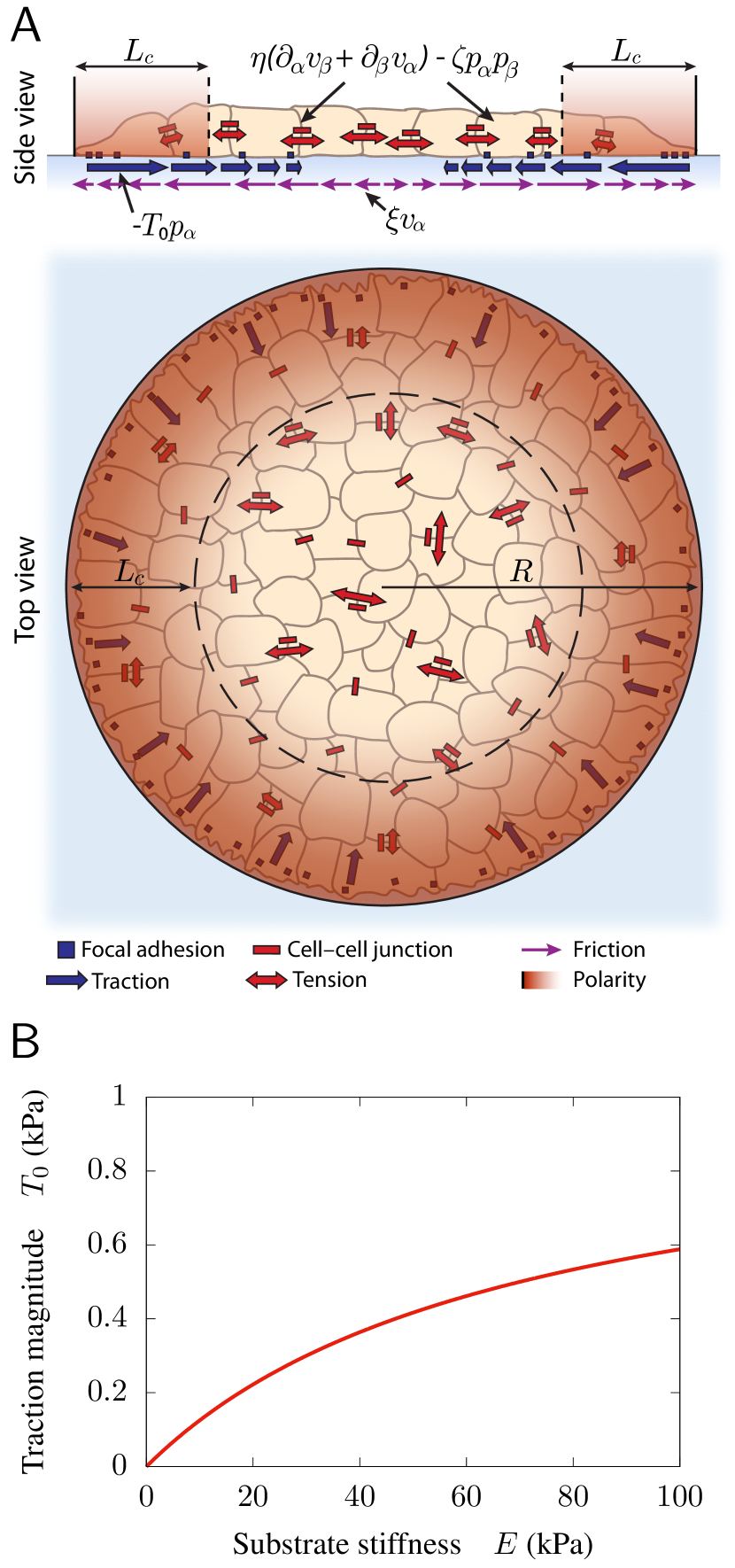
We base our analysis on a continuum active polar fluid model for the spreading of an epithelial monolayer, which is thus described in terms of a polarity field and a velocity field in two dimensions Blanch-Mercader et al. (2017); Pérez-González et al. (2019). We neglect cell proliferation as well as the bulk elasticity of the cell monolayer, which eventually limit the spreading process Serra-Picamal et al. (2012); Basan et al. (2013); Recho et al. (2016); Yabunaka and Marcq (2017). Tissue spreading is driven by the traction forces exerted by cells close to the monolayer edge, which polarize perpendicularly to the edge to migrate towards free space. In contrast, the inner region of the monolayer remains essentially unpolarized, featuring much weaker and transient traction forces Pérez-González et al. (2019) (Fig. 1A). Hence, we take a free energy for the polarity field that favors the unpolarized state in the bulk, with a restoring coefficient , and we impose a normal and maximal polarity as a boundary condition at the monolayer edge. In addition, the polar free energy includes a cost for polarity gradients, with the Frank constant of nematic elasticity in the one-constant approximation de Gennes and Prost (1993). Altogether,
| (1) |
We assume that the polarity field is set by flow-independent mechanisms, so that it follows a purely relaxational dynamics, and that it equilibrates fast compared to the spreading dynamics Pérez-González et al. (2019). Hence, , which yields
| (2) |
where is the characteristic length with which the polarity modulus decays from at the monolayer edge to at the center (red shade in Fig. 1A).
Then, force balance imposes
| (3) |
where is the stress tensor of the monolayer, and is the external traction force density acting on it. We relate these forces to the polarity and velocity fields via the following constitutive equations for a compressible active polar fluid Pérez-González et al. (2019); Oriola et al. (2017):
| (4a) | ||||
| (4b) | ||||
Here, is the monolayer viscosity, and is the cell-substrate friction coefficient. Respectively, is the active stress coefficient accounting for the contractility of polarized cells, and is the contact active force coefficient accounting for the maximal traction stress exerted by polarized cells on the substrate, , with the monolayer height (Fig. 1A).
To model cellular response to substrate properties, we assume that the parameters of cell-substrate interactions depend on substrate stiffness. Tissue cells tend to exert larger traction forces on stiffer substrates Discher et al. (2005); Ladoux and Nicolas (2012); Gupta et al. (2016); Saez et al. (2005); Ghibaudo et al. (2008); Ladoux et al. (2010); Trichet et al. (2012); Elosegui-Artola et al. (2014); Gupta et al. (2015); Li et al. (2015); Saez et al. (2010). Many studies have explained this response by simply assuming that intracellular force is exerted on an in-series connection of two linear elastic media, namely the substrate and the attached cellular structures, with Young’s modulus and , respectively Walcott and Sun (2010); Zemel et al. (2010); Marcq et al. (2011); Trichet et al. (2012); Sens (2013); Gupta et al. (2015). Following them, we take cell-substrate forces that depend on substrate stiffness as (Fig. 1B)
| (5) |
where and are the maximal active traction stress and friction coefficient on an infinitely stiff substrate, respectively.
This minimal approach neglects some aspects of the cellular response to the mechanical properties of the substrate. First, we focus on purely elastic substrates; we do not consider substrate viscoelasticity, which also affects cellular and tissue forces Murrell et al. (2011); Chaudhuri et al. (2015); Imai et al. (2015); Zheng et al. (2017); Bennett et al. (2018); Charrier et al. (2018). Second, we do not explicitly account for the substrate deformation field, which could mediate long-range elastic interactions between cells Angelini et al. (2010); Reinhart-King et al. (2008). However, a dependence very similar to Eq. 5 and Fig. 1B was obtained when accounting for the non-local substrate elasticity Banerjee and Marchetti (2012). Finally, cell-cell and cell-substrate adhesions are coupled through the actin cytoskeleton, and hence they may exhibit mechanical crosstalk. However, the influence of substrate stiffness on cell-cell interactions Guo et al. (2006); Ng et al. (2012); Lembong et al. (2017); Ladoux and Mège (2017); Pérez-González et al. (2019) remains poorly understood. Thus, for the sake of simplicity, we assume a stiffness-independent intercellular contractility .
| Symbol | Description | Estimate |
|---|---|---|
| monolayer height | m Pérez-González et al. (2019) | |
| nematic length | m Blanch-Mercader et al. (2017); Pérez-González et al. (2019) | |
| maximal traction | kPa Blanch-Mercader et al. (2017); Pérez-González et al. (2019) | |
| intercellular contractility | kPa Pérez-González et al. (2019) | |
| maximal friction coefficient | kPas/m2 Cochet-Escartin et al. (2014) | |
| monolayer viscosity | MPas Blanch-Mercader et al. (2017); Pérez-González et al. (2019) | |
| minimal hydrodynamic screening length | m () | |
| characteristic cellular stiffness | kPa Douezan et al. (2012) | |
| stiffness gradient | kPa/mm Sunyer et al. (2016) | |
| stiffness offset (soft edge) | kPa Sunyer et al. (2016) |
III Results and discussion
III.1 Wetting transition
In this section, we study the effect of substrate stiffness on the tissue wetting transition. We consider a circular cell monolayer spreading radially (Fig. 1A), such as those extending from spheroidal cell aggregates Beaune et al. (2014). In addition to a maximal normal polarity at the edge, , we impose a stress-free boundary condition, , with the monolayer radius. Then, neglecting cell-substrate viscous friction (), the model Eqs. 2 to 5 can be solved analytically Pérez-González et al. (2019). Thus, we obtain the spreading velocity , and hence the spreading parameter Beaune et al. (2014) . For , which is the case in most experiments, it reads
| (6) |
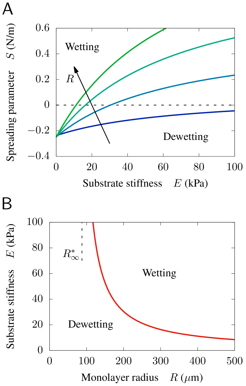
The spreading parameter increases with substrate stiffness (Fig. 2A). Therefore, the competition between intercellular contractility and stiffness-dependent traction forces entails the existence of a critical substrate stiffness
| (7) |
above which the tissue spreads (, wetting) and below which it retracts (, dewetting). The spreading parameter also increases with tissue size (Fig. 2A), and hence the critical stiffness decreases with monolayer radius (Fig. 2B). In Eq. 7, is the critical radius for tissue wetting unveiled in our previous work Pérez-González et al. (2019), which emerges from the competition between bulk and contact active forces. Here, the critical radius decreases with substrate stiffness,
| (8) |
asymptotically tending to for large stiffness (Fig. 2B).
In the following, we compare our prediction (Eq. 7) to published experimental results. For example, for cell monolayers of m, and the values of traction and contractility in Table 1, Pérez-González et al. observed wetting only on substrates of kPa Pérez-González et al. (2019), suggesting that kPa. Thus, comparing to Eq. 7, we estimate that kPa for the cell monolayers of Ref. Pérez-González et al. (2019). In another study, Douezan et al. reported kPa and kPa for cell aggregates of radius m Douezan et al. (2012). Introducing these values into Eq. 7, we infer m. Taking m, this value suggests that the intercellular contractility and the maximal traction stress are of the same order for the cell aggregates of Ref. Douezan et al. (2012). In conclusion, our result explains the experimental observation of a wetting transition induced by substrate stiffness Douezan et al. (2012); Douezan and Brochard-Wyart (2012); Pérez-González et al. (2019), and it predicts that the critical stiffness depends on tissue size. Testing this prediction requires new experiments that, in addition to varying substrate stiffness, would systematically vary tissue size.
III.2 Tissue durotaxis
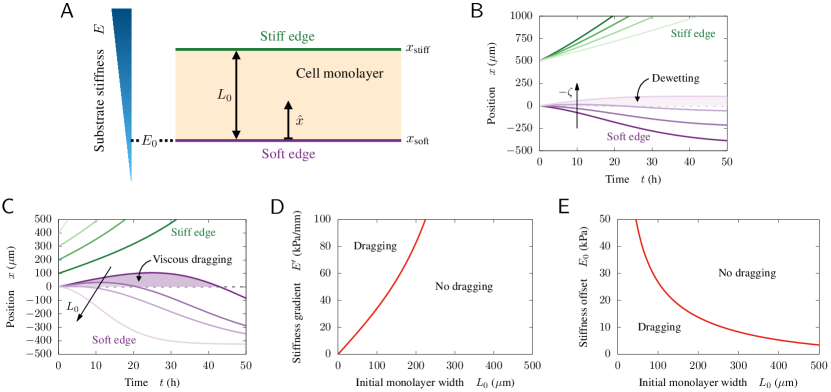
Motivated by recent experiments Sunyer et al. (2016), we consider a rectangular cell monolayer spreading on a substrate with a linear stiffness gradient
| (9) |
We use the terms “stiff edge” and “soft edge” to denote the tissue interfaces at the stiffer and softer sides of the substrate, respectively (Fig. 3A). As previously, we impose a normal maximal polarity at the edges, , and stress-free boundary conditions, . To simulate tissue spreading, we analytically obtain the polarity profile from Eq. 2 and we numerically solve the force balance equation, Eqs. 3 and 4 with Eqs. 5 and 9, to obtain the velocity profile at each time step. We use a finite differences scheme on a grid of points and a time step s. Then, we evolve the interface positions according to and , from initial conditions and , respectively.
III.2.1 Different wetting states at each monolayer edge
For small intercellular contractility , active traction forces drive tissue spreading, which is faster on the stiffer side. The soft edge moves towards decreasing stiffness; therefore, its active traction progressively decreases. As a consequence, the soft edge slows down, tending to stop at the substrate position for which contractile tension balances traction forces (Fig. 3B, darker lines).
A larger contractility, however, may overcome the active traction force at the soft edge but not at the stiff edge. In this case, the stiff edge advances but the soft edge retracts (Fig. 3B, lightest lines). Thus, by simultaneously wetting on the stiff side and dewetting from the soft side, the tissue migrates directionally up the stiffness gradient. Finally, a sufficiently large contractility would induce the retraction of both edges, thus causing monolayer dewetting. Overall, these results show that tissue durotaxis can emerge from the different wetting dynamics of the soft and stiff monolayer edges.
III.2.2 Interface dragging by hydrodynamic force transmission
The previous durotactic mechanism is at work regardless of tissue size. For monolayers wider than the hydrodynamic screening length , viscous stresses do not fully transmit throughout the tissue because cell-substrate friction screens hydrodynamic interactions at distances larger than Blanch-Mercader et al. (2017). Therefore, in the limit , the soft and stiff edges move entirely independently. In contrast, for narrower monolayers, , both interfaces are strongly coupled.
In the following, we focus on hydrodynamic interactions between the tissue interfaces. To avoid the wetting effects explained above, we set . In narrow monolayers, the active tractions on each edge generate flows that span the entire tissue, thus affecting the motion of the other edge. The viscous force transmitted this way can overcome the active traction at the soft edge, outcompeting its spreading tendency and thereby dragging it towards increasing stiffness. This dragging effect is transient; the interfaces eventually decouple when the monolayer becomes too wide to sustain edge-to-edge force transmission (Fig. 3C, darker lines). Accordingly, monolayers that are initially too wide do not experience dragging at all (Fig. 3C, lighter lines). In conclusion, tissue durotaxis can result from interface dragging due to the transmission of viscous forces between the two monolayer edges.
To derive the conditions for interface dragging, we consider the limit in which the width of the polarized boundary layer of cells is much smaller than the total tissue width, , which is generally the case in experiments Blanch-Mercader et al. (2017); Pérez-González et al. (2019); Sunyer et al. (2016). In this limit, the active forces are accumulated at the edges, and therefore enter as boundary conditions:
| (10) |
Here, and , with given in Eq. 9 and given in Eq. 5. Then, force balance reads
| (11) |
which can be solved analytically. From the solution, we determine that the initial velocity of the soft edge is positive (dragging) whenever the following condition is fulfilled:
| (12) |
Here, and . Thus, in combination with Eqs. 5 and 9, Eq. 12 specifies the condition for interface dragging in terms of the stiffness gradient , the stiffness offset , and the initial monolayer width . Interface dragging is predicted for monolayers narrow enough to sustain edge-to-edge force transmission. In addition, the monolayer must be on a sufficiently steep stiffness gradient, and on a sufficiently soft region of the substrate to have the required difference in active traction between both edges (Fig. 3D-E).
III.2.3 Discussion
In recent experiments, Sunyer et al. observed asymmetric tissue spreading on a gradient of substrate stiffness Sunyer et al. (2016). Our model reproduces this observation, as we illustrate in Fig. 3B (darker lines). Moreover, on a very soft region of a substrate with a very large stiffness gradient, Sunyer et al. also observed one example of directed tissue migration up the gradient Sunyer et al. (2016), consistent with our predictions. Here, we have unveiled two mechanisms for such a directed migration, which can be distinguished by their collective character. First, tissue durotaxis based on simultaneous wetting and dewetting (Fig. 3B, lightest lines) needs not be collective in nature; force transmission between edges can take place but is not required. In contrast, durotaxis based on interface dragging (Fig. 3C, darker lines) is a collective effect since it relies on edge-to-edge force transmission. In the experiments by Sunyer et al., durotaxis emerges from intercellular force transmission Sunyer et al. (2016), so that both mechanisms might be at play. Therefore, further experiments are required to disentangle the contributions of these two durotaxis mechanisms.
Sunyer et al. measured traction forces of equal magnitude on the stiff and soft edges. Hence, their model assumed that cells exert equal active forces on both edges. They then explained durotaxis based on the different substrate deformations caused by these equal forces Sunyer et al. (2016). However, for almost static cell monolayers, other studies measured larger tractions on stiffer substrates Saez et al. (2010). This suggests that the active, static contribution of the traction forces increases with substrate stiffness. Hence, here we assumed that cells exert larger active forces on the stiff edge. Thus, in our model, durotaxis is driven by differences in active traction between the tissue edges. Equal total tractions might then result from an additional regulation of cellular forces by stiffness gradients, as well as from the addition of active tractions and viscous friction forces on the substrate, both of which are maximal at the tissue edges and increase with substrate stiffness. Future work could address this point quantitatively by fitting the predictions of our model to experimental data.
Finally, previous mechanical models of tissue durotaxis treated the cell monolayer as an elastic medium, thus imposing full transmission of force through the tissue Banerjee and Marchetti (2011); Sunyer et al. (2016); González-Valverde and García-Aznar (2018); Escribano et al. (2018). Instead, given that tissue spreading occurs over time scales of several hours Blanch-Mercader et al. (2017), at which the tissue should have a fluid behavior Gonzalez-Rodriguez et al. (2012); Wyatt et al. (2016); Khalilgharibi et al. (2016), we model the cell monolayer as a viscous medium. Thus, our model can suitably account for wetting effects and hydrodynamic interactions in tissue spreading. In particular, we show that these two intrinsic features of fluid media can naturally give rise to durotaxis, which needs not rely on long-range elastic interactions across the tissue. Moreover, we show that the screening of hydrodynamic interactions at long distances gives rise to size-dependent effects such as viscous dragging. Looking for these effects in future experiments may help discriminate between the elastic and viscous behavior of spreading tissues.
IV Conclusions
We have studied how substrate stiffness affects the spreading of epithelial tissues. We have extended an active polar fluid model for tissue spreading to incorporate the dependence of cellular traction forces on substrate stiffness. This way, we have shown how substrate stiffness induces a wetting transition between a droplet-like cell aggregate and a spreading monolayer, explaining experimental observations Douezan et al. (2012); Pérez-González et al. (2019). We have predicted that the critical stiffness for tissue wetting decreases with tissue size. Further experiments are required to test this prediction. Moreover, we have also explained how gradients of substrate stiffness may give rise to collective cell migration towards increasing stiffness, a behavior known as tissue durotaxis. We have detailed two mechanisms for tissue durotaxis, one based on different wetting states and the other on hydrodynamic interactions between the tissue interfaces. Both mechanisms can coexist, but they can be discriminated because the latter is lost for sufficiently wide cell monolayers. Thus, further systematic experiments are required to assess the relative contributions of the two durotactic mechanisms that we have unveiled. Overall, our results show how the adaptation of cellular traction forces to substrate stiffness impacts the collective migration and the active wetting properties of epithelial tissues.
Acknowledgements.
We thank Carlos Pérez-González for discussions and for designing Fig. 1A. We thank Carles Blanch-Mercader, Raimon Sunyer, Xavier Trepat, and the members of Trepat’s lab for discussions. R.A. acknowledges support from Fundació “La Caixa” and from the Human Frontiers of Science Program (LT000475/2018-C). R.A. thanks Jacques Prost and acknowledges EMBO (Short Term Fellowship ASTF 365-2015), The Company of Biologists (Development Travelling Fellowship DEVTF-151206), and Fundació Universitària Agustí Pedro i Pons for supporting visits to Institut Curie. R.A. and J.C. acknowledge the MINECO under project FIS2016-78507-C2-2-P and Generalitat de Catalunya under project 2014-SGR-878.References
- Friedl and Gilmour (2009) Peter Friedl and Darren Gilmour, “Collective cell migration in morphogenesis, regeneration and cancer,” Nat. Rev. Mol. Cell Biol. 10, 445–457 (2009).
- Hakim and Silberzan (2017) Vincent Hakim and Pascal Silberzan, “Collective cell migration: a physics perspective,” Reports Prog. Phys. 80, 076601 (2017).
- Gonzalez-Rodriguez et al. (2012) David Gonzalez-Rodriguez, Karine Guevorkian, Stéphane Douezan, and Françoise Brochard-Wyart, “Soft Matter Models of Developing Tissues and Tumors,” Science 338, 910–917 (2012).
- Beaune et al. (2014) Grégory Beaune, Tomita Vasilica Stirbat, Nada Khalifat, Olivier Cochet-Escartin, Simon Garcia, Vasily Valérïévitch Gurchenkov, Michael P Murrell, Sylvie Dufour, Damien Cuvelier, and Françoise Brochard-Wyart, “How cells flow in the spreading of cellular aggregates,” Proc. Natl. Acad. Sci. U. S. A. 111, 8055–8060 (2014).
- Morita et al. (2017) Hitoshi Morita, Silvia Grigolon, Martin Bock, S.F. Gabriel Krens, Guillaume Salbreux, and Carl-Philipp Heisenberg, “The Physical Basis of Coordinated Tissue Spreading in Zebrafish Gastrulation,” Dev. Cell 40, 354–366.e4 (2017).
- Wallmeyer et al. (2018) Bernhard Wallmeyer, Sarah Trinschek, Sargon Yigit, Uwe Thiele, and Timo Betz, “Collective Cell Migration in Embryogenesis Follows the Laws of Wetting,” Biophys. J. 114, 213–222 (2018).
- Ryan et al. (2001) P L Ryan, R A Foty, J Kohn, and M S Steinberg, “Tissue spreading on implantable substrates is a competitive outcome of cell-cell vs. cell-substratum adhesivity,” Proc. Natl. Acad. Sci. U. S. A. 98, 4323–4327 (2001).
- Douezan et al. (2011) Stéphane Douezan, Karine Guevorkian, Randa Naouar, Sylvie Dufour, Damien Cuvelier, and Françoise Brochard-Wyart, “Spreading dynamics and wetting transition of cellular aggregates,” Proc. Natl. Acad. Sci. U. S. A. 108, 7315–7320 (2011).
- Ravasio et al. (2015) Andrea Ravasio, Anh Phuong Le, Thuan Beng Saw, Victoria Tarle, Hui Ting Ong, Cristina Bertocchi, René-Marc Mège, Chwee Teck Lim, Nir S. Gov, and Benoit Ladoux, “Regulation of epithelial cell organization by tuning cell-substrate adhesion,” Integr. Biol. 7, 1228–1241 (2015).
- Smeets et al. (2016) Bart Smeets, Ricard Alert, Jiří Pešek, Ignacio Pagonabarraga, Herman Ramon, and Romaric Vincent, “Emergent structures and dynamics of cell colonies by contact inhibition of locomotion,” Proc. Natl. Acad. Sci. U. S. A. 113, 14621–14626 (2016).
- Pérez-González et al. (2019) Carlos Pérez-González, Ricard Alert, Carles Blanch-Mercader, Manuel Gómez-González, Tomasz Kolodziej, Elsa Bazellieres, Jaume Casademunt, and Xavier Trepat, “Active wetting of epithelial tissues,” Nat. Phys. 15, 79–88 (2019).
- Discher et al. (2005) Dennis E Discher, Paul Janmey, and Yu-Li Wang, “Tissue Cells Feel and Respond to the Stiffness of Their Substrate,” Science 310, 1139–1143 (2005).
- Ladoux and Nicolas (2012) Benoit Ladoux and Alice Nicolas, “Physically based principles of cell adhesion mechanosensitivity in tissues,” Reports Prog. Phys. 75, 116601 (2012).
- Gupta et al. (2016) Mukund Gupta, Bryant Doss, Chwee Teck Lim, Raphael Voituriez, and Benoit Ladoux, “Single cell rigidity sensing: a complex relationship between focal adhesion dynamics and large-scale actin cytoskeleton remodeling,” Cell Adh. Migr. 10, 554–567 (2016).
- Douezan and Brochard-Wyart (2012) S Douezan and F Brochard-Wyart, “Dewetting of cellular monolayers,” Eur. Phys. J. E 35, 34 (2012).
- Douezan et al. (2012) Stéphane Douezan, Julien Dumond, and Françoise Brochard-Wyart, “Wetting transitions of cellular aggregates induced by substrate rigidity,” Soft Matter 8, 4578–4583 (2012).
- Guo et al. (2006) Wei-hui Guo, Margo T Frey, Nancy A Burnham, and Yu-li Wang, “Substrate Rigidity Regulates the Formation and Maintenance of Tissues,” Biophys. J. 90, 2213–2220 (2006).
- Barriga et al. (2018) Elias H. Barriga, Kristian Franze, Guillaume Charras, and Roberto Mayor, “Tissue stiffening coordinates morphogenesis by triggering collective cell migration in vivo,” Nature 554, 523–527 (2018).
- Angelini et al. (2010) Thomas E. Angelini, Edouard Hannezo, Xavier Trepat, Jeffrey J. Fredberg, and David A. Weitz, “Cell Migration Driven by Cooperative Substrate Deformation Patterns,” Phys. Rev. Lett. 104, 168104 (2010).
- Ng et al. (2012) Mei Rosa Ng, Achim Besser, Gaudenz Danuser, and Joan S. Brugge, “Substrate stiffness regulates cadherin-dependent collective migration through myosin-II contractility,” J. Cell Biol. 199, 545–563 (2012).
- Saez et al. (2007) A. Saez, M. Ghibaudo, A. Buguin, P. Silberzan, and B. Ladoux, “Rigidity-driven growth and migration of epithelial cells on microstructured anisotropic substrates,” Proc. Natl. Acad. Sci. U. S. A. 104, 8281–8286 (2007).
- Edwards and Schwarz (2011) Carina M. Edwards and Ulrich S. Schwarz, “Force Localization in Contracting Cell Layers,” Phys. Rev. Lett. 107, 128101 (2011).
- Lange and Fabry (2013) Janina R. Lange and Ben Fabry, “Cell and tissue mechanics in cell migration,” Exp. Cell Res. 319, 2418–2423 (2013).
- Ladoux and Mège (2017) Benoit Ladoux and René-Marc Mège, “Mechanobiology of collective cell behaviours,” Nat. Rev. Mol. Cell Biol. 18, 743–757 (2017).
- Barriga and Mayor (2018) Elias H. Barriga and Roberto Mayor, “Adjustable viscoelasticity allows for efficient collective cell migration,” Semin. Cell Dev. Biol. (2018), 10.1016/j.semcdb.2018.05.027.
- Sunyer et al. (2016) Raimon Sunyer, Vito Conte, Jorge Escribano, Alberto Elosegui-Artola, Anna Labernadie, Léo Valon, Daniel Navajas, José Manuel García-Aznar, José J. Muñoz, Pere Roca-Cusachs, and Xavier Trepat, “Collective cell durotaxis emerges from long-range intercellular force transmission,” Science 353, 1157–1161 (2016).
- Blanch-Mercader et al. (2017) C. Blanch-Mercader, R. Vincent, E. Bazellières, X. Serra-Picamal, X. Trepat, and J. Casademunt, “Effective viscosity and dynamics of spreading epithelia: a solvable model,” Soft Matter 13, 1235–1243 (2017).
- Serra-Picamal et al. (2012) Xavier Serra-Picamal, Vito Conte, Romaric Vincent, Ester Añón, Dhananjay T. Tambe, Elsa Bazellieres, James P. Butler, Jeffrey J. Fredberg, and Xavier Trepat, “Mechanical waves during tissue expansion,” Nat. Phys. 8, 628–634 (2012).
- Basan et al. (2013) Markus Basan, Jens Elgeti, Edouard Hannezo, Wouter-Jan Rappel, and Herbert Levine, “Alignment of cellular motility forces with tissue flow as a mechanism for efficient wound healing,” Proc. Natl. Acad. Sci. U. S. A. 110, 2452–2459 (2013).
- Recho et al. (2016) Pierre Recho, Jonas Ranft, and Philippe Marcq, “One-dimensional collective migration of a proliferating cell monolayer,” Soft Matter 12, 2381–2391 (2016).
- Yabunaka and Marcq (2017) Shunsuke Yabunaka and Philippe Marcq, “Cell growth, division, and death in cohesive tissues: A thermodynamic approach,” Phys. Rev. E 96, 022406 (2017).
- de Gennes and Prost (1993) Pierre-Gilles de Gennes and Jacques Prost, The Physics of Liquid Crystals, 2nd ed. (Oxford University Press, 1993).
- Oriola et al. (2017) David Oriola, Ricard Alert, and Jaume Casademunt, “Fluidization and Active Thinning by Molecular Kinetics in Active Gels,” Phys. Rev. Lett. 118, 088002 (2017).
- Saez et al. (2005) Alexandre Saez, Axel Buguin, Pascal Silberzan, and Benoît Ladoux, “Is the Mechanical Activity of Epithelial Cells Controlled by Deformations or Forces?” Biophys. J. 89, L52–L54 (2005).
- Ghibaudo et al. (2008) Marion Ghibaudo, Alexandre Saez, Léa Trichet, Alain Xayaphoummine, Julien Browaeys, Pascal Silberzan, Axel Buguin, and Benoît Ladoux, “Traction forces and rigidity sensing regulate cell functions,” Soft Matter 4, 1836–1843 (2008).
- Ladoux et al. (2010) Benoit Ladoux, Ester Anon, Mireille Lambert, Aleksandr Rabodzey, Pascal Hersen, Axel Buguin, Pascal Silberzan, and René-Marc Mège, “Strength Dependence of Cadherin-Mediated Adhesions,” Biophys. J. 98, 534–542 (2010).
- Trichet et al. (2012) Léa Trichet, Jimmy Le Digabel, Rhoda J Hawkins, Sri Ram Krishna Vedula, Mukund Gupta, Claire Ribrault, Pascal Hersen, Raphaël Voituriez, and Benoît Ladoux, “Evidence of a large-scale mechanosensing mechanism for cellular adaptation to substrate stiffness,” Proc. Natl. Acad. Sci. U. S. A. 109, 6933–6938 (2012).
- Elosegui-Artola et al. (2014) Alberto Elosegui-Artola, Elsa Bazellières, Michael D Allen, Ion Andreu, Roger Oria, Raimon Sunyer, Jennifer J Gomm, John F Marshall, J Louise Jones, Xavier Trepat, and Pere Roca-Cusachs, “Rigidity sensing and adaptation through regulation of integrin types,” Nat. Mater. 13, 631–637 (2014).
- Gupta et al. (2015) Mukund Gupta, Bibhu Ranjan Sarangi, Joran Deschamps, Yasaman Nematbakhsh, Andrew Callan-Jones, Felix Margadant, René-Marc Mège, Chwee Teck Lim, Raphaël Voituriez, and Benoît Ladoux, “Adaptive rheology and ordering of cell cytoskeleton govern matrix rigidity sensing,” Nat. Commun. 6, 7525 (2015).
- Li et al. (2015) Jianjun Li, Dong Han, and Ya-Pu Zhao, “Kinetic behaviour of the cells touching substrate: the interfacial stiffness guides cell spreading,” Sci. Rep. 4, 3910 (2015).
- Saez et al. (2010) A Saez, E Anon, M Ghibaudo, O du Roure, J-M Di Meglio, P Hersen, P Silberzan, A Buguin, and B Ladoux, “Traction forces exerted by epithelial cell sheets,” J. Phys. Condens. Matter 22, 194119 (2010).
- Walcott and Sun (2010) Sam Walcott and Sean X Sun, “A mechanical model of actin stress fiber formation and substrate elasticity sensing in adherent cells,” Proc. Natl. Acad. Sci. U. S. A. 107, 7757–7762 (2010).
- Zemel et al. (2010) A Zemel, F Rehfeldt, A E X Brown, D E Discher, and S A Safran, “Optimal matrix rigidity for stress fiber polarization in stem cells,” Nat. Phys. 6, 468–473 (2010).
- Marcq et al. (2011) Philippe Marcq, Natsuhiko Yoshinaga, and Jacques Prost, “Rigidity Sensing Explained by Active Matter Theory,” Biophys. J. 101, L33–L35 (2011).
- Sens (2013) Pierre Sens, “Rigidity sensing by stochastic sliding friction,” Europhys. Lett. 104, 38003 (2013).
- Murrell et al. (2011) Michael Murrell, Roger Kamm, and Paul Matsudaira, “Substrate Viscosity Enhances Correlation in Epithelial Sheet Movement,” Biophys. J. 101, 297–306 (2011).
- Chaudhuri et al. (2015) Ovijit Chaudhuri, Luo Gu, Max Darnell, Darinka Klumpers, Sidi A Bencherif, James C Weaver, Nathaniel Huebsch, and David J Mooney, “Substrate stress relaxation regulates cell spreading,” Nat. Commun. 6, 6365 (2015).
- Imai et al. (2015) Misako Imai, Kazuya Furusawa, Takeomi Mizutani, Kazushige Kawabata, and Hisashi Haga, “Three-dimensional morphogenesis of MDCK cells induced by cellular contractile forces on a viscous substrate,” Sci. Rep. 5, 14208 (2015).
- Zheng et al. (2017) Ji Yun Zheng, Siew Ping Han, Yi-Jen Chiu, Ai Kia Yip, Nicolas Boichat, Shi Wen Zhu, Jun Zhong, and Paul Matsudaira, “Epithelial Monolayers Coalesce on a Viscoelastic Substrate through Redistribution of Vinculin,” Biophys. J. 113, 1585–1598 (2017).
- Bennett et al. (2018) Mark Bennett, Marco Cantini, Julien Reboud, Jonathan M Cooper, Pere Roca-Cusachs, and Manuel Salmeron-Sanchez, “Molecular clutch drives cell response to surface viscosity,” Proc. Natl. Acad. Sci. U. S. A. 115, 1192–1197 (2018).
- Charrier et al. (2018) Elisabeth E. Charrier, Katarzyna Pogoda, Rebecca G. Wells, and Paul A. Janmey, “Control of cell morphology and differentiation by substrates with independently tunable elasticity and viscous dissipation,” Nat. Commun. 9, 449 (2018).
- Reinhart-King et al. (2008) Cynthia A Reinhart-King, Micah Dembo, and Daniel A Hammer, “Cell-cell mechanical communication through compliant substrates,” Biophys. J. 95, 6044–6051 (2008).
- Banerjee and Marchetti (2012) Shiladitya Banerjee and M. Cristina Marchetti, “Contractile Stresses in Cohesive Cell Layers on Finite-Thickness Substrates,” Phys. Rev. Lett. 109, 108101 (2012).
- Lembong et al. (2017) Josephine Lembong, Benedikt Sabass, and Howard A Stone, “Calcium oscillations in wounded fibroblast monolayers are spatially regulated through substrate mechanics,” Phys. Biol. 14, 045006 (2017).
- Cochet-Escartin et al. (2014) Olivier Cochet-Escartin, Jonas Ranft, Pascal Silberzan, and Philippe Marcq, “Border Forces and Friction Control Epithelial Closure Dynamics,” Biophys. J. 106, 65–73 (2014).
- Banerjee and Marchetti (2011) S. Banerjee and M. C. Marchetti, “Substrate rigidity deforms and polarizes active gels,” Europhys. Lett. 96, 28003 (2011).
- González-Valverde and García-Aznar (2018) Ismael González-Valverde and José Manuel García-Aznar, “Mechanical modeling of collective cell migration: An agent-based and continuum material approach,” Comput. Methods Appl. Mech. Eng. 337, 246–262 (2018).
- Escribano et al. (2018) Jorge Escribano, Raimon Sunyer, María Teresa Sánchez, Xavier Trepat, Pere Roca-Cusachs, and José Manuel García-Aznar, “A hybrid computational model for collective cell durotaxis,” Biomech. Model. Mechanobiol. 17, 1037–1052 (2018).
- Wyatt et al. (2016) Tom Wyatt, Buzz Baum, and Guillaume Charras, “A question of time: tissue adaptation to mechanical forces,” Curr. Opin. Cell Biol. 38, 68–73 (2016).
- Khalilgharibi et al. (2016) Nargess Khalilgharibi, Jonathan Fouchard, Pierre Recho, Guillaume Charras, and Alexandre Kabla, “The dynamic mechanical properties of cellularised aggregates,” Curr. Opin. Cell Biol. 42, 113–120 (2016).