Thermo-mechanical pain:
the signaling role of heat dissipation in biological tissues
Abstract
Abstract: Mechanical algesia is an important process for the preservation of living organisms, allowing potentially life-saving reflexes or decisions when given body parts are stressed. Yet, its various underlying mechanisms remain to be fully unravelled. Here, we quantitatively discuss how the detection of painful mechanical stimuli by the human central nervous system may, partly, rely on thermal measurements. Indeed, most fractures in a body, including microscopic ones, release some heat, which diffuses in the surrounding tissues. Through this physical process, the thermo-sensitive TRP proteins, that translate abnormal temperatures into action potentials, shall be sensitive to damaging mechanical inputs. The implication of these polymodal receptors in mechanical algesia has been regularly reported, and we here provide a physical explanation for the coupling between thermal and mechanical pain. In particular, in the human skin, we show how the neighbouring neurites of a broken collagen fiber can undergo a sudden thermal elevation that ranges from a fraction to tens of degrees. As this theoretical temperature anomaly lies in the sensibility range of the TRPV3 and TRPV1 cation channels, known to trigger action potentials in the neural system, a degree of mechanical pain can hence be generated.
Introduction:
on rupture and energy dissipation
The growth of mechanical damages through a body is an irreversible thermodynamic process Rice (1978). Indeed, when a fracture progresses by a given surface unit, it dissipates a specific amount of energy, that is referred to, by rupture physicists, as the energy release rate, expressed in J m-2. In most engineering materials (e.g., Hahn et al. (1972)), this quantity, denoted , is well studied, since it characterizes the loading necessary for a crack to propagate Griffith (1921). For instance, it is in the order of J m-2 in weak glasses Wiederhorn (1967) and can reach kJ m-2 in the strongest media, as titanium Sastry et al. (1981) or steel Huang and Altstetter (1991).
When it comes to biological tissues, this energy release rate can also be estimated, and was notably measured to be about J m-2 in the human hand skin Pereira et al. (1997). An important question, then, is how is this dissipated mechanical energy received and felt by the human body?
Most generally, there are many possible ways for it to be transformed, ranging from its storage as surface potential energy on the walls of the new fractures Rice and Drucker (1967) to its emission to the far field as mechanical Morrissey and Rice (1998) or electromagnetic Pallares et al. (2012) waves, that is, sound and luminescence. It was, in particular, shown that a significant part of the mechanical input is converted into heat close to the damage Rice and Levy (1969); Fuller et al. (1975); Toussaint et al. (2016); Vincent-Dospital et al. (2020a), as the rupture of stretched atomic and molecular bonds is prone to generate a local and incoherent -thermal- atomic motion.
The related elevations in temperature have been measured in various synthetic solids (e.g., Fuller et al. (1975); Toussaint et al. (2016); Palumbo et al. (2017)), and are believed to be more than only a side effect of the fracturing process. Indeed, from its positive feedback on the dynamics of rupture, it was pointed out as a likely cause for the brittleness of matter Carbone and Persson (2005); Vincent-Dospital et al. (2020b) and for the instability of some seismic faults Rice (2006); Wibberley and Shimamoto (2005); Sulem and Famin (2009).
We here propose that, in the human body, this damage-induced heat is to be sensed by the neuronal network, and may hence explain a degree of coupling between the thermal and mechanical pain, which has been regularly suspected (e.g., Wang and Woolf (2005); Caterina and Park (2006); Goodman (2003)).
I Thermo-mechanical nociception
The perception of pain (i.e., nociception or algesia) arises from the bio-electrical signals (referred to as action potentials) that sensory neurons send from the aggressed body part to the nervous system (e.g, Oaklander and Siegel (2005)). To initiate such messages, the dolorous inputs, being mechanical, thermal or chemical, need to be converted accordingly, at the surface of sensory neurites (i.e., the extensions of neurons cell bodies).
We will here focus, as an example, on the nociception in the human skin. The TRPs proteins (Transient Receptor Potential cation channels) are notably believed to be responsible for the reporting of the temperature of the skin, and that of other body parts, to the nervous system Wang and Woolf (2005); Tóth et al. (2014). In particular, TRPV3 send action potentials between and C, with an activation intensity that is gradual with temperature Singh et al. (2019); Xu et al. (2002), leading to a harmless perception of warmth. The feel of a more intense, potentially more noxious, heat occurs when TRPV1 is activated, at higher skin temperatures above C. Other TRP proteins manage the detection of a cool skin, such as TRPM8 which activates below C. The physico-chemical mechanism to translate heat into current is, in all cases, believed to be a temperature dependant shifts in the TRPs voltage-dependent activation curves Nilius et al. (2005), that is, in their ability to pass ions through a neuron membrane depending on the balance of charge on each side of this membrane.
The role of the TRP proteins is, however, not limited to thermal sensing, and some are known to be sensitive to chemical aggression, responding for instance to abnormal pH, to capsaïcine (i.e., the component of chilli pepper that is felt as hot), to menthol (that is felt as cold), or to arachnid acids Wang and Woolf (2005); Caterina and Park (2006). They are hence often referred to as polymodal nociceptors. Similarly, a growing suspicion seems to have risen that these sensors could also be involved in the feeling of mechanical pain Wang and Woolf (2005); Caterina and Park (2006); Goodman (2003). While the latter is complex, and shall rely on many types of nociceptors other than the TRPs Hill and Bautista (2020), the detection of thermal and mechanical inputs has indeed been shown to be somewhat coupled. In particular, the pain threshold of human subjects was reported to be a decreasing function of the ambient temperature Culp et al. (1989), and, in rats, the drug-induced inhibition of TRPV1 and TRPV3 has proven to reduce mechanical hyperalgesia Walker et al. (2003); Pomonis et al. (2003); McGaraughty et al. (2017) (i.e., the increased sensibility to mechanical pain after a first stimulus).
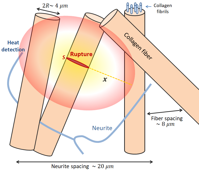
We suggest that this apparent coupling may be explained by the actual (physical) coupling between mechanical damage and heat dissipation. It is for instance well known that burns can be induced by friction on the skin (e.g., Al-Qattan et al. (2010)), due to the heat that is there generated. Similarly, a micro-crack of the epidermis or the dermis, that is caused by some mechanical input, is to release some heat in the surrounding tissues, and this heat may well be detected by the skin thermal nociceptors, as illustrated in Fig. 1.
II Temperature elevation around a broken collagen fiber
As a major structural cutaneous constituent, let us consider a collagen fiber, which has a typical radius Verhaegen et al. (2012) m. While it is itself composed of many fibrils and proteins Buehler (2006), the failure of this unit is likely a characteristic step Zohdi (2007) in any wider damage of the surrounding matter, as it is the case for engineered fibrous materials (e.g., Toussaint et al. (2016)). As per the energy release rate of skin Pereira et al. (1997), the rupture of this fiber shall dissipate an energy nJ, that is here assumed to be mainly converted into heat on the atomic scale, which then diffuses S. Carslaw and Jaeger (1959) to the surrounding skin molecules. For simplicity, we suppose that the collagen fiber break is brutal, that is, with a rupture velocity comparable to that of sound in skin Freund (1972); Goss and Dunn (1980), m s-1. We can then, for various times after the fracture that are superior enough to ns, compute the temperature rise around the damage, by integrating the Fourier heat diffusion kernel S. Carslaw and Jaeger (1959) over the broken surface :
| (1) |
In this equation, is the integration distance between a given point where the temperature is computed, and the various infinitesimal heat sources of , that have an elementary surface . In addition, the heat conductivity and volumetric heat capacity of skin are respectively denoted and , whose values are about J m-1 s-1 K-1 and MJ K-1 m-3 Cohen (1977).
If the rise in temperature described by Eq. (1) can be captured by the human neuronal system, it could then be treated as mechanical pain. In a healthy skin, the density of neurites in the torso may be estimated Oaklander et al. (1998) to be about mm-2, a quantity from which we derive an order of magnitude for the maximum distance between the surface of a broken collagen bundle and that of a neuronal receptor: m. Interestingly, such an approximate maximum distance is similar to the typical gap between the surfaces of two collagen fibers, which was measured Verhaegen et al. (2012) in average to be about m. Thus, if only two contiguous fibers were to break in response to a mechanical stimuli, one of it would, probably, be rather close (that is, in the micrometer range) to a neurite.
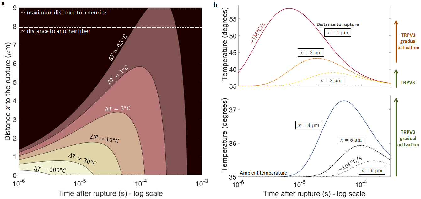
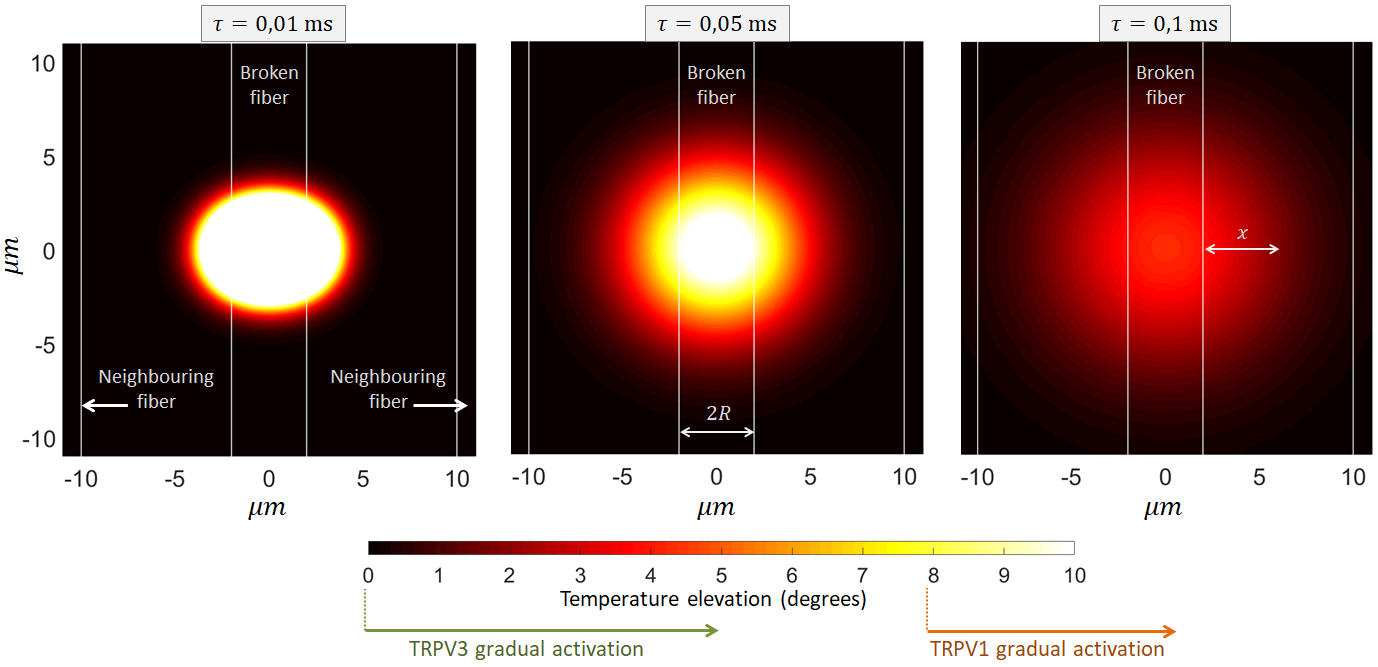
Following this simple model, we show, in Fig. 2, the evolution of the temperature predicted by Eq. (1), at various distances up to m perpendicularly to the broken fiber surface, being the normal internal skin temperature. We use C. Typical surface skin temperatures can indeed be measured (e.g., Otsuka et al. (2002)) to lie between C and C, with an internal one that should be slightly higher, transiting to about C (e.g., Saxena and Arya (1981)). Of course, a lower skin temperature (for instance at extremities) means that a stronger thermal anomaly would be needed for the TRPs threshold to be reached. Additionally to Fig. 2, we show, in Fig. 3, the related spatial temperature maps at three given times after the fracture.
Close to the rupture plane, that is, for m, modelled temperatures superior to that of the activation of TRPV1 (C) are quickly reached, in about s. A painful message can thus be triggered. More conservatively, if the thermal transducers are further away from the rupture point ( to m), they undergo a temperature elevation of half a degree to a few degrees, about ms after the damage. While this quantity is not enough to trigger TRPV1, and not vastly outside the range of the normal temperature oscillations of the human skin Shusterman et al. (1997), it could still be perceived as some abnormally sudden and localised heat by the brain. TRPV3 was indeed shown to be rather sensitive to small temperature changes around the normal body temperature Singh et al. (2019); Xu et al. (2002), and with a more intense response the faster these changes are Xu et al. (2002). Here, as shown in Fig. 2, we expect very high heating rates ranging from k∘C s-1 to M∘C s-1.
III Discussion
III.1 About the skin model simplicity
Before discussing further the preceding results, and their implication for the feeling of pain, let us acknowledge the simplicity of the model we have considered.
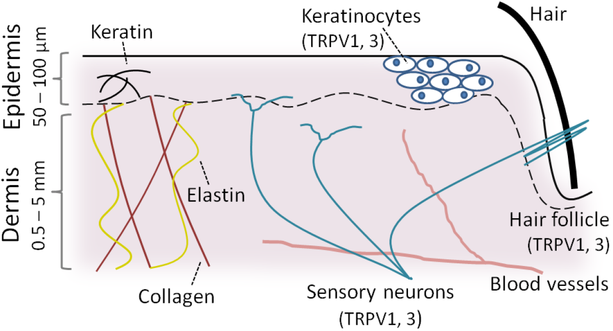
In our derivation, we have notably assumed thin, evenly distributed, neurites, that are all thermo-sensitive. This might of course be rather simplified, considering the various types of cutaneous neurons (e.g., Hill and Bautista (2020)) and their respective densities in different body parts Oaklander and Siegel (2005). In addition, the expression of the TRPs in the skin (and their role in thermo-sensing) is not limited to its sensory neurons, as they also appear in other cells, notably in the keratinocyte cells Wang and Woolf (2005); Moqrich et al. (2005); Yang et al. (2017) of the epidermis. Most of TRPV3 has actually been found in these keratinocyte cells, although it was detected in sensory neurons Xu et al. (2002).
Figure 4 shows a typical schematics of the human skin, and, by comparison, our conceptual model shown in Fig. 1 is of course only a proxy for the actual cutaneous tissue.
We have notably supposed the thermal properties of skin to be homogeneous. For instance, the TRP proteins reside within the cell membranes, and the heat capacity and conductivity of these lipid bilayers (Bastos et al., 2019) are to slightly differ from the surrounding, aqueous, environment. The extra heat could also be preferably conducted inside the collagen network rather than outside of it. Overall, however, the various components of skin shall have comparable thermal properties. Note also that other conduction laws, more refined than plain Fourier diffusion, may be considered in the modelling of heat transfer in skin (e.g., Hooshmand et al. (2015)).
We have, additionally, used a rather homogeneous approach in our mechanical description of skin. In particular, we have looked into the rupture of a single collagen fiber, and it is to be mentioned that the rupture or the frictional contacts of other structural skin proteins, such as keratin and elastin (e.g., see Fig. 4), may be also involved in the generation of heat. Yet, collagen accounts for about % of the dry mass of the fat-free cutaneous tissue McGrath and Uitto (2010), and is thus likely involved in most of its significant damages. Most generally, of course, any damage may release some heat burst in the skin, but only that of the toughest (i.e., structural) elements shall generate significant thermal anomalies when rupturing. We have assumed that the energy release rate of collagen was similar to a measured mean skin property Pereira et al. (1997) (i.e., J m-2). Such an assumption for may actually be conservative. Indeed, collagen being a main structural component of skin, it is likely to exhibit a mechanical toughness which is higher than the mean property of the full tissue, which would result in an even larger temperature perturbation than the one estimated here. The considered order of magnitude for is also in line with the energy release rate of a vast majority of polymeric fibers Porter et al. (2013), and is thus physically coherent. Note however that the internal friction of the collagen fibers (that is, whithout an actual rupture, but which is also prone to release some local heat burst) should play an important role in the cutaneous strength Yang et al. (2015). Our focus on the rupture of a simple unit thus remains a simplified view.
Overall, the simple approach we followed should lead to reasonable first order estimates of the temperature bursts related to microscopic damages in skin, on which we base the following points of discussion.
III.2 A thermal anomaly on the edge of detectability
The modelled damage-induced temperature bursts (i.e., Figs 2 and 3) reaching the neurons in less than a millisecond, they could in theory trigger pain reflexes, whose characteristic delays are orders of magnitude bigger, and mainly arise from the two-way travel time of the bio-electrical signals from the neurites to the central nervous system (e.g, Oaklander and Siegel (2005)).
The time interval during which the temperature elevation holds at a given location is, however, also of importance. In our case, it is of the order of ms and less (see Fig. 2), and a question stands on the response time of in situ TRPs proteins. Recent laboratory studies Yao et al. (2010); Liu and Qin (2019), applying fast temperature rise to patch clamp samples, indeed suggest that the TRPs reach a steady current emission in times as large as a few milliseconds. It may imply that the temperature signal we have here described should, in practice, be low-pass filtered. While it could stand as a predicament of the theory we propose, a early (transient) response from the proteins channels could still be enough information to be interpreted as pain by the central nervous system.
Furthermore, the response of nociceptors is known to be multifactor, depending for instance in ions concentration and voltage, and the behavior of TRP proteins are known to hold a complex hysteresis. They have notably shown Liu and Qin (2016) an improved response time to temperature jumps after a first excitation. Overall, and while an activation time longer than the millisecond is likely in regard to the current state in the art, the actual responsiveness of in situ channels has not been measured, in particular as a nontrivial amplification of transients can occur in cell signaling (e.g., Iverson et al. (2020)).
When it comes to the strength of the temperature anomaly, only part, rather than the whole, of the released energy could be transformed into heat, leading to an equivalent reduction in our computed temperatures. And, if collagen fibers were to slowly creep rather than brutally snap, more time would be given to the thermal diffusion to evacuate the thus progressively generated heat, so that would also be significantly smaller Vincent-Dospital et al. (2020b). As an example, if only % of was to be dissipated into heat, the local temperature elevation would be twice less than what is shown in Figs. 2 and 3, and the threshold for TRPV1 activation would barely be reached, even close to the damage. In practice, this conversion efficiency in organic tissues is not known, and would benefit from some experimental characterisation. However, in a soft polymer (which skin partly is), Vincent-Dospital et al. (2020a) have shown it to be close to %, so that this value, although not conservative, is not unsound.
The preceding points suggest that the time and amplitude spans of our modelled anomaly are truly at the limit of the acknowledge sensitivity of TRPs. However, we have here only considered a microscopic lesion. While the rupture of a single fiber is likely representative of the orders of magnitude at stake in this very local phenomenon, larger traumas, in particular if not limited to collagen bundles, could be accompanied with stronger and longer thermal anomalies. There, the spatial extent of the warmed-up neurites may also play some role in the nociception (e.g., Oaklander and Siegel (2005)).
III.3 Hyperalgesia rather than algesia
Interestingly, the suspected involvement of TRPs Walker et al. (2003); Pomonis et al. (2003); McGaraughty et al. (2017) in mechanical sensibility was reported for hyperalgesia (i.e., the increased sensibility to mechanical pain after a first stimulus) rather than for algesia itself, and we propose that it could be explained by our computed temperatures. Indeed, as we suspect that the energy that is converted into heat lies at the limit of the thermal nociceptors sensibility, the damaged tissues might need to already be inflamed (that is, warmer) for a noxious signal to be generated by a microscopic rupture. Such an effect can, backwardly, be considered as the detection of a sub-threshold background signal (from an inflammation) thanks to a noisy spike (from the rupture of a fiber), and this sort of noise benefice for the nervous system is regularly considered (e.g., Faisal et al. (2008)). Alternatively, but not excludingly, the limitation of the TRPs action to hyperalgesia could arise from the hysteresis in these channels response. It has for instance been shown Liu and Qin (2019); Liu et al. (2011) that TRVP3 needs a first activation at a noxious heat level before responding to temperature changes in a more normal range, which our model covers.
III.4 On membrane stretching and other mechano-nociception
We should here restate that the thermo-mechanical pain process that we have described shall certainly not exhaustively account for any sense of mechanical pain. It is rather an explanation to its coupling with the ambiant temperature Culp et al. (1989) and the involvement of TRP proteins. Other mechano-nociceptors are however likely at play (e.g., Hill and Bautista (2020)): for instance, the well named Piezo channels, which opening is believed to be directly related to the cell membranes strain, and which could contribute to noxious mechanical sensing Murthy et al. (2018).
Interestingly, as another explanation to the mechanical sensitivity of TRPs, it was proposed Liu and Montell (2015) that the stretching of cell membranes could similarly force the opening of their thermal nociceptors. Of course, such a view and that we have developed are not exclusive, as the activation of a channel may, in practice, be polymodal, that is, the thermal responsiveness of a TRP could be enhanced by its abnormal strain.
Studies having studied the biothermomechanics of skin also proposed Xu and Lu (2009); Yin et al. (2020) a pain pathway based on the thermal stress in cutaneous tissues heated with external sources. Indeed, high temperature gradients are prone to cause an inhomogeneous thermal expansion of the skin, and thus, to cause thermal stresses high enough to active its mechano-nociceptors. This view of thermo-mechanical coupling is different than the one we have here discussed. It refers to temperature anomalies leading to stress perturbations, while we suggest that stress-related damages lead to temperature anomalies. It could however stand as a secondary pain mechanism in the framework of our theory, with the rupture-induced thermal bursts causing local stress perturbations. Yet, with a temperature elevation of about C (see Fig. 2), a skin thermal expansion coefficient of about K-1 Yin et al. (2020) and a Young modulus between and MPa Yin et al. (2020), such stress perturbation should be at least an order of magnitude less than the likely local stress (MPa Miyazaki and Hayashi (1999)) having caused a damage in the first place. In our context, it shall thus not be significant.
III.5 Concluding remarks
Finally, note that other and similar phenomena than those we have discussed could also be at play in thermo-mechanical sensing. For instance, it was shown (e.g., Yang et al. (2015)), that the important toughness of skin is explained by the reorientation and the sliding of collagen fibrils in a stretched skin. The related friction between these collagen units, which occurs before their actual rupture, is also to generate some heat bursts. By contrast, the vasoconstriction in a compressed body would be prone to induce a local cooling Rubinstein and Sessler (1990), which could also be sensed.
The main concepts we have here discussed, while we have focused on the example of skin, are also general enough to stand for both somatic (that is, related to the skin, tissues and muscles) and visceral (i.e., related to internal organs) pain, in which the TRPs are likely involved, and for which collagen is also a key structural component Sikandar and Dickenson (2012).
Let us conclude by amusingly pointing out that temperature monitoring has regularly been used by material scientists, including the authors of the present manuscript, to monitor the ongoing damage of engineered solids (e.g., Toussaint et al. (2016); Pallares et al. (2012); Palumbo et al. (2017)), and, in these experiments, we might have unknowingly mimicked our own biology.
Such experiments, often focusing on the characterisation of the light emission around propagating fractures, would be a natural next step in the investigation of the hereby presented theory. While mechanical tests on single collagen fibers are possible (e.g., Miyazaki and Hayashi (1999)), it might be challenging to reach, with an infrared camera, the resolution needed to characterise a damage at this fiber level (i.e., a sub-millisecond time and micrometric space resolution). However, studies of larger scale injuries should be more easily accessible, such as the thermo-mechanical studies of the tearing, the cutting, or the puncturing of mammal skin.
Acknowledgements and conflicts of interest
The authors acknowledge the support of the IRP France-Norway D-FFRACT, and of the Universities of Strasbourg and Oslo. They declare no competing financial interests in the publishing of this work. We also thank Charlotte Kruckow for her insightful suggestions.
References
- Rice [1978] J. R. Rice. Thermodynamics of the quasi-static growth of Griffith cracks. Journal of the Mechanics and Physics of Solids, 26(2):61 – 78, 1978. doi: 10.1016/0022-5096(78)90014-5.
- Hahn et al. [1972] G. T. Hahn, M. F. Kanninen, and A. R. Rosenfield. Fracture toughness of materials. Annual Review of Materials Science, 2(1):381–404, 1972. doi: 10.1146/annurev.ms.02.080172.002121.
- Griffith [1921] A. Griffith. The Phenomena of Rupture and Flow in Solids. Philosophical Transactions of the Royal Society of London A: Mathematical, Physical and Engineering Sciences, 221(582-593):163–198, January 1921. ISSN 1471-2962. doi: 10.1098/rsta.1921.0006.
- Wiederhorn [1967] S. M. Wiederhorn. Influence of water vapor on crack propagation in soda-lime glass. Journal of the American Ceramic Society, 50(8):407–414, 1967. doi: 10.1111/j.1151-2916.1967.tb15145.x.
- Sastry et al. [1981] S. M. L. Sastry, R. J. Lederich, and B. B. Rath. Subcritical crack-growth under sustained load in Ti-6AI-6V-2Sn. Metallurgical Transactions A, 12(1):83–94, Jan 1981. ISSN 1543-1940. doi: 10.1007/BF02648512.
- Huang and Altstetter [1991] J. H. Huang and C. J. Altstetter. Internal hydrogen-induced subcritical crack growth in austenitic stainless steels. Metallurgical Transactions A, 22(11):2605–2618, Nov 1991. ISSN 1543-1940. doi: 10.1007/BF02851354.
- Pereira et al. [1997] B. P. Pereira, P. W. Lucas, and T. Swee-Hin. Ranking the fracture toughness of thin mammalian soft tissues using the scissors cutting test. Journal of Biomechanics, 30(1):91 – 94, 1997. ISSN 0021-9290. doi: 10.1016/S0021-9290(96)00101-7.
- Rice and Drucker [1967] J. R. Rice and D. C. Drucker. Energy changes in stressed bodies due to void and crack growth. International Journal of Fracture Mechanics, 3:19–27, 1967. ISSN 1573-2673. doi: 10.1007/BF00188642.
- Morrissey and Rice [1998] J. W. Morrissey and J. R. Rice. Crack front waves. Journal of the Mechanics and Physics of Solids, 46(3):467 – 487, 1998. ISSN 0022-5096. doi: 10.1016/S0022-5096(97)00072-0.
- Pallares et al. [2012] G. Pallares, C. L. Rountree, L. Douillard, F. Charra, and E. Bouchaud. Fractoluminescence characterization of the energy dissipated during fast fracture of glass. Europhysics Letters, 99(2):28003, 2012.
- Rice and Levy [1969] J. R. Rice and N. Levy. Local heating by plastic deformation at a crack tip. Physics of Strength and Plasticity, pages 277–293, 1969.
- Fuller et al. [1975] K. N. G. Fuller, P. G. Fox, and J. E. Field. The temperature rise at the tip of fast-moving cracks in glassy polymers. Proceedings of the Royal Society of London A: Mathematical, Physical and Engineering Sciences, 341(1627):537–557, 1975. ISSN 0080-4630. doi: 10.1098/rspa.1975.0007.
- Toussaint et al. [2016] R. Toussaint, O. Lengliné, S. Santucci, T. Vincent-Dospital, M. Naert-Guillot, and K. J. Måløy. How cracks are hot and cool: a burning issue for paper. Soft Matter, 12:5563–5571, 2016. doi: 10.1039/C6SM00615A.
- Vincent-Dospital et al. [2020a] T. Vincent-Dospital, R. Toussaint, S. Santucci, L. Vanel, D. Bonamy, L. Hattali, A. Cochard, K. J. Måløy, and E. G. Flekkøy. How heat controls fracture: the thermodynamics of creeping and avalanching cracks. Soft Matter, 2020a. doi: 10.1039/d0sm010. accepted.
- Palumbo et al. [2017] D. Palumbo, R. De Finis, F. Ancona, and U. Galietti. Damage monitoring in fracture mechanics by evaluation of the heat dissipated in the cyclic plastic zone ahead of the crack tip with thermal measurements. Engineering Fracture Mechanics, 181:65 – 76, 2017. ISSN 0013-7944. doi: 10.1016/j.engfracmech.2017.06.017.
- Carbone and Persson [2005] G. Carbone and B. N. J. Persson. Hot cracks in rubber: Origin of the giant toughness of rubberlike materials. Phys. Rev. Lett., 95:114301, Sep 2005. doi: 10.1103/PhysRevLett.95.114301.
- Vincent-Dospital et al. [2020b] T. Vincent-Dospital, R. Toussaint, A. Cochard, K. J. Måløy, and E. G. Flekkøy. Thermal weakening of cracks and brittle-ductile transition of matter: A phase model. Physical Review Materials, 02 2020b. doi: 10.1103/PhysRevMaterials.4.023604.
- Rice [2006] J. R. Rice. Heating and weakening of faults during earthquake slip. Journal of Geophysical Research: Solid Earth, 111(B5), 2006. doi: 10.1029/2005JB004006.
- Wibberley and Shimamoto [2005] C. Wibberley and T. Shimamoto. Earthquake slip weakening and asperities explained by fluid pressurization. Nature, 436:689–92, 09 2005.
- Sulem and Famin [2009] J. Sulem and V. Famin. Thermal decomposition of carbonates in fault zones: Slip-weakening and temperature-limiting effects. Journal of Geophysical Research: Solid Earth, 114(B3), 2009. doi: 10.1029/2008JB006004.
- Wang and Woolf [2005] H. Wang and C. J. Woolf. Pain TRPs. Neuron, 46(1):9 – 12, 2005. ISSN 0896-6273. doi: 10.1016/j.neuron.2005.03.011.
- Caterina and Park [2006] M. J. Caterina and U. Park. Chapter 4 TRPV1: A polymodal sensor in the nociceptor terminal. In The Nociceptive Membrane, volume 57 of Current Topics in Membranes, pages 113 – 150. Academic Press, 2006. doi: 10.1016/S1063-5823(06)57003-6.
- Goodman [2003] M. B. Goodman. Sensation is painless. Trends in Neurosciences, 26(12):643 – 645, 2003. ISSN 0166-2236. doi: 10.1016/j.tins.2003.09.013.
- Verhaegen et al. [2012] P. D.H.M. Verhaegen, J. Van Marle, A. Kuehne, H. J. Schouten, E. A. Gaffney, P. K. Maini, E. Middelkoop, and P. P.M. Van Zuijlen. Collagen bundle morphometry in skin and scar tissue: a novel distance mapping method provides superior measurements compared to fourier analysis. Journal of Microscopy, 245(1):82–89, 2012. doi: 10.1111/j.1365-2818.2011.03547.x.
- Oaklander et al. [1998] A.-L. Oaklander, K. Romans, S. Horasek, A. Stocks, P. Hauer, and R. A. Meyer. Unilateral postherpetic neuralgia is associated with bilateral sensory neuron damage. Annals of Neurology, 44(5):789–795, 1998. doi: 10.1002/ana.410440513.
- Oaklander and Siegel [2005] A.-L. Oaklander and S. M. Siegel. Cutaneous innervation: Form and function. Journal of the American Academy of Dermatology, 53:1027–1037, 2005. doi: 10.1016/j.jaad.2005.08.049.
- Tóth et al. [2014] B. I. Tóth, A. Oláh, A. Gábor Szöllősi, and T. Bíró. TRP channels in the skin. British journal of pharmacology, 171:2568 – 2581, 2014. doi: 10.1113/jphysiol.2005.088377.
- Singh et al. [2019] A. K. Singh, L. L. McGoldrick, L. Demirkhanyan, M. Leslie, E. Zakharian, and A. I. Sobolevsky. Structural basis of temperature sensation by the TRP channel TRPV3. Nature Structural and Molecular Biology, 26:994–998, 2019. doi: 10.1038/s41594-019-0318-7.
- Xu et al. [2002] H. Xu, I. S. Ramsey, S. A. Kotecha, M. M. Moran, J. A. Chong, D. Lawson, P. Ge, J. Lilly, I. Silos-Santiago, Y. Xie, P. S. DiStefano, R. Curtis, and D. E. Clapham. TRPV3 is a calcium-permeable temperature-sensitive cation channel. Nature, 418:181–186, 2002. doi: 10.1038/nature00882.
- Nilius et al. [2005] B. Nilius, K. Talavera, G. Owsianik, J. Prenen, G. Droogmans, and T. Voets. Gating of TRP channels: a voltage connection. The Journal of physiology, 567:35 – 44, 2005. doi: 10.1113/jphysiol.2005.088377.
- Hill and Bautista [2020] R. Z. Hill and D. M. Bautista. Getting in touch with mechanical pain mechanisms. Trends in Neurosciences, 43:311 – 325, 2020. ISSN 0166-2236. doi: 10.1016/j.tins.2020.03.004.
- Culp et al. [1989] W. J. Culp, J. Ochoa, M. Cline, and R. Dotson. Heat and mechanical hyperalgesia induced by capsaisin: cross modality threshold modulation in human C nociceptors. Brain, 112(5):1317–1331, 10 1989. ISSN 0006-8950. doi: 10.1093/brain/112.5.1317.
- Walker et al. [2003] K. M. Walker, L. Urban, S. J. Medhurst, S. Patel, M. Panesar, A. J. Fox, and P. McIntyre. The vr1 antagonist capsazepine reverses mechanical hyperalgesia in models of inflammatory and neuropathic pain. Journal of Pharmacology and Experimental Therapeutics, 304(1):56–62, 2003. ISSN 0022-3565. doi: 10.1124/jpet.102.042010.
- Pomonis et al. [2003] J. D. Pomonis, J. E. Harrison, L. Mark, D. R. Bristol, K. J. Valenzano, and K. Walker. A novel, orally effective vanilloid receptor 1 antagonist with analgesic properties. in vivo characterization in rat models of inflammatory and neuropathic pain. Journal of Pharmacology and Experimental Therapeutics, 306(1):387–393, 2003. ISSN 0022-3565. doi: 10.1124/jpet.102.046268.
- McGaraughty et al. [2017] S. McGaraughty, K. L Chu, J. Xu, L. Leys, R. J. Radek, M. J. Dart, A. Gomtsyan, R. G. Schmidt, P. R. Kym, and J.-D. Brederson. TRPV3 modulates nociceptive signaling through peripheral and supraspinal sites in rats. Journal of neurophysiology, 118:904–916, 2017. ISSN 1522-1598. doi: 10.1152/jn.00104.2017.
- Al-Qattan et al. [2010] M. M. Al-Qattan, K. Al-Zahrani, B. Al-Shanawani, and N. Al-Arfaj. Friction Burn Injuries to the Dorsum of the Hand After Car and Industrial Accidents: Classification, Management, and Functional Recovery. Journal of Burn Care and Research, 31(4):610–615, 07 2010. ISSN 1559-047X. doi: 10.1097/BCR.0b013e3181e4d6b9.
- Buehler [2006] M. J. Buehler. Nature designs tough collagen: Explaining the nanostructure of collagen fibrils. Proceedings of the National Academy of Sciences, 103(33):12285–12290, 2006. ISSN 0027-8424. doi: 10.1073/pnas.0603216103.
- Zohdi [2007] T.I. Zohdi. A computational framework for network modeling of fibrous biological tissue deformation and rupture. Computer Methods in Applied Mechanics and Engineering, 196(31):2972 – 2980, 2007. ISSN 0045-7825. doi: 10.1016/j.cma.2006.06.015. Computational Bioengineering.
- S. Carslaw and Jaeger [1959] H. S. Carslaw and J. C. Jaeger. Conduction of Heat in Solids. Oxford: Clarendon Press, 1959.
- Freund [1972] L. B. Freund. Crack propagation in an elastic solid subjected to general loading. Journal of the Mechanics and Physics of Solids, 20(3):129 – 152, 1972. ISSN 0022-5096. doi: 10.1016/0022-5096(72)90006-3.
- Goss and Dunn [1980] S. A. Goss and F. Dunn. Ultrasonic propagation properties of collagen. Physics in Medicine and Biology, 25(5):827–837, sep 1980. doi: 10.1088/0031-9155/25/5/001.
- Cohen [1977] M. L. Cohen. Measurement of the thermal properties of human skin. a review. Journal of Investigative Dermatology, 69(3):333 – 338, 1977. ISSN 0022-202X. doi: 10.1111/1523-1747.ep12507965.
- Otsuka et al. [2002] K. Otsuka, S. Okada, M. Hassan, and T. Togawa. Imaging of skin thermal properties with estimation of ambient radiation temperature. IEEE Engineering in Medicine and Biology Magazine, 21(6):49–55, 2002. doi: 10.1109/MEMB.2002.1175138.
- Saxena and Arya [1981] V.P. Saxena and D. Arya. Steady-state heat distribution in epidermis, dermis and subdermal tissues. Journal of Theoretical Biology, 89(3):423 – 432, 1981. ISSN 0022-5193.
- Shusterman et al. [1997] V. Shusterman, K. P. Anderson, and O. Barnea. Spontaneous skin temperature oscillations in normal human subjects. American Journal of Physiology-Regulatory, Integrative and Comparative Physiology, 273(3):R1173–R1181, 1997. doi: 10.1152/ajpregu.1997.273.3.R1173.
- Moqrich et al. [2005] A. Moqrich, S. W. Hwang, T. J. Earley, M. J. Petrus, A. N. Murray, K. S. R. Spencer, M. Andahazy, G. M. Story, and A. Patapoutian. Impaired thermosensation in mice lacking TRPV3, a heat and camphor sensor in the skin. Science, 307(5714):1468–1472, 2005. ISSN 0036-8075. doi: 10.1126/science.1108609.
- Yang et al. [2017] P. Yang, J. Feng, J. Luo, M. Madison, and H. Hu. A Critical Role for TRP Channels in the Skin, chapter 6, pages 95–111. CRC Press / Taylor and Francis, 2017. doi: 10.4324/9781315152837-6.
- Bastos et al. [2019] A. R. N. Bastos, C. D. S. Brites, P. A. Rojas-Gutierrez, C. DeWolf, R. A. S. Ferreira, J. A. Capobianco, and L. D. Carlos. Thermal properties of lipid bilayers determined using upconversion nanothermometry. Advanced Functional Materials, 29(48):1905474, 2019. doi: 10.1002/adfm.201905474.
- Hooshmand et al. [2015] P. Hooshmand, A. Moradi, and B. Khezry. Bioheat transfer analysis of biological tissues induced by laser irradiation. International Journal of Thermal Sciences, 90:214 – 223, 2015. ISSN 1290-0729. doi: 10.1016/j.ijthermalsci.2014.12.004.
- McGrath and Uitto [2010] J. A. McGrath and J. Uitto. Anatomy and Organization of Human Skin, chapter 3, pages 1–53. John Wiley and Sons, Ltd, 2010. ISBN 9781444317633. doi: 10.1002/9781444317633.ch3.
- Porter et al. [2013] D. Porter, J. Guan, and F. Vollrath. Spider silk: Super material or thin fibre? Advanced Materials, 25(9):1275–1279, 2013. doi: 10.1002/adma.201204158.
- Yang et al. [2015] W. Yang, V. R. Sherman, B. Gludovatz, E. Schaible, P. Stewart, R. O. Ritchie, and M. A. Meyers. On the tear resistance of skin. Nature Communications, 6:6649, 2015. ISSN 2041-1723. doi: 10.1038/ncomms7649.
- Yao et al. [2010] J. Yao, B. Liu, and F. Qin. Kinetic and energetic analysis of thermally activated TRPV1 channels. Biophysical journal, 99:1743–1753, 2010. ISSN 1542-0086. doi: 10.1016/j.bpj.2010.07.022.
- Liu and Qin [2019] B. Liu and F. Qin. Patch-clamp combined with fast temperature jumps to study thermal TRP channels. In TRP Channels, volume 1987 of Methods in Molecular Biology. Humana, New York, NY, 2019. doi: 10.1007/978-1-4939-9446-5˙9.
- Liu and Qin [2016] B. Liu and F. Qin. Use dependence of heat sensitivity of vanilloid receptor TRPV2. Biophysical journal, 110:1523–1537, 2016. ISSN 1542-0086. doi: 10.1016/j.bpj.2016.03.005.
- Iverson et al. [2020] E. Iverson, M. Yang, H. Zhang, and J. H. McCoy. Nontrivial amplification below the threshold for excitable cell signaling. Phys. Rev. E, 102:032409, Sep 2020. doi: 10.1103/PhysRevE.102.032409.
- Faisal et al. [2008] A. A. Faisal, L. P. J. Selen, and D. M. Wolpert. Noise in the nervous system. Nature Reviews Neuroscience, 9:292–303, 2008. ISSN 1471-0048. doi: 10.1038/nrn2258.
- Liu et al. [2011] Beiying Liu, Jing Yao, Michael X. Zhu, and Feng Qin. Hysteresis of gating underlines sensitization of TRPV3 channels. Journal of General Physiology, 138(5):509–520, 10 2011. ISSN 0022-1295. doi: 10.1085/jgp.201110689.
- Murthy et al. [2018] S. E. Murthy, M. C. Loud, I. Daou, K. L. Marshall, F. Schwaller, J. Kühnemund, A. G. Francisco, W. T. Keenan, A. E. Dubin, G. R. Lewin, and A. Patapoutian. The mechanosensitive ion channel Piezo2 mediates sensitivity to mechanical pain in mice. Science Translational Medicine, 10(462), 2018. ISSN 1946-6234. doi: 10.1126/scitranslmed.aat9897.
- Liu and Montell [2015] C. Liu and C. Montell. Forcing open TRP channels: Mechanical gating as a unifying activation mechanism. Biochemical and Biophysical Research Communications, 460(1):22 – 25, 2015. ISSN 0006-291X. doi: 10.1016/j.bbrc.2015.02.067.
- Xu and Lu [2009] F. Xu and T. Lu. Skin biothermomechanics: Modeling and experimental characterization. Advances in Applied Mechanics, 43:147–248, 2009.
- Yin et al. [2020] Y. Yin, M. Li, Y. Li, and J. Song. Skin pain sensation of epidermal electronic device/skin system considering non-fourier heat conduction. Journal of the Mechanics and Physics of Solids, 138:103927, 2020. ISSN 0022-5096. doi: 10.1016/j.jmps.2020.103927.
- Miyazaki and Hayashi [1999] H. Miyazaki and K. Hayashi. Tensile tests of collagen fibers obtained from the rabbit patellar tendon. Biomedical Microdevices, 2:151–157, 1999. ISSN 1572-8781. doi: 10.1023/A:1009953805658.
- Rubinstein and Sessler [1990] E.-H. Rubinstein and Daniel I. Sessler. Skin-surface Temperature Gradients Correlate with Fingertip Blood Flow in Humans. Anesthesiology: The Journal of the American Society of Anesthesiologists, 73(3):541–545, 09 1990. ISSN 0003-3022.
- Sikandar and Dickenson [2012] S. Sikandar and A. H. Dickenson. Visceral pain: the ins and outs, the ups and downs. Current opinion in supportive and palliative care, 6:17–26, 2012. doi: 10.1097/SPC.0b013e32834f6ec9.