Comparative Resonant Inelastic X-ray Scattering Study of Ca2RuO4 and Ca3Ru2O7
Abstract
We present a combined oxygen -egde x-ray absorption spectroscopy (XAS) and resonant inelastic x-ray scattering (RIXS) study of the bilayer ruthenate Ca3Ru2O7. Our RIXS experiments on Ca3Ru2O7 were carried out on the overlapping in-plane and inner apical oxygen resonances, which are distinguishable from the outer apical one. Comparison to equivalent oxygen -edge spectra recorded on band-Mott insulating Ca2RuO4 is made. In contrast to Ca2RuO4 spectra, which contain excitations linked to Mott physics, Ca3Ru2O7 spectra feature only intra- ones that do not directly involve the Coulomb energy scale. As found in Ca2RuO4, we resolve two intra- excitations in Ca3Ru2O7. Moreover, the lowest lying excitation in Ca3Ru2O7 shows a significant dispersion, revealing a collective character differently from what is observed in Ca2RuO4. Theoretical modelling supports the interpretation of this lowest energy excitation in Ca3Ru2O7 as a magnetic transverse mode with multi-particle character, whereas the corresponding excitation in Ca2RuO4 is assigned to combined longitudinal and transverse spin modes. These fundamental differences are discussed in terms of the inequivalent magnetic ground-state manifestations in Ca2RuO4 and Ca3Ru2O7.
I Introduction
Transition metal (TM) oxides with 4 valence electrons often exhibit unconventional magnetic and electronic properties. These are dictated by the competition of comparable energy scales set by local interactions, including the Hund’s rule and crystal field (CF) terms, together with intrinsic spin-orbit coupling (SOC) of TM ions. By entangling the electron spin to the shape of the electronic cloud in the crystal, SOC makes the electronic spin-orbital states highly sensitive to the inter-site connectivity and effective dimensionality of the underlying lattice. One of the most important consequences is the possibility to tune the relative strength of competing magnetic interactions by varying the effective dimensionality in layered materials. Calcium-based ruthenates of the Ruddlesden-Popper family Can+1RunO3n+1 offer one of the richest playgrounds with a great variety of phenomenology. The bilayer compound Ca3Ru2O7 and its derivatives have been the subject of intense investigations due to a multitude of interesting low-temperature properties, such as spin-valve and giant magnetoresistance effects Karpus et al. (2004); Lin et al. (2005); Bao et al. (2008); Kikugawa et al. (2010); Zhu et al. (2016); Xing et al. (2018); Sow et al. (2019). It has been established that Ca3Ru2O7 undergoes a magnetic transition at K and an electronic transition at K Cao et al. (2003); Yoshida et al. (2004); Ohmichi et al. (2004). The latter transition is sometimes referred to as a metal-insulator transition even though the system remains (semi) metallic down to base temperature Yoshida et al. (2004); Ohmichi et al. (2004). Those referring to a metal-insulator transition are probably motivated by the steep up-rise of the out-of-plane resistivity Kikugawa et al. (2010). Another reason is the lattice response across . Cooling below generates a -axis lattice parameter compression and, through the Poisson’s relation, an in-plane lattice parameter enhancement Yoshida et al. (2005). This resembles what happens at the metal-insulator transition (350 K) of Ca2RuO4 Nakatsuji et al. (2004); Takenaka et al. (2017). There, the -axis compression leads to an almost fully occupied orbital and a Mott-gap opening in the half-filled and bands. However, the effect in Ca3Ru2O7 is much smaller (0.1% and 1% compression of the lattice parameter in Ca3Ru2O7 and Ca2RuO4, respectively) Yoshida et al. (2005); Friedt et al. (2001). The fact that both Ca3Ru2O7 and Ca2RuO4 undergo similar -axis compressive transitions but end up with different ground states makes comparative studies interesting. In addition to the electronic properties, the magnetic ground states of these two compounds differ as well. Whereas Ca2RuO4 displays a G-type antiferromagnetic state below K Braden et al. (1998), the in-plane magnetic moments in Ca3Ru2O7 order ferromagnetically, leading to an A-type antiferromagnetic state Yoshida et al. (2005). This difference in the in-plane magnetic order implies that the interaction within the layers plays an important role for the magnetic ground state of these compounds. The investigation of the magnetic and orbital degrees of freedom and their excitation spectrum therefore offers a view on the complex interplay between different energy scales relevant for the ground state. In this respect, recent spectroscopic and neutron scattering measurements demonstrated that the magnetic ordering in Ca2RuO4 may sustain both longitudinal and transverse magnon modes with a large anisotropy gap, which reflects the impact of broken tetragonal symmetry in combination with SOC Jain et al. (2017); Souliou et al. (2017); Das et al. (2018).

In this paper, we present a combined oxygen -edge x-ray absorption spectroscopy (XAS) and resonant inelastic x-ray scattering (RIXS) study of Ca3Ru2O7 and compare it to previously published work on Ca2RuO4 Fatuzzo et al. (2015); Das et al. (2018), with the aim to investigate the distinctive fingerprints of the magnetic state in the single and bilayer compounds. With this methodology, the Ru 4 orbitals are accessed indirectly through their hybridization with oxygen orbitals. This indirect approach has routinely been applied to different TM oxides Bisogni et al. (2012); Moretti Sala et al. (2014); Lu et al. (2018); Pincini et al. (2019).
Our study demonstrates that in Ca3Ru2O7, only the two lowest intra- excitations are observed, whereas in Ca2RuO4, the Mott insulating ground state produces a set of excitations within the subspace, which consists of two low-energy and two mid/high-energy structures. An important difference – the main observation reported here – is that the lowest lying excitation exhibits a clear dispersive character in Ca3Ru2O7. This marked collective behavior is not found with the corresponding excitation in Ca2RuO4. The fundamentally different magnetic ground states of Ca2RuO4 and Ca3Ru2O7 are therefore manifested in the excitation spectrum, both within the and between the and sectors. We discuss this within the theoretical framework of fast collision approximation for the RIXS cross-section Ament et al. (2007, 2011). Taking into account the different magnetic ground states of Ca2RuO4 and Ca3Ru2O7, qualitative agreement is obtained. Moreover, we analyze the nature of the lowest lying intra- excitation. In this fashion, we show that this excitation is magnetic in both compounds, but with fundamentally different natures. In Ca2RuO4, the lowest lying excitation is consistent with composite longitudinal amplitude and transverse spin modes, whereas in Ca3Ru2O7 it has a dominant transverse spin nature.
These results provide decisive evidence for the capability of oxygen -edge RIXS in probing the complex structure of electronic excitations in 4 ruthenates. Particularly, it is confirmed that the low-energy spin/orbital modes are also directly accessible in virtue of modest SOC Lu et al. (2018). Such elementary excitations reflect the balance among competing interactions, being therefore crucial for revealing the origin of emergent phases and for determining the low-energy Hamiltonian in layered ruthenates, where magnetic interactions are no longer dictated by a global spin SU(2) symmetry alone.
II Methods
High quality single crystals of Ca3Ru2O7 were grown by the floating zone techniques Fukazawa et al. (2000); Nakatsuji and Maeno (2001), aligned ex situ by x-ray LAUE and cleaved in situ using the top-post method. XAS and RIXS Ament et al. (2011) measurements were carried out at the ADRESS beamline Ghiringhelli et al. (2006); Strocov et al. (2010) of the Swiss Light Source (SLS) at the Paul Scherrer Institut. The scattering geometry is indicated in Fig. 1(a). A fixed angle of 130∘ between incident and scattered light was used. In-plane momentum is varied by controlling the incident photon angle . In this work, the reciprocal space is indexed in tetragonal notation. Grazing and normal incidence conditions refer to and , respectively. Linear vertical (LV) and horizontal (LH) light polarizations were used to probe the oxygen -edge at which an energy resolution of 22.5 meV (Gaussian standard deviation ) was obtained. Elastic scattering throughout this work is modelled by a Gaussian lineshape with set by the energy resolution. The presented data is collected at the base temperature K unless otherwise indicated.
III Results
The oxygen -edge XAS spectra taken with LH light polarization on Ca2RuO4 and Ca3Ru2O7 are shown in Figs. 1(b) and (c). For Ca2RuO4, the apical and planar oxygen resonances are disentangled by using LH light near normal or grazing conditions respectively Fatuzzo et al. (2015); Das et al. (2018). The first and second pre-edges, indicated by dashed and solid vertical lines, correspond respectively to the resonances at the apical and planar oxygen sites, from which hybridization with Ru orbitals takes place. In the case of Ca3Ru2O7, there are three oxygen sites: one planar O(p) and two apical, outer O(a1) and inner O(a2) – see Fig. 1(a). Compared to Ca2RuO4, these oxygen sites are harder to distinguish in the XAS spectra of Ca3Ru2O7 as the O(p) and O(a2) sites have similar CF environments. Similarly to other layered oxides Moretti Sala et al. (2014); Chen et al. (1992), we assign the first pre-edge of Ca3Ru2O7 to the O(a1) site. In the normal ( = 0∘) condition, this resonance appears as a shoulder (528.3 eV) on the second pre-edge (528.9 eV) that is assigned to the O(p) and O(a2) sites. The reduced splitting of the oxygen -pre-edges is also known from the XAS study of the Ruddlesden-Popper Sr1+nRunO3n+1 series with and 3 Malvestuto et al. (2011, 2013). Eventually, for cubic SrRuO3, the two pre-edges merge together and only one feature is observed Guedes et al. (2012). The features at higher energies correspond to resonances probing the O orbitals hybridized with the unoccupied Ru states.
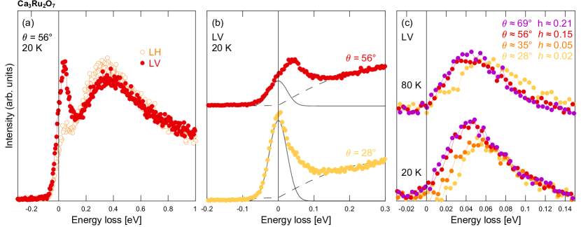
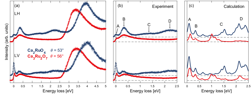
For our RIXS study of Ca3Ru2O7, we have focused entirely on the most intense oxygen -pre-edge that probes the planar and inner apical sites. In Figs. 2(a) and (b), spectra recorded with LV and LH light are compared to the corresponding planar spectra of Ca2RuO4. First, we notice that the ”block” of -excitations in Ca3Ru2O7 around 3.5 eV is consistently shifted to lower energies relatively to what is found in Ca2RuO4. Another noticeable difference is that among the four ”low” energy excitations reported Das et al. (2018) for Ca2RuO4 [labelled as A,B,C, and D in Fig. 2(b)], only the two lowest (A and B) are found in Ca3Ru2O7. The B excitation of bilayer Ca3Ru2O7 has a significantly smaller amplitude and is much broader than in Ca2RuO4. However, for both compounds, the B excitation is more intense when probed with LH polarization, see Fig. 3(a).
The lowest lying excitation (labelled as A) is overlapping with the elastic line and careful analysis is required to separate these two contributions. Elastic scattering is most pronounced near the specular condition, therefore the A excitation appears as a shoulder on the energy loss side – see Fig. 3(b). Near grazing condition, the situation is reversed and the elastic scattering appears as a shoulder on the left side of the A excitation peak. To model the elastic contribution, we use a Gaussian profile with the linewidth set by the energy resolution. In this fashion, it is possible to extract the A excitation by subtracting the elastic component as well as the contributions from the B excitation and background, as illustrated in Figs. 3(b) and (c). As the incidence angle – and hence the in-plane momentum transfer – is varied, the A excitation is dispersing to a lower energy away from the zone center. Finally, the A excitation persists at least up to 80 K, as shown in Fig. 3(c).
The momentum dependence of the A and B excitations extracted from the Ca3Ru2O7 data are compared in Fig. 4 to the corresponding excitations in Ca2RuO4. For the B excitation, the peak position is defined as the maximum obtained from the derivative of the spectrum, since the peak is extremely broad. Within the energy resolution of this experiment, no momentum dependence can be resolved for this excitation in Ca3Ru2O7. The situation is different in Ca2RuO4, where a small upward dispersion away from the zone center is detected for the B excitation Das et al. (2018). Most pronounced differences are observed for the A excitation. The strong dispersion found in Ca3Ru2O7 is completely absent in Ca2RuO4. Additionally, the excitation is located at significantly higher energies in Ca2RuO4 at around 80 meV compared to 55 meV in Ca3Ru2O7.
IV Discussion
To discuss the XAS spectra, we first summarize the interpretation of the Ca2RuO4 data published recently Fatuzzo et al. (2015); Das et al. (2018). The exact mechanism behind the Mott insulating state of Ca2RuO4 has long been under discussion and various theoretical models have been proposed Koga et al. (2004); Liebsch and Ishida (2007); Gorelov et al. (2010). In this context, the Ca2RuO4 XAS results strongly support the explanation via a complete orbital polarization with the almost fully occupied orbital. Indeed, the XAS spectra, shown in Fig.1(b), are in perfect accordance with this picture. For example, near normal incidence, the LH polarized light promotes a core electron into the O orbital that hybridizes with Ru orbitals at the apical and Ru orbitals at the planar site. Because the fully occupied Ru orbital is not available for absorption, the intensity at this resonance is strongly suppressed and a pronounced response is only observed at the apical oxygen resonance. For near grazing incidence the situation is reversed and the stronger XAS response flips to the planar resonance. Comparing the Ca2RuO4 and Ca3Ru2O7 spectra, the differences in crystal structure and orbital occupation become apparent. Due to the two nonequivalent apical oxygen sites in Ca3Ru2O7, the apical feature splits and the outer apical one is only visible as a shoulder to the strong planar resonance that overlaps with the inner apical resonance. Taking into account the relative intensities of the two features, the XAS results suggest a different orbital occupation than in Ca2RuO4, with an only partially filled . This partial occupation is also in accordance with the reduced -axis compression in Ca3Ru2O7 compared to Ca2RuO4 Yoshida et al. (2005); Friedt et al. (2001).

Next, we turn to discuss the RIXS spectra. The fact that completely different oxygen -edge RIXS spectra are observed for Ca2RuO4 and Ca3Ru2O7 is a beautiful example of how ground state fingerprints are encoded into the excitations. In principle, the CF environment around an in-plane oxygen should be similar for Ca2RuO4 and Ca3Ru2O7. Yet, the RIXS excitation spectra are fundamentally different for these two compounds. In Ca2RuO4, a sequence of excitations has been identified in the sector, which are separated from the higher energy features in the energy range 3–5 eV Das et al. (2018); Gretarsson et al. (2019) – see Fig. 2(a). In particular, two broad excitations located around 1 eV and 2 eV, labelled as C and D, are linked to the energy scales of Hund’s coupling and Coulomb interaction responsible for the Mott insulating ground state. In semi-metallic Ca3Ru2O7 by contrast, these excitations are completely absent – see Fig. 2(b). Even within the lower energy sector, pronounced differences are identified. Although two excitations (labelled as A and B) – with similar energy scales – are resolved for both compounds, they appear to have a fundamentally different nature. In Ca3Ru2O7 the lowest lying excitation is clearly dispersive, whereas in Ca2RuO4 no dispersion was resolved for the corresponding branch.
To gain insight into the microscopic picture behind these excitations, the RIXS response was modelled for both compounds, and compared to the experimental spectra in Fig. 2. We used the fast collision approximation Ament et al. (2007, 2011) of the RIXS cross-section describing the light-induced excitation – and subsequent absorption – of an electron from the O level into the level, for both LV and LH incoming polarization. Full detailed description of this approach is reported in Appendices A and B. The RIXS intensity was calculated via exact diagonalization of a model Hamiltonian defined on a cluster of two ruthenium sites connected by one planar oxygen site along an in-plane direction. The bond bending due to the rotation of the octahedra around the axis is allowed. The ruthenium-site Hamiltonian is defined on the subspace and consists of three terms: (1) CF splitting between the and , orbitals, (2) SOC , and (3) Coulomb interaction, which is expanded into intra-orbital and inter-orbital Hubbard interactions of strengths and , respectively. Inter-orbital Hund’s coupling as well as the pair-hopping term, are both of strength . Material specific values eV, eV, eV, and eV Mizokawa et al. (2001); Veenstra et al. (2014); Fatuzzo et al. (2015); Han and Millis (2018), are used to evaluate the model for both Ca2RuO4 and Ca3Ru2O7. Similar values of , and have been used for DMFT calculations Sutter et al. (2017) of Ca2RuO4 and are comparable to those used in modelling the spin-excitation dispersion observed by neutron scattering Jain et al. (2017), RIXS spectra Das et al. (2018), as well as magnetic anisotropy Porter et al. (2018). Here, we point out that small differences from previous estimates of the microscopic parameters are fully awaited since, in our description, the oxygen degrees of freedom are explicitly included, and this may lead to a renormalization of the local interaction terms. To take into account the different ground states, Ca2RuO4 is modelled with an antiferromagnetic (AFM) in-plane interaction, whereas we consider an extra exchange field to stabilize the ferromagnetic (FM) ground state and spins along the in-plane easy axis in Ca3Ru2O7 Braden et al. (1998); Yoshida et al. (2005). Henceforth, we will also refer to the Ca2RuO4 and Ca3Ru2O7 bonds as AFM and FM, respectively, corresponding to the G-type and A-type AFM structures.
In Fig. 2(c) the calculated RIXS responses in LH and LV polarizations are presented for both ground states, showing a good overall agreement with the experimental spectra in Fig. 2(b). In both cases four distinct excitations are evident, with approximate energy losses of 0.08, 0.4, 1–1.5 and 2 eV. In the FM case, we observe an overall decrease of the peak intensities. This effect is even more pronounced for excitations above 1 eV.
The origin of the four features for Ca2RuO4 has already been assigned in a previous work, and we recall it here for convenience Das et al. (2018). We point out that, in the present simulation, the RIXS intensity has been evaluated by fully taking into account the scattering geometry in Fig. 1(a), and that the LV and LH spectra have been obtained by averaging the spectra over two orthogonal in-plane bond directions of the cluster.
Excitations A, B, C, and D in Ca2RuO4 have been interpreted on the basis of the multiplet structure of the configuration of the Ru4+ ion. In particular, they are associated to transitions within low-energy spin-orbital configurations, which have one doubly occupied orbital (doublon), or two doubly occupied orbitals. In the framework of the ionic picture, structures C and D have been assigned to driven spin-state transitions between and states in the single- and two-doublon sectors Das et al. (2018). The suppression of the weight associated to the features above 1 eV in the case of a FM configuration is a consequence of the Pauli blocking of those intra- transitions. This mechanism may justify the lowered intensity of C and D structures in the FM background of Ru-O planes in Ca3Ru2O7.
In Ca2RuO4, the lowest energy features A and B are associated to spin-orbital excitations within the subspace of the multiplets, whose energies are determined by the relative strength of the CF potential and the SOC. Even though they are not fully resolved experimentally due to limited energy resolution, those excitations are accessible in oxygen -edge because of the SOC in the Ru shell which strongly hybridizes states with O orbitals. In particular, the B structure has been attributed to multiple transitions to the highest energy spin-orbital sector, while the A structure has been generically associated to composite magnetic transitions within the lowest-energy sector. We observe that, in the FM case, features occurring at a similar energy scale are observed, and we want to elucidate the possible spin-orbital (magnetic) origin of these excitations, with a special focus on the lowest A feature.
Beforehand, we observe that the lowest lying excitation A has an energy scale typical of both optical phonons and magnons. The strong dispersion of this excitation near the zone center is, however, atypical for optical phonons. In Sr2RuO4, where complete phonon dispersions have been calculated and probed by neutron scattering Braden et al. (2007); Wang et al. (2010), optical phonons are in fact found at and meV. None of them has a dispersion around the zone center compatible with what we observe in Ca3Ru2O7. This is demonstrated in Fig. 4, where the relevant optical Sr2RuO4 phonon dispersion – measured by neutron scattering – is shown in grey. In comparison to the A excitation measured in Ca3Ru2O7, this optical phonon is non-dispersive. Moreover, the 70 meV optical phonon found in Sr2RuO4 stems from vibrations of the apical oxygen whereas we are probing on the in-plane oxygen site. On this basis, assigning a magnetic origin to the lowest lying excitation appears the most plausible interpretation. We stress that it is not unusual to observe magnetic excitations beyond the magnetic ordered state due to the persistence of short-range magnetic correlations. In cuprates and iron pnictides, paramagnon excitations are found deep into the magnetically disordered state Dean et al. (2013); Monney et al. (2016); Pelliciari et al. (2019). Observing no significant temperature dependence of the A excitation dispersion is therefore expected.
To further verify the magnetic origin of the A excitations in Ca2RuO4 and Ca3Ru2O7, and to reveal their distinct nature, we evaluated the dynamic spin structure factors , and spin-spin dynamic spin structure factors , , [Fig. 5(b),(c),(e),(f)] for and , which are the only viable values for the momentum transfer of our Ru-O-Ru cluster. Here, we point out that the ground state is made by magnetic moments that are aligned in the Ru plane. For convenience, we refer to , for a spin mode excitation that is collinear, perpendicular in-plane, perpendicular out-of-plane with respect to the orientation of the ordered magnetic moments in the ground state, respectively.
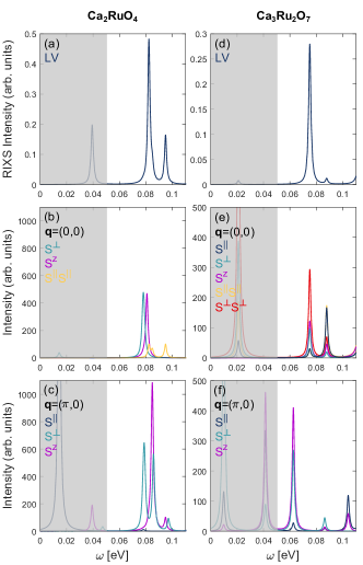
Let us start with the AFM case in Fig. 5(a)–(c). The comparison of the low-energy part of the RIXS spectrum for the AFM configuration with the calculated spin structure factors at allows to associate the dominant excitation in the RIXS spectrum to features with transverse single spin modes, i.e. and , and longitudinal two-spin correlation functions. This is consistent with the previous interpretation of the A excitation as evidence of composite excitations such as longitudinal (Higgs) two-particle and transverse bimagnon modes Souliou et al. (2017); Das et al. (2018). In particular, from the analysis we observe that the lowest energy spin excitations occur at about 20 and 40 meV – see Fig. 5(b),(c), mainly through single spin flip at each Ru site.The former energy scale is related to the effective single ion anisotropy due to the interplay of spin-orbit and crystal field potential. A distinctive aspect of the magnetic ground state is that, due to the spin-orbit coupling and crystal field potential, there is neither rotational nor parity conservation for the local spin. The resulting ground state is then a quantum superposition of several components. Specifically, it consists of dominant exchange driven anisotropic antiferromagnetic correlations, and it also includes states corresponding to the variation of amplitude and direction of the local magnetic moments with respect to the easy axis. This peculiar character of the ground state allows to have a significant spectral weight associated to high-energy excitations, corresponding to the RIXS active states close to 80 meV – see Fig. 5(a). Taking into account the energy profile of the dynamic spin response, we deduce that the modes at about 80 meV have a multi-particle spin character, as they are accessible by means of both single transverse and double longitudinal spin excitations. Our results also predict the existence of a lower energy feature in the RIXS spectrum, having similar character, at 40 meV, in an energy range which is not detectable in the present experiment.
The FM case offers a similar result, since the lowest A feature may also be associated to magnetic excitations. Notably in this case, the dominant excitation occurs at slightly lower energy, and corresponds to mainly transverse spin excitations, of single- and or two-particle type . Moreover, according to the simulation, the existence of a very weak feature located at 20 meV is also predicted.
Having identified the nature of the magnetic excitations associated to the lowest RIXS feature in both the AFM and FM ground states, one can also estimate the bandwidth of the continuum of the corresponding collective modes propagating along the path. Comparing the relevant at and shows that, in the AFM case, magnetic peaks are located approximately in the same energy range at different wave vectors. On the contrary, in the FM configuration, the peaks associated to the single spin excitations are shifted to lower energies by 20-30 meV, when going from to . This is in accordance with what is observed in the experimental spectra – see Fig. 4. We also carried out the calculation of the local and two-site orbital angular momentum correlation functions, which reveal that the A peak in the AFM case has significant orbital contribution, while it is substantially suppressed in the FM case. This is consistent with the observation that the FM ground state has a different orbital pattern Forte et al. (2010) when compared to the AFM configuration. The doublon can have a stronger tendency to occupy different orbitals on neighbouring Ru sites in the FM case. Moreover, the spin-orbit coupling tends to align the orbital moments; since the Ru spins are also ferromagnetically correlated. This implies that orbital variations can be suppressed in the low-energy spin sector. Here, we argue that the lack of the orbital component in the targeted excitation allows to have a larger effective exchange, which results in an enhancement of the bandwidth as we find in the cluster analysis.
V Conclusions
In summary, we have carried out a combined oxygen -edge XAS and RIXS study of Ca3Ru2O7. Our results are compared to (i) equivalent experimental results previously obtained on single layer Ca2RuO4 and (ii) local cluster modelling of Ca3Ru2O7 and Ca2RuO4. In particular, the oxygen -edge RIXS spectra are fundamentally different in Ca3Ru2O7 and Ca2RuO4, reflecting their different ground states. Whereas in Ca2RuO4 a set of excitations within the subspace, consisting of two low-energy and two mid/high-energy structures, is observed, only the two lowest intra- excitations have a significant amplitude in Ca3Ru2O7. This effect is captured by the local cluster modelling taking into account the different in-plane magnetic couplings. Finally, we demonstrated that the lowest lying intra- excitation in Ca3Ru2O7 is dispersing, revealing its collective origin. We argue based on the exact dispersion and comparison to spin correlation function computations, that this excitation is magnonic rather than phononic in nature. In fact, it is suggested to be dominantly a transverse mode with multi-particle character, which is indirectly allowed at the oxygen -edge through substantial SOC of Ru ions.
VI Appendix A: Model Hamiltonian
We report here the details of the microscopic model describing the energy levels and wave functions of the considered Ru-O-Ru cluster. The examined Hamiltonian Cuoco et al. (2006a, b) is expressed as:
| (1) |
The first term in Eq. 1 is the kinetic operator describing the Ru-O connectivity:
| (2) |
where is the creation operator for an electron with spin at the site in the orbital of the sector (, , ), while is the annihilation operator of an electron with spin at the site in the orbital of the (, , ) space of the oxygen. Hopping amplitudes include all the allowed symmetry terms according to the Slater-Koster rules Slater and Koster (1954); Brzezicki et al. (2015) for a given bond connecting a ruthenium to an oxygen atom along, say, the direction. We allow for the relative rotation of the oxygen octahedra sorrounding the Ru site, assuming that the Ru-O-Ru bond can form an angle =(180°-). The case with = 0°corresponds to the tetragonal undistorted bond, while a nonvanishing value of arises when the RuO6 octahedra are rotated of the corresponding angle around the axis.
The second term is the Coulomb interaction, which is expressed in terms of Kanamori parameters , , and as follows
| (3) |
where , are the on site charge for spin and the spin operators for the orbital, respectively. () is the intra (inter)- orbital Coulomb repulsion, is the Hund coupling, and the pair hopping term. Due to the invariance for rotations in the orbital space, the following relations hold: , .
The part of the Hamiltonian is the crystalline field potential, controlling the symmetry lowering from cubic to tetragonal one, due to the compression of RuO6 octahedra along the axis.:
| (4) |
The SOC Hamiltonian reads as
| (5) |
Due to the cubic CF terms in RuO6 octahedra separating the lower from the unoccupied levels, stands for the angular momentum operator projected onto the subspace. Its components have the following expression in terms of orbital fermionic operators:
| (6) |
Finally, in Eq. 1 is a an effective exchange field which pins the magnetization at the Ru sites to be in the () plane for the FM ground state:
| (7) |
VII APPENDIX B: Calculation of the RIXS cross section
The RIXS intensity is described by the Kramers-Heisenberg relation
| (8) |
where and and stand for the energy and momentum transferred by the scattered photon, and and for the incoming and outgoing light polarization vectors. We adopt the dipole and fast collision approximation, in which the RIXS scattering amplitude is reduced to
| (9) |
where is the effective RIXS scattering operator describing two subsequent dipole transitions, and is the core-hole broadening. In the oxygen -edge RIXS, the dipole transitions create an O 1 core hole and extra valence electron in a 2 obital and viceversa, and the scattering operator has the following expression:
| (10) |
where is the orbital and the sum over the different spin states is assumed. Matrix elements are then evaluated among oxygen valence states in Eq. 9. Notably, the valence electron in a 2 obital hybridizes and interacts with the Ru electrons.
In the adopted experimental scattering geometry, the dependence upon the incident angle and scattering angle between the incoming/outgoing polariization vectors is:
| (11) | |||||
Here the coordinate frame corresponds to the tetragonal axis frame . Since the outgoing polarization is not resolved, the RIXS intensity is obtained by summing up incoherently over all the three polarization directions .
VIII Acknowlegdements
The experimental work was performed at the ADRESS beamline of the SLS at the Paul Scherrer Institut, Villigen PSI, Switzerland. We thank the ADRESS beamline staff for technical support. K.v.A., M.H., Q.W, L.D., O.I., J.C. acknowledge support by the Swiss National Science Foundation. M.C. and F.F. acknowledge support by the project ”Two-dimensional Oxides Platform for SPIN-orbitronics nanotechnology (TOPSPIN)” funded by the MIUR (PRIN) Bando 2017 - Grant 20177SL7HC. Y.S. is funded by the Swedish Research Council (VR) with a Starting Grant (No.2017-05078). Work at the Paul Scherrer Institut has been funded by the Swiss National Science Foundation through the Sinergia network Mott Physics Beyond the Heisenberg (MPBH) model (SNSF Research grant numbers CRSII2_141962 and CRSII2_160765).
References
- Karpus et al. (2004) J. F. Karpus, R. Gupta, H. Barath, S. L. Cooper, and G. Cao, Phys. Rev. Lett. 93, 167205 (2004).
- Lin et al. (2005) X. N. Lin, Z. X. Zhou, V. Durairaj, P. Schlottmann, and G. Cao, Phys. Rev. Lett. 95, 017203 (2005).
- Bao et al. (2008) W. Bao, Z. Q. Mao, Z. Qu, and J. W. Lynn, Phys. Rev. Lett. 100, 247203 (2008).
- Kikugawa et al. (2010) N. Kikugawa, A. Winfried Rost, C. William Hicks, A. John Schofield, and A. Peter Mackenzie, J. Phys. Soc. Jpn. 79, 024704 (2010).
- Zhu et al. (2016) M. Zhu, J. Peng, T. Zou, K. Prokes, S. D. Mahanti, T. Hong, Z. Q. Mao, G. Q. Liu, and X. Ke, Phys. Rev. Lett. 116, 216401 (2016).
- Xing et al. (2018) H. Xing, L. Wen, C. Shen, J. He, X. Cai, J. Peng, S. Wang, M. Tian, Z.-A. Xu, W. Ku, Z. Mao, and Y. Liu, Phys. Rev. B 97, 041113(R) (2018).
- Sow et al. (2019) C. Sow, R. Numasaki, G. Mattoni, S. Yonezawa, N. Kikugawa, S. Uji, and Y. Maeno, Phys. Rev. Lett. 122, 196602 (2019).
- Cao et al. (2003) G. Cao, L. Balicas, Y. Xin, J. E. Crow, and C. S. Nelson, Phys. Rev. B 67, 184405 (2003).
- Yoshida et al. (2004) Y. Yoshida, I. Nagai, S.-I. Ikeda, N. Shirakawa, M. Kosaka, and N. Môri, Phys. Rev. B 69, 220411(R) (2004).
- Ohmichi et al. (2004) E. Ohmichi, Y. Yoshida, S. I. Ikeda, N. Shirakawa, and T. Osada, Phys. Rev. B 70, 104414 (2004).
- Yoshida et al. (2005) Y. Yoshida, S.-I. Ikeda, H. Matsuhata, N. Shirakawa, C. H. Lee, and S. Katano, Phys. Rev. B 72, 054412 (2005).
- Nakatsuji et al. (2004) S. Nakatsuji, V. Dobrosavljević, D. Tanasković, M. Minakata, H. Fukazawa, and Y. Maeno, Phys. Rev. Lett. 93, 146401 (2004).
- Takenaka et al. (2017) K. Takenaka, Y. Okamoto, T. Shinoda, N. Katayama, and Y. Sakai, Nature Communications 8, 14102 (2017).
- Friedt et al. (2001) O. Friedt, M. Braden, G. André, P. Adelmann, S. Nakatsuji, and Y. Maeno, Phys. Rev. B 63, 174432 (2001).
- Braden et al. (1998) M. Braden, G. André, S. Nakatsuji, and Y. Maeno, Phys. Rev. B 58, 847 (1998).
- Jain et al. (2017) A. Jain, M. Krautloher, J. Porras, G. H. Ryu, D. P. Chen, D. L. Abernathy, J. T. Park, A. Ivanov, J. Chaloupka, G. Khaliullin, B. Keimer, and B. J. Kim, Nat. Phys. 13, 633 (2017).
- Souliou et al. (2017) S.-M. Souliou, J. Chaloupka, G. Khaliullin, G. Ryu, A. Jain, B. J. Kim, M. Le Tacon, and B. Keimer, Phys. Rev. Lett. 119, 067201 (2017).
- Das et al. (2018) L. Das, F. Forte, R. Fittipaldi, C. G. Fatuzzo, V. Granata, O. Ivashko, M. Horio, F. Schindler, M. Dantz, Y. Tseng, D. E. McNally, H. M. Rønnow, W. Wan, N. B. Christensen, J. Pelliciari, P. Olalde-Velasco, N. Kikugawa, T. Neupert, A. Vecchione, T. Schmitt, M. Cuoco, and J. Chang, Phys. Rev. X 8, 011048 (2018).
- Fatuzzo et al. (2015) C. G. Fatuzzo, M. Dantz, S. Fatale, P. Olalde-Velasco, N. E. Shaik, B. Dalla Piazza, S. Toth, J. Pelliciari, R. Fittipaldi, A. Vecchione, N. Kikugawa, J. S. Brooks, H. M. Rønnow, M. Grioni, C. Rüegg, T. Schmitt, and J. Chang, Phys. Rev. B 91, 155104 (2015).
- Bisogni et al. (2012) V. Bisogni, L. Simonelli, L. J. P. Ament, F. Forte, M. Moretti Sala, M. Minola, S. Huotari, J. van den Brink, G. Ghiringhelli, N. B. Brookes, and L. Braicovich, Phys. Rev. B 85, 214527 (2012).
- Moretti Sala et al. (2014) M. Moretti Sala, M. Rossi, S. Boseggia, J. Akimitsu, N. B. Brookes, M. Isobe, M. Minola, H. Okabe, H. M. Rønnow, L. Simonelli, D. F. McMorrow, and G. Monaco, Phys. Rev. B 89, 121101(R) (2014).
- Lu et al. (2018) X. Lu, P. Olalde-Velasco, Y. Huang, V. Bisogni, J. Pelliciari, S. Fatale, M. Dantz, J. G. Vale, E. C. Hunter, J. Chang, V. N. Strocov, R. S. Perry, M. Grioni, D. F. McMorrow, H. M. Rønnow, and T. Schmitt, Phys. Rev. B 97, 041102(R) (2018).
- Pincini et al. (2019) D. Pincini, L. S. I. Veiga, C. D. Dashwood, F. Forte, M. Cuoco, R. S. Perry, P. Bencok, A. T. Boothroyd, and D. F. McMorrow, Phys. Rev. B 99, 075125 (2019).
- Ament et al. (2007) L. J. P. Ament, F. Forte, and J. van den Brink, Phys. Rev. B 75, 115118 (2007).
- Ament et al. (2011) L. J. P. Ament, M. van Veenendaal, T. P. Devereaux, J. P. Hill, and J. van den Brink, Rev. Mod. Phys. 83, 705 (2011).
- Fukazawa et al. (2000) H. Fukazawa, S. Nakatsuji, and Y. Maeno, Physica B 281, 613 (2000).
- Nakatsuji and Maeno (2001) S. Nakatsuji and Y. Maeno, Journal of Solid State Chemistry 156, 26 (2001).
- Ghiringhelli et al. (2006) G. Ghiringhelli, A. Piazzalunga, C. Dallera, G. Trezzi, L. Braicovich, T. Schmitt, V. N. Strocov, R. Betemps, L. Patthey, X. Wang, and M. Grioni, Review of Scientific Instruments 77, 113108 (2006).
- Strocov et al. (2010) V. Strocov, T. Schmitt, U. Flechsig, T. Schmidt, A. Imhof, Q. Chen, J. Raabe, R. Betemps, D. Zimoch, J. Krempasky, et al., Journal of synchrotron radiation 17, 631 (2010).
- Chen et al. (1992) C. T. Chen, L. H. Tjeng, J. Kwo, H. L. Kao, P. Rudolf, F. Sette, and R. M. Fleming, Phys. Rev. Lett. 68, 2543 (1992).
- Malvestuto et al. (2011) M. Malvestuto, E. Carleschi, R. Fittipaldi, E. Gorelov, E. Pavarini, M. Cuoco, Y. Maeno, F. Parmigiani, and A. Vecchione, Phys. Rev. B 83, 165121 (2011).
- Malvestuto et al. (2013) M. Malvestuto, V. Capogrosso, E. Carleschi, L. Galli, E. Gorelov, E. Pavarini, R. Fittipaldi, F. Forte, M. Cuoco, A. Vecchione, and F. Parmigiani, Phys. Rev. B 88, 195143 (2013).
- Guedes et al. (2012) E. B. Guedes, M. Abbate, K. Ishigami, A. Fujimori, K. Yoshimatsu, H. Kumigashira, M. Oshima, F. C. Vicentin, P. T. Fonseca, and R. J. O. Mossanek, Phys. Rev. B 86, 235127 (2012).
- Koga et al. (2004) A. Koga, N. Kawakami, T. M. Rice, and M. Sigrist, Phys. Rev. Lett. 92, 216402 (2004).
- Liebsch and Ishida (2007) A. Liebsch and H. Ishida, Phys. Rev. Lett. 98, 216403 (2007).
- Gorelov et al. (2010) E. Gorelov, M. Karolak, T. O. Wehling, F. Lechermann, A. I. Lichtenstein, and E. Pavarini, Phys. Rev. Lett. 104, 226401 (2010).
- Braden et al. (2007) M. Braden, W. Reichardt, Y. Sidis, Z. Mao, and Y. Maeno, Phys. Rev. B 76, 014505 (2007).
- Gretarsson et al. (2019) H. Gretarsson, H. Suzuki, H. Kim, K. Ueda, M. Krautloher, B. J. Kim, H. Yavaş, G. Khaliullin, and B. Keimer, Phys. Rev. B 100, 045123 (2019).
- Mizokawa et al. (2001) T. Mizokawa, L. H. Tjeng, G. A. Sawatzky, G. Ghiringhelli, O. Tjernberg, N. B. Brookes, H. Fukazawa, S. Nakatsuji, and Y. Maeno, Phys. Rev. Lett. 87, 077202 (2001).
- Veenstra et al. (2014) C. N. Veenstra, Z.-H. Zhu, M. Raichle, B. M. Ludbrook, A. Nicolaou, B. Slomski, G. Landolt, S. Kittaka, Y. Maeno, J. H. Dil, I. S. Elfimov, M. W. Haverkort, and A. Damascelli, Phys. Rev. Lett. 112, 127002 (2014).
- Han and Millis (2018) Q. Han and A. Millis, Phys. Rev. Lett. 121, 067601 (2018).
- Sutter et al. (2017) D. Sutter, C. Fatuzzo, S. Moser, M. Kim, R. Fittipaldi, A. Vecchione, V. Granata, Y. Sassa, F. Cossalter, G. Gatti, M. Grioni, H. M. Rønnow, N. C. Plumb, C. E. Matt, M. Shi, M. Hoesch, T. K. Kim, T. R. Chang, H. T. Jeng, C. Jozwiak, A. Bostwick, E. Rotenberg, A. Georges, T. Neupert, and J. Chang, Nat. Comm. 8, 15176 (2017).
- Porter et al. (2018) D. G. Porter, V. Granata, F. Forte, S. Di Matteo, M. Cuoco, R. Fittipaldi, A. Vecchione, and A. Bombardi, Phys. Rev. B 98, 125142 (2018).
- Wang et al. (2010) Y. Wang, J. J. Wang, J. E. Saal, S. L. Shang, L.-Q. Chen, and Z.-K. Liu, Phys. Rev. B 82, 172503 (2010).
- Dean et al. (2013) M. P. M. Dean, G. Dellea, R. S. Springell, F. Yakhou-Harris, K. Kummer, N. B. Brookes, X. Liu, Y.-J. Sun, J. Strle, T. Schmitt, L. Braicovich, G. Ghiringhelli, I. Božović, and J. P. Hill, Nature Materials 12, 1019 EP (2013).
- Monney et al. (2016) C. Monney, T. Schmitt, C. E. Matt, J. Mesot, V. N. Strocov, O. J. Lipscombe, S. M. Hayden, and J. Chang, Phys. Rev. B 93, 075103 (2016).
- Pelliciari et al. (2019) J. Pelliciari, K. Ishii, Y. Huang, M. Dantz, X. Lu, P. Olalde-Velasco, V. N. Strocov, S. Kasahara, L. Xing, X. Wang, C. Jin, Y. Matsuda, T. Shibauchi, T. Das, and T. Schmitt, Communications Physics 2, 139 (2019).
- Forte et al. (2010) F. Forte, M. Cuoco, and C. Noce, Phys. Rev. B 82, 155104 (2010).
- Cuoco et al. (2006a) M. Cuoco, F. Forte, and C. Noce, Phys. Rev. B 74, 195124 (2006a).
- Cuoco et al. (2006b) M. Cuoco, F. Forte, and C. Noce, Phys. Rev. B 73, 094428 (2006b).
- Slater and Koster (1954) J. C. Slater and G. F. Koster, Physical Review 94, 1498 (1954).
- Brzezicki et al. (2015) W. Brzezicki, C. Noce, A. Romano, and M. Cuoco, Phys. Rev. Lett. 114, 247002 (2015).