New Light on Cortical Neuropeptides and Synaptic Network Plasticity
Abstract
Neuropeptides, members of a large and evolutionarily ancient family of proteinaceous cell-cell signaling molecules, are widely recognized as extremely potent regulators of brain function and behavior. At the cellular level, neuropeptides are known to act mainly via modulation of ion channel and synapse function, but functional impacts emerging at the level of complex cortical synaptic networks have resisted mechanistic analysis. New findings from single-cell RNA-seq transcriptomics now illuminate intricate patterns of cortical neuropeptide signaling gene expression and new tools now offer powerful molecular access to cortical neuropeptide signaling. Here we highlight some of these new findings and tools, focusing especially on prospects for experimental and theoretical exploration of peptidergic and synaptic networks interactions underlying cortical function and plasticity.
Highlights
-
•
New findings expand the evidence for involvement of local neuropeptide signaling in cortical synaptic network plasticity.
-
•
Single-cell transcriptomes indicate that every cortical neuron secretes at least one neuropeptide and displays at least one neuropeptide receptor.
-
•
Transcriptomic neurotaxonomies predict cortical peptidergic network architectures.
-
•
New genetic tools open transcriptomic network predictions to experimental test.
-
•
New genetic tools open neuropeptide roles in synaptic network plasticity to experimental and theoretical exploration
©2020, This manuscript version is made available under the CC-BY-NC-SA 4.0 license.
http://creativecommons.org/licenses/by-nc-sa/4.0/
Introduction
Neuropeptides are diffusible cell-cell signaling molecules that act in brain as powerful regulators of social valence, sleep, appetite, anxiety, stress response, pain perception and memory [1, 2, 3, 4, 5, 6]. Many neuropeptides were first discovered as neuroendocrine hormones regulating growth, reproduction, gut function or other bodily processes, but later shown to also signal strictly between clearly non-neuroendocrine neurons [5]. Others, such as the opioid neuropeptides, were discovered during molecular and physiological explorations focused on central nervous system perceptual and cognitive impacts [7].
Well over one hundred genes encoding primary neuropeptide signaling proteins, both precursors and receptors, are present in the human genome. A high degree of sequence conservation across these genes - spanning the entire animal kingdom - argues strongly for very ancient evolutionary origins of peptidergic signaling [8, 9, 10]. There is even evidence that neuropeptides may have coordinated the behavior of slow-moving, ancestral metazoans before the advent of neurons and specialized, fast synaptic connections [10, 11]. Though most animal species today rely primarily upon fast synaptic transmission using recycling small-molecule neurotransmitters (with rapid re-uptake or degradation) for their speedy behaviors, neuropeptides remain critical to adaptive nervous system function, acting via slower biochemical coupling as modulators of ion channel and synaptic function [5, 12, 13, 14, 15]. It now appears likely that most (and perhaps all) present-day neurons both secrete and respond to at least one neuropeptide, in addition to a small-molecule synaptic neurotransmitter, and that such co-release may be critical to synaptic network function [5, 16, 17, 18, 19].
Transcriptomic and physiological evidence now points to a view of mammalian cerebral cortex as a stack of fast synaptic and slow peptidergic modulatory networks that interconnect neurons of highly differentiated synaptic and neuromodulatory connection affinities. Here we’ll summarize some of this evidence and remark upon a few of the new molecular, anatomical, physiological and theoretical tools now bringing new light to these highly diverse cortical networks and their interactions. Finally, we’ll mention ways that interests converging from neuroscience and computer science disciplines may deepen our understanding of memory engram formation by deep synaptic networks, be they natural or artificial. We begin here with a brief overview of the present understanding of peptidergic signaling mechanisms.
The neuropeptide signaling canon in brief
Active neuropeptide (NP) molecules (small peptides usually consisting of 3-36 amino acids) are produced within source neurons by enzymatic fragmentation and covalent modification of neuropeptide precursor protein (NPP) gene products, stored in large, dense-cored vesicles and then secreted in response to calcium influx during neuronal activity [1, 3, 6]. NP molecules, once secreted, persist for many minutes in brain interstitial spaces, long enough for paracrine diffusion over distances embracing hundreds of potential target neurons [1, 20, 21, 22, 23]. In contrast to synaptic signaling, where small molecule transmitter actions are restricted by rapid re-uptake or degradation processes to just one very closely apposed postsynaptic neuronal site, a neuropeptide is “broadcast” over much longer, though still limited, ranges set by release site anatomy, quantities secreted, diffusion physics and slow degradation processes. Such paracrine neuropeptide signaling may nonetheless precisely address specific neurons within a broadcast volume: only neurons expressing receptors “cognate to” (i.e., selective for) a particular secreted peptide molecule will respond [1, 3, 5]. Consider an analogy with radio or television broadcasting, where many receivers generally exist within a given broadcast area, but only those sets tuned to the specific frequency of a particular station respond to the broadcast. Neuropeptide signal connectivity is thus surely determined in large part by cell-specific patterns of differential ligand and receptor gene expression.
Most neuropeptide receptors are encoded by members of the very large superfamily of G-protein-coupled receptor (GPCR) genes [1, 3]. GPCRs are selective, high-affinity receptors distinguished by characteristic seven-transmembrane-segment atomic structures and signal transduction involving heterotrimeric G-proteins (hence the terminology) [24, 25]. Most GPCR genes encode olfactory receptors [26, 27], but mouse and human genomes still comprise over 350 genes encoding GPCRs selective for diverse endogenous ligands. At least one hundred of these are neuropeptide-selective (NP-GPCRs), while dozens more are selective for other widely studied neuromodulators including dopamine, serotonin, norepinephrine and endocannabinoids [28].
Though GPCRs are highly diverse in ligand specificity, they are much less diverse in downstream signaling actions. While GPCR signaling has many additional facets and complexities [1, 29, 30, 31, 32], the primary actions of most known neuronal GPCRs are mediated by the intracellular second messengers cyclic AMP, ionic calcium, and/or phosphoinositide (PI) lipid metabolites. These second messengers, in turn, act via regulation of protein kinases, directly upon ion channels, or upon gene expression to impact neuronal firing and synaptic transmission on a wide range of time scales. Primary effects of the GPCRs expressed in cortex fall into just three major categories distinguished by G-protein alpha subunit (G) family: the Gi/o family (i/o) inhibits cAMP production, the Gs family (s) stimulates cAMP production, and the Gq/11 family (q) stimulates phospholipase C activity to generate PI metabolites and amplify calcium signaling [33]. For most NP-GPCR genes, the primary G family (e.g., i/o, s or q/11) is now known [34] and provides a useful first-order prediction of the encoded GPCR’s signal transduction impact. Though this basic trichotomy is certainly an oversimplification, such predictions are useful to form presently fragmentary information about actions of neuropeptides and diverse other GPCR-mediated modulatory ligands into experimentally testable hypotheses. Broad conservation of GPCR sequences also makes it reasonable to tentatively generalize mechanisms and impacts of neuropeptide signaling across different neuropeptides, brain regions and animal phyla [8, 35]. We’ll now touch upon new opportunities to evaluate and leverage such generalizations.
New light on mechanisms and effects of cortical neuropeptide signaling
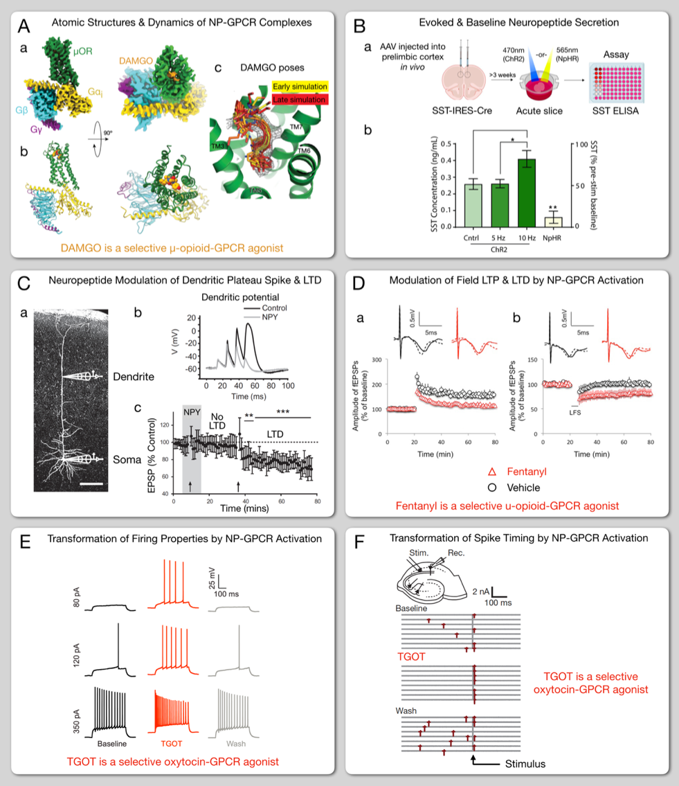
Our understanding of cortical synaptic network plasticity is now benefitting from advances in elucidating basic molecular mechanisms and effects of neuropeptide signaling. One overarching advance, exemplified in Fig. 1A, has been the determination of atomic structures of a rapidly growing number of GPCRs [24, 45, 36], coupled with structure-based molecular dynamic simulations of interaction of GPCRs with ligands and heterotrimeric G proteins [46]. Besides providing major insights into the structural basis of NP-GPCR function, these new results are now enabling the development of genetically encoded sensors and effectors targeting specific neuropeptide ligand-receptor systems, as discussed in the section “New physiological tools“ below.
In combination with new genetically encoded sensors and effectors, neuron-type-specific gene expression driver lines and viral vector injection methods, as exemplified in Fig. 1B, and emerging gene editing methods now provide means to target neuropeptide signaling in specific genetically and anatomically defined neuron subsets within intact cortical tissues. Such means seem certain to help resolve troublesome uncertainties as to function that have resulted until now from the well-established but poorly defined presence of dauntingly large numbers of peptidergic ligands and receptors in nearly every brain region. Figures 1C-F represent ways that more traditional electrophysiological and pharmacological tools also continue to provide new insight into neuropeptide signaling impacts, focusing especially on indications of neuropeptide involvement in modulation of synaptic plasticity. Combinations of new genetic tools with advanced physiological and pharmacological methods are well matched to the substantial remaining challenges of understanding peptidergic modulation of cellular, synaptic and network function and plasticity.
New light on the patterning of neuropeptide precursor and receptor gene expression
Neuron type diversity, first recognized by pioneering nineteenth century microscopists, is a fundamental element of nervous system organization [47, 48, 49, 50, 51]. Over recent decades, differential immunostaining for various neuropeptides (e.g., vasoactive intestinal peptide, somatostatin, neuropeptide Y, substance P, and cholecystokinin) has come into wide use for discriminating anatomically and functionally distinct neuron types [52]. More recently, in situ hybridization and microarray gene expression data have established that mRNA transcripts encoding these five NPPs and many other NPP and NP-GPCR genes are expressed in large numbers of cortical neurons [53, 54]. Expression data combining whole-genome depth with single-cell resolution have been lacking, however, and almost unimaginable until recently. This lack has hindered exploration of the full extent of cortical neuropeptide gene expression and made it difficult to design robust experiments to develop and test cogent hypotheses regarding the network consequences of cortical neuropeptide signaling. Single-cell RNA-seq methods have now revolutionized the study of differential gene expression in brain [48, 51, 55, 56, 57]. We recently reported a neuropeptide-focused analysis [33] of a body of such data acquired from over 23,000 individual mouse cortical neurons [55]. Our analysis supports the surprising conclusion that all (or very nearly all) cortical neurons express at least one NPP gene at an extremely high level, and at least one NP-GPCR gene. The extreme abundance of NPP transcripts in most cortical neurons can be considered prima facie evidence that the corresponding NP products are produced, secreted and functionally important [33]. Furthermore, the vast majority of individual cortical neurons actually express more than one (sometimes as many as ten!) distinct NPP and NP-GPCR genes.
![[Uncaptioned image]](/html/2004.07975/assets/Table1.png)
Table 1, adapted from reference [33], tabulates a total of 58 NP-GPCR genes that are expressed at substantial levels in mouse sensory and motor cortical areas, along with identities of corresponding cognate NPP genes (i.e., NPP genes for which the expected neuropeptide product matches the ligand selectivity of a given NP-GPCR). Of the 58 NP-GPCR genes, the 34 in column A are cognate to an NPP gene expressed at very high levels in the same cortical areas, suggesting the possibility of locally intracortical neuropeptide signaling. This suggestion is especially compelling for the GABAergic interneurons, since most have only short local axons, making it unlikely that their secreted NP products could act at very remote sites. Conversely, very low expression within a given cortical locale of NPP genes cognate to the 24 column B NP-GPCR genes suggests that the corresponding 24 receptors will never activate in response to locally generated neuropeptide. The column B NP-GPCRs nonetheless remain strong candidates for activation by neuropeptides released by afferent axons projecting from distant subcortical or cortical structures, where the relevant cognate NPP genes are in fact expressed. Single-cell RNA-seq data have also enabled the precise data-driven clustering of large and diverse neuron populations into neurotaxonomies representing tractably small numbers of neuron classes, subclasses and types [48, 50, 51, 55, 56, 57, 58]. Such transcriptomic neurotaxonomies become very useful if one assumes that the individual neurons within each type cluster share similar single-neuron physiological dynamics, patterns of synaptic and neuromodulatory connectivity to other neuron types, and play similar roles in adaptive network function. Though further critical examination of this neurotaxonomy premise is necessary and ongoing [48, 58], it seems reasonable and very likely to reward further experimental and theoretical pursuit [51, 56].
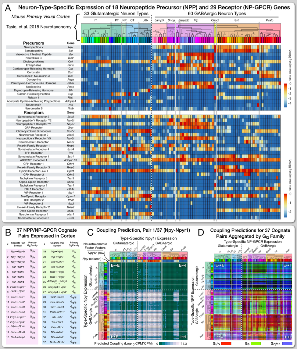
Figure 2 represents results, described more fully in reference [33], where we examined patterns of neuropeptide precursor and receptor gene expression in mouse neocortex through the powerful prism of transcriptomic neurotaxonomy. Fig. 2A illustrates our finding that expression profiles of 18 NPP and 29 NP-GPCR genes suggest that each transcriptomic neuron type expresses its own unique combination of these 47 NP genes. This finding is consistent with the well-established utility of neuropeptides as molecular markers of neuron type [52] but extends to a considerably deeper neurotaxonomy. Indeed, we have presented a machine-learning analysis indicating that a neuron-type clustering based solely on differential expression of these 47 genes matches deep clustering results based on genome-wide clustering [55] and does so with extraordinary precision in comparison to other, similarly small gene sets [33].
Recalling our radio broadcast metaphor, a neuron expressing a given NPP gene and another neuron expressing a cognate NP-GPCR gene can be cast as a directed communication channel, where “tuning” of both the “transmitter” and the “receiver” are established by each neuron’s gene expression profile. Broadcasts on different channels might communicate separate messages simultaneously to receivers tuned to separate channels within the same geographical reception area. The 18 NPP and 29 NP-GPCR genes whose differential expression profiles are represented in Fig. 2A comprise the 37 different cognate NPP/NP-GPCR pairs tabulated in Fig. 2B, where each cognate pair can be considered a distinct communication channel, with addressing fixed by gene expression profiles. Figure 2C is a prediction of one 93 x 93 coupling matrix or “molecular connectome” derived from expression profiles for one single cognate NPP/NP-GPCR pair, Npy-Npyr1, amongst the 93 neuron types represented in Fig. 2A. Since the data of Fig. 2A actually cover 37 analogous cognate pairs, they actually predict of a stack of 37 neuron-type-based peptidergic coupling matrices (all 37 of which are laid out for both mouse visual and motor cortical areas in reference [33]). Fig. 2D aggregates the entire stack of 37 coupling matrices for visual cortex, factored by the three G signal transduction families and their expected second-messenger impacts [33]. Coupling matrices like those of Fig. 2C and 2D can be understood as experimentally testable predictions of dense peptidergic modulatory networks. The experimental work needed to test such predictions and explore their ramifications is now feasible given the ongoing emergence of tools like those summarized in the following section.
New physiological tools
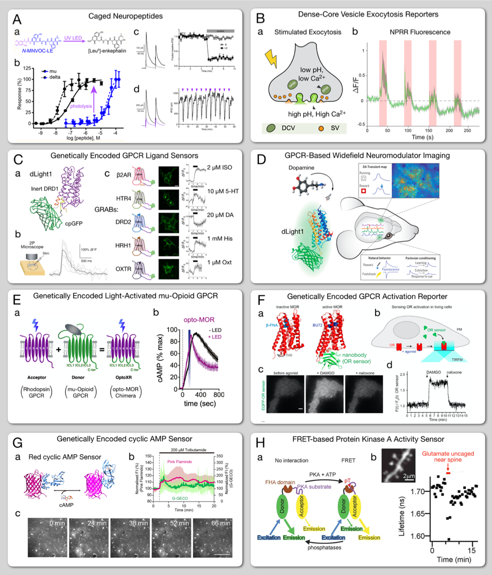
Figure 3 illustrates a small sampling of the bounty of newly available tools for the anatomically precise and neuron-type-specific real-time control and measurement of cortical neuropeptide signaling in single-cells in synaptic network context. These and many other emerging effectors and sensors are well-suited to application in fluorescence imaging experiments – conducted in brain slices to optimize analytic interpretation and/or in vivo to optimize physiological and behavioral context. Such experimentation may be guided by coupling matrix predictions like those of figure 2C and 2D or by many other old or new physiological or behavioral hypotheses.
The eight panels of Figure 3 are ordered by stages of NP signaling from peptide release to receptor binding to downstream intracellular signal transduction events. Figure 3A demonstrates a UV-activatable caged neuropeptide [59]. Figure 3B represents a neuropeptide vesicle exocytosis reporter [60]. Figure 3C exemplifies similar GPCR-based ligand sensors developed independently by two research groups [61, 62]. Given structural similarities across the parent monoamine- and neuropeptide-selective GPCRs, both groups have validated these sensor platforms for growing numbers of NP-GPCRs. Figure 3D illustrates results from an imaging application of one of these sensors to visualize dopamine release in vivo [61]. Figure 3E represents genetically encoded, light-activated GPCRs [63, 64]. Figure 3F illustrates a genetically encoded reporter of GPCR activation [65]. Figure 3G represents design and both cell culture and in vivo imaging applications of a fluorescent-protein-based cAMP sensor [66]. Figure 3H illustrates a protein kinase A activity sensor [67].
All of these genetically encoded tools are suitable for use in combination with neuron-type-specific and/or anatomically specific expression and labeling strategies (e.g., via Cre driver lines and/or viral vectors) to explore peptide responses of specific neuron types and to test type-specific coupling predictions like those illustrated in Fig. 2C and 2D. These and many other genetically encoded tools seem certain to lead in the near future to still sharper delineation of neuropeptide involvement in modulating forms of synaptic, membrane and network plasticity like those sampled in Figs. 1C-F. They also seem likely to bring new light to presently enigmatic but critically important issues of neuropeptide secretion and diffusion dynamics in situ within the intricate confines of complex brain tissues.
Neuropeptides and synaptic learning rules
Studies of long-lasting forms of plasticity at individual synapses, e.g., long-term potentiation (LTP), long-term depression (LTD) or, more generally, spike-timing-dependent plasticity (STDP), form the foundation for many of today’s models for the formation of memory engrams in synaptic networks [68, 69, 40]. There is also now a growing body of evidence to suggest that synaptic learning rules (e.g., STDP curves) are themselves subject to parametric adjustment by neuroactive ligands acting via GPCRs [13, 70]. Such local learning rules, replicated throughout synaptic networks, are now widely hypothesized as “modulated STDP” mechanisms to provide a basis for long-lasting network adaptations and the formation of meaningful memory engrams [71, 72, 73]. Many lines of evidence, in addition to those already touched upon here, suggest that neuropeptides acting via NP-GPCRs may play central roles in the modulation of synaptic learning rules [2, 4].
Grappling the dynamic peptidergic and synaptic network interactions underlying learning and memory is certain to require theoretical and computational approaches. Work at the presently very active and fertile intersection of synaptic learning studies in neuroscience and machine learning in computer science [72, 74, 75, 76, 77] is salient here. Efforts in neuroscience and computer science to model or engineer learning in deep neural networks (be they biological or artificial) share the hard problem of individually adjusting very large numbers of “synaptic weights” (so called in both fields) to adapt network function according to experience. This problem is called “credit assignment” because “credit” (or “blame”!) must be assigned correctly to guide strengthening (or weakening) of particular individual synapses dependent upon their contributions to success (or failure) in a given perceptual, mnemonic or motor task. Neuroscientists struggle with credit assignment as they search for rules of synaptic plasticity adequate to explain biological learning. Computer scientists are driven by a quest for greater computational efficiency in training artificial neural networks and the suspicion that evolution may have found better solutions to the credit assignment problem than those conceived so far by human inventors.
The box entitled “Neuronal networks as labeled multidigraphs” delineates a model that schematizes ways that studies of neuromodulatory and synaptic network interplay might use high-resolution neurotaxonomies like those now emerging from single-cell transcriptomes [48, 49, 51, 55, 57, 78] and now emerging from delineation of synaptic connectomes based on high-throughput electron microscopy [79, 80, 81] and viral tracing methods (e.g., [82]). This multidigraph model, representing a stack of distinct modulatory and synaptic networks pinned together at neuron-type identity nodes, was originally developed in the context of our work on prediction of cortical neuropeptide networks from transcriptomic data [33]. Here we represent a slightly more general treatment, potentially applicable to the wide range of broadcast neuromodulators acting via high-affinity GPCRs, including monoamines (dopamine, serotonin, etc.), spillover small-molecule transmitters (glutamate, GABA, ACh) and endocannabinoids in addition to neuropeptides. This neurotaxonomic model might also be abstracted beyond coverage of strictly local volume-diffusion-based regimes to encompass modulatory signaling extended via axons projecting between distant brain regions, while still preserving the notion of addressing via differential gene expression. We expect that such neurotaxonomic models may be critical for both experimental and theoretical attempts to draw lines from modulated STDP mechanisms to credit assignment in biological memory engram formation. We also imagine that models involving analogues to such broadcast modulation of local synaptic learning rules may attract new attention from computer scientists seeking to engineer more efficient credit assignment schemes.
Box: Neuronal networks as labeled multidigraphs
The language and tools of combinatorial graph theory provide a natural framework to model and analyze synaptic and modulatory signalling networks. A labeled multidigraph is a directed graph where multiple labeled connections between nodes are allowed. These connections can represent different types of directed action between node pairs. The nodes of the graph typically represent the neurons in the network, and the different kinds of actions between the nodes are encoded by the edges between them. We denote by the neuronal network interactions, where is a set of neurons and is a set of connections, where each connection is described by representing an edge of type from node to node . The type of the edge, , models attributes of a connection such as excitatory, inhibitory, or modulatory, and denotes the weight of the connection .
Modeling Dynamics:
We assume that each neuron has an internal state that is available only to the neuron itself within the network. This internal state may be as simple as the scalar membrane potential in the case of leaky integrate-and-fire neuron models, or in more complex models it may hold a list of cellular properties, including levels or states of modulated target proteins.
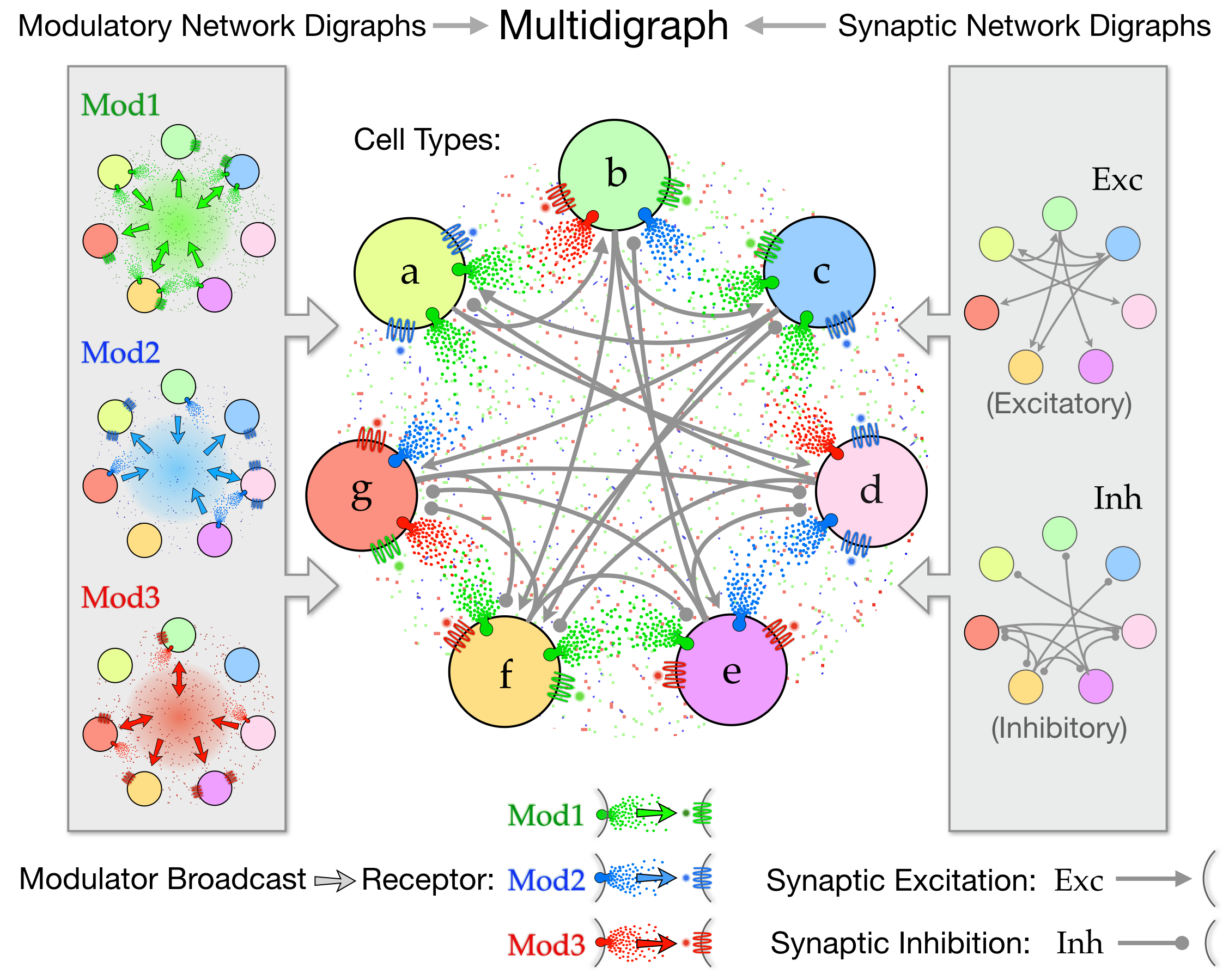 Figure 4: Synaptic and broadcast communication in a neuronal network can be modeled as a multidigraph. and denote the cell type and the output type, respectively.
Each neuron generates outputs (e.g., synaptic and modulatory) based on its state, which are observed by other neurons connected to the source neuron by the corresponding edge type. Specifically, output of neuron at time , , is generated by
(1)
where is the state of neuron at time and is an output-specific emission function. This output is observed by neuron through the connection weight . Let represent synaptic communication so that, when , corresponds to the synaptic weight between neurons and , and corresponds to spike output of neuron . Similarly, let be a set of broadcast signaling pathways so that, when , the slower and smoother nature of broadcast communication is reflected in .
Importantly, we posit here that broadcast modulatory impacts can be modeled neurotaxonomically, that is, as summing across individual cells on a source-cell-type by receiving-cell-type basis, with edge weights set by patterns of source- and receptor-protein gene expression [33]. Such neuron-type-level specificity may be ideally suited to theoretical exploration of network homeostasis and plasticity. By expanding upon the approach of Bellec et al. [83] and switching to a continuous-time notation, we summarize neuronal dynamics as
(2)
where is the state and we have suppressed the time dependence of the variables and communication delays for simplicity of notation, denotes the transformation governing neuronal dynamics, and denote cell types, neuron is of type , and denotes external input to neuron . In its simplest form, the diffusive nature of broadcast signaling suggests that the weights between cell types can be considered as averages over cells of the same type: , adding extra structure to a simpler weighted synaptic model.
Temporal credit assignment: Many real world tasks require action on the time scale of synaptic communication, necessitating efficient learning of and , and suggesting an auxiliary role for modulatory signaling (and , ). Inspired by results implicating dopamine modulation of spike-timing-dependent plasticity (STDP) in credit assignment [72, 84, 85], we hypothesize that other forms and patterns of broadcast modulatory signaling may also promote efficient training of spiking networks. While an overarching temporal credit assignment theory for multidigraphs does not yet exist, approaches enabling credit assignment at multiple time scales [83, 85, 86, 87] can provide natural solutions for incorporating the rich and flexible repertoire of broadcast modulation. Finally, devising approximate, surrogate gradients [77, 83, 86, 88] for the non-differentiable spiking signal enables efficient in-silico experiments and brings the fields of computational neuroscience and machine learning closer.
Figure 4: Synaptic and broadcast communication in a neuronal network can be modeled as a multidigraph. and denote the cell type and the output type, respectively.
Each neuron generates outputs (e.g., synaptic and modulatory) based on its state, which are observed by other neurons connected to the source neuron by the corresponding edge type. Specifically, output of neuron at time , , is generated by
(1)
where is the state of neuron at time and is an output-specific emission function. This output is observed by neuron through the connection weight . Let represent synaptic communication so that, when , corresponds to the synaptic weight between neurons and , and corresponds to spike output of neuron . Similarly, let be a set of broadcast signaling pathways so that, when , the slower and smoother nature of broadcast communication is reflected in .
Importantly, we posit here that broadcast modulatory impacts can be modeled neurotaxonomically, that is, as summing across individual cells on a source-cell-type by receiving-cell-type basis, with edge weights set by patterns of source- and receptor-protein gene expression [33]. Such neuron-type-level specificity may be ideally suited to theoretical exploration of network homeostasis and plasticity. By expanding upon the approach of Bellec et al. [83] and switching to a continuous-time notation, we summarize neuronal dynamics as
(2)
where is the state and we have suppressed the time dependence of the variables and communication delays for simplicity of notation, denotes the transformation governing neuronal dynamics, and denote cell types, neuron is of type , and denotes external input to neuron . In its simplest form, the diffusive nature of broadcast signaling suggests that the weights between cell types can be considered as averages over cells of the same type: , adding extra structure to a simpler weighted synaptic model.
Temporal credit assignment: Many real world tasks require action on the time scale of synaptic communication, necessitating efficient learning of and , and suggesting an auxiliary role for modulatory signaling (and , ). Inspired by results implicating dopamine modulation of spike-timing-dependent plasticity (STDP) in credit assignment [72, 84, 85], we hypothesize that other forms and patterns of broadcast modulatory signaling may also promote efficient training of spiking networks. While an overarching temporal credit assignment theory for multidigraphs does not yet exist, approaches enabling credit assignment at multiple time scales [83, 85, 86, 87] can provide natural solutions for incorporating the rich and flexible repertoire of broadcast modulation. Finally, devising approximate, surrogate gradients [77, 83, 86, 88] for the non-differentiable spiking signal enables efficient in-silico experiments and brings the fields of computational neuroscience and machine learning closer.
Conclusions
Single-cell RNA-seq transcriptomes are now offering new and surprising perspectives on cortical neuropeptide signaling. While the likelihood of intracortical neuropeptide signaling was recognized some time ago (e.g., [89]) and it has long been known that dozens of NP signaling genes are expressed in cortex, it now appears that all cortical neurons express at least one NPP and at least one NP-GPCR gene. Transcriptomic and anatomical data now strongly suggest that local broadcast of NP products interconnects every cortical neuron by a tall stack of overlapping neuron-type-specific peptidergic modulator networks. Patterns of NP gene expression are moreover highly coherent with the deep neurotaxonomies now materializing from genome-wide single-cell transcriptomes. This coherence joins with new opportunities for experimental access offered by transcriptomic neurotaxonomy to make transcriptomic predictions of cortical NP networks highly amenable to experimental test and theoretical exploration. The coherence of transcriptomic neuron type with NP signaling gene expression also suggests that connections between the differentiation of neuron types and differentiation of neuropeptide networks are ancient and fundamental to brain function.
Many longstanding open questions about roles of neuropeptide signaling in the functional homeostasis and plasticity of cortical synaptic networks are re-framed by these new transcriptomic perspectives. In this writing, we have attempted to highlight new results that frame old questions into new hypotheses and to touch upon fundamental advances and new tools that are now making such hypotheses specific and testable. We look forward very eagerly to watching this progress promote better understanding of the homeostasis, adaptation and memory-related plasticity of cortical synaptic networks, and to seeing what new principles of cortical computation such understanding brings from present dark corners into the light!
Acknowledgements
The authors are grateful to Drs. Scott Owen, Rohan Gala, Forrest Collman and Christof Koch, and to Leila Elabbady for many helpful discussions and comments.
Funding
We wish to thank the Allen Institute founder, Paul G. Allen, for his vision, encouragement and support.
References
- [1] Stephen D Meriney and Erika Fanselow. Synaptic Transmission, chapter Neuropeptide Transmitters. Academic Press, 2019.
- [2] CR Gøtzsche and DPD Woldbye. The role of npy in learning and memory. Neuropeptides, 55:79–89, 2016.
- [3] Ariel Y Deutch, Andrea Giuffrida, and James L Roberts. Nonclassic signaling in the brain. In From Molecules to Networks, pages 239–255. Elsevier, 2014.
- [4] Éva Borbély, Bálint Scheich, and Zsuzsanna Helyes. Neuropeptides in learning and memory. Neuropeptides, 47(6):439–450, 2013.
- [5] Anthony N Van Den Pol. Neuropeptide transmission in brain circuits. Neuron, 76(1):98–115, 2012.
- [6] J Peter H Burbach. What are neuropeptides? In Neuropeptides, pages 1–36. Springer, 2011.
- [7] EJ Simon. In search of the opiate receptor. The American journal of the medical sciences, 266(3):160–168, 1973.
- [8] Maurice R Elphick, Olivier Mirabeau, and Dan Larhammar. Evolution of neuropeptide signalling systems. Journal of Experimental Biology, 221(3), 2018.
- [9] Gáspár Jékely. Global view of the evolution and diversity of metazoan neuropeptide signaling. Proceedings of the National Academy of Sciences, 110(21):8702–8707, 2013.
- [10] Frederique Varoqueaux and Dirk Fasshauer. Getting nervous: an evolutionary overhaul for communication. Annual review of genetics, 51:455–476, 2017.
-
[11]
Carolyn L Smith, Thomas S Reese, Tzipe Govezensky, and Rafael A Barrio.
Coherent directed movement toward food modeled in trichoplax, a
ciliated animal lacking a nervous system.
Proceedings of the National Academy of Sciences,
116(18):8901–8908, 2019.
* Evidence for the coordination of behavior by petidergic signaling in a animal that may be the closest modern counterpart to the ancient common ancestor of all of today’s animals that possess neurons and synapses. - [12] Monika Liguz-Lecznar, Joanna Urban-Ciecko, and Malgorzata Kossut. Somatostatin and somatostatin-containing neurons in shaping neuronal activity and plasticity. Frontiers in neural circuits, 10:48, 2016.
- [13] Farzan Nadim and Dirk Bucher. Neuromodulation of neurons and synapses. Current opinion in neurobiology, 29:48–56, 2014.
- [14] David A McCormick and Michael P Nusbaum. Editorial overview: neuromodulation: tuning the properties of neurons, networks and behavior. Current opinion in neurobiology, 29:iv, 2014.
- [15] Cornelia I Bargmann. Beyond the connectome: how neuromodulators shape neural circuits. Bioessays, 34(6):458–465, 2012.
- [16] Elizabeth C Cropper, Jian Jing, Ferdinand S Vilim, and Klaudiusz R Weiss. Peptide cotransmitters as dynamic, intrinsic modulators of network activity. Frontiers in neural circuits, 12:78, 2018.
- [17] Adam J Granger, Michael L Wallace, and Bernardo L Sabatini. Multi-transmitter neurons in the mammalian central nervous system. Current opinion in neurobiology, 45:85–91, 2017.
- [18] Michael P Nusbaum, Dawn M Blitz, and Eve Marder. Functional consequences of neuropeptide and small-molecule co-transmission. Nature Reviews Neuroscience, 18(7):389, 2017.
- [19] Erik Svensson, John Apergis-Schoute, Geoffrey Burnstock, Michael P Nusbaum, David Parker, and Helgi B Schiöth. General principles of neuronal co-transmission: Insights from multiple model systems. Frontiers in neural circuits, 12:117, 2019.
- [20] Alán Alpár, Marco Benevento, Roman A Romanov, Tomas Hökfelt, and Tibor Harkany. Hypothalamic cell diversity: non-neuronal codes for long-distance volume transmission by neuropeptides. Current opinion in neurobiology, 56:16–23, 2019.
- [21] Dick R Nässel. Neuropeptide signaling near and far: how localized and timed is the action of neuropeptides in brain circuits? Invertebrate neuroscience, 9(2):57, 2009.
- [22] Bice Chini, Matthijs Verhage, and Valery Grinevich. The action radius of oxytocin release in the mammalian cns: from single vesicles to behavior. Trends in pharmacological sciences, 38(11):982–991, 2017.
- [23] Gareth Leng and Mike Ludwig. Neurotransmitters and peptides: whispered secrets and public announcements. The Journal of physiology, 586(23):5625–5632, 2008.
-
[24]
William I Weis and Brian K Kobilka.
The molecular basis of g protein–coupled receptor activation.
Annual review of biochemistry, 87:897–919, 2018.
* A landmark update on the atomic structural and molecular dynamic bases of signal transduction by GPCRs. The work reviewed is enabling development of genetically encoded tools that are likely to be critical to future explorations of cortical neuropeptide signaling. - [25] Viktoriya Syrovatkina, Kamela O Alegre, Raja Dey, and Xin-Yun Huang. Regulation, signaling, and physiological functions of g-proteins. Journal of molecular biology, 428(19):3850–3868, 2016.
- [26] Xinmin Zhang and Stuart Firestein. The olfactory receptor gene superfamily of the mouse. Nature neuroscience, 5(2):124–133, 2002.
- [27] Linda Buck and Richard Axel. A novel multigene family may encode odorant receptors: a molecular basis for odor recognition. Cell, 65(1):175–187, 1991.
- [28] Demetrios K Vassilatis, John G Hohmann, Hongkui Zeng, Fusheng Li, Jane E Ranchalis, Marty T Mortrud, Analisa Brown, Stephanie S Rodriguez, John R Weller, Abbie C Wright, et al. The g protein-coupled receptor repertoires of human and mouse. Proceedings of the National Academy of Sciences, 100(8):4903–4908, 2003.
- [29] Vsevolod V Gurevich and Eugenia V Gurevich. Gpcr signaling regulation: the role of grks and arrestins. Frontiers in pharmacology, 10:125, 2019.
- [30] Bertil Hille, Eamonn J Dickson, Martin Kruse, Oscar Vivas, and Byung-Chang Suh. Phosphoinositides regulate ion channels. Biochimica Et Biophysica Acta (BBA)-Molecular and Cell Biology of Lipids, 1851(6):844–856, 2015.
- [31] Dasiel Oscar Borroto-Escuela and Kjell Fuxe. Oligomeric receptor complexes and their allosteric receptor-receptor interactions in the plasma membrane represent a new biological principle for integration of signals in the cns. Frontiers in molecular neuroscience, 12:230, 2019.
- [32] Daniel Wacker, Raymond C Stevens, and Bryan L Roth. How ligands illuminate gpcr molecular pharmacology. Cell, 170(3):414–427, 2017.
-
[33]
Stephen J Smith, Uygar Sümbül, Lucas T Graybuck, Forrest Collman,
Sharmishtaa Seshamani, Rohan Gala, Olga Gliko, Leila Elabbady, Jeremy A
Miller, Trygve E Bakken, et al.
Single-cell transcriptomic evidence for dense intracortical
neuropeptide networks.
eLife, 8, 2019.
** The original presentation of our results regarding the expression of NPP and NP-GPCR genes by mouse cortical neurons. This publication also delineates adjacency matrix predictions for the entire stack of neuropeptide networks predicted by the 37 cognate NPP/NP-GPCR pairs tabulated here in Fig. 2B. - [34] Jane F Armstrong, Elena Faccenda, Simon D Harding, Adam J Pawson, Christopher Southan, Joanna L Sharman, Brice Campo, David R Cavanagh, Stephen PH Alexander, Anthony P Davenport, et al. The iuphar/bps guide to pharmacology in 2020: extending immunopharmacology content and introducing the iuphar/mmv guide to malaria pharmacology. Nucleic acids research, 48(D1):D1006–D1021, 2020.
- [35] Gáspár Jékely, Sarah Melzer, Isabel Beets, Ilona C Grunwald Kadow, Joris Koene, Sara Haddad, and Lindy Holden-Dye. The long and the short of it–a perspective on peptidergic regulation of circuits and behaviour. Journal of Experimental Biology, 221(3):jeb166710, 2018.
- [36] Antoine Koehl, Hongli Hu, Shoji Maeda, Yan Zhang, Qianhui Qu, Joseph M Paggi, Naomi R Latorraca, Daniel Hilger, Roger Dawson, Hugues Matile, et al. Structure of the -opioid receptor–g i protein complex. Nature, 558(7711):547–552, 2018.
- [37] Nigel C Dao, Dakota F Brockway, and Nicole A Crowley. In vitro optogenetic characterization of neuropeptide release from prefrontal cortical somatostatin neurons. Neuroscience, 419:1–4, 2019.
- [38] Tanya L Daigle, Linda Madisen, Travis A Hage, Matthew T Valley, Ulf Knoblich, Rylan S Larsen, Marc M Takeno, Lawrence Huang, Hong Gu, Rachael Larsen, et al. A suite of transgenic driver and reporter mouse lines with enhanced brain-cell-type targeting and functionality. Cell, 174(2):465–480, 2018.
- [39] Leila Haery, Benjamin E Deverman, Katherine S Matho, Ali Cetin, Kenton Woodard, Connie Cepko, Karen I Guerin, Meghan A Rego, Ina Ersing, Susanna Bachle, et al. Adeno-associated virus technologies and methods for targeted neuronal manipulation. Frontiers in Neuroanatomy, 13:93, 2019.
- [40] Gina G Turrigiano. The dialectic of hebb and homeostasis. Philosophical Transactions of the Royal Society B: Biological Sciences, 372(1715):20160258, 2017.
- [41] Trevor J Hamilton, Sara Xapelli, Sheldon D Michaelson, Matthew E Larkum, and William F Colmers. Modulation of distal calcium electrogenesis by neuropeptide y1 receptors inhibits neocortical long-term depression. Journal of Neuroscience, 33(27):11184–11193, 2013.
- [42] Hai Tian, Yueming Xu, Fucun Liu, Guowei Wang, and Sanjue Hu. Effect of acute fentanyl treatment on synaptic plasticity in the hippocampal ca1 region in rats. Frontiers in pharmacology, 6:251, 2015.
- [43] Natasha N Tirko, Katherine W Eyring, Ioana Carcea, Mariela Mitre, Moses V Chao, Robert C Froemke, and Richard W Tsien. Oxytocin transforms firing mode of ca2 hippocampal neurons. Neuron, 100(3):593–608, 2018.
- [44] Scott F Owen, Sebnem N Tuncdemir, Patrick L Bader, Natasha N Tirko, Gord Fishell, and Richard W Tsien. Oxytocin enhances hippocampal spike transmission by modulating fast-spiking interneurons. Nature, 500(7463):458–462, 2013.
- [45] Haaris Ahsan Safdari, Shubhi Pandey, Arun K Shukla, and Somnath Dutta. Illuminating gpcr signaling by cryo-em. Trends in cell biology, 28(8):591–594, 2018.
- [46] Serdar Durdagi, Berna Dogan, Ismail Erol, Gülru Kayık, and Busecan Aksoydan. Current status of multiscale simulations on gpcrs. Current opinion in structural biology, 55:93–103, 2019.
- [47] Alexander Shakeel Bates, Jasper Janssens, Gregory SXE Jefferis, and Stein Aerts. Neuronal cell types in the fly: single-cell anatomy meets single-cell genomics. Current opinion in neurobiology, 56:125–134, 2019.
- [48] Gord Fishell and Adam Kepecs. Interneuron types as attractors and controllers. Annual review of neuroscience, 43, 2019.
- [49] Graziana Gatto, Kelly Megan Smith, Sarah Elizabeth Ross, and Martyn Goulding. Neuronal diversity in the somatosensory system: bridging the gap between cell type and function. Current opinion in neurobiology, 56:167–174, 2019.
- [50] Maria Antonietta Tosches and Gilles Laurent. Evolution of neuronal identity in the cerebral cortex. Current opinion in neurobiology, 56:199–208, 2019.
- [51] Hongkui Zeng and Joshua R Sanes. Neuronal cell-type classification: challenges, opportunities and the path forward. Nature Reviews Neuroscience, 18(9):530, 2017.
- [52] Christopher L Rees, Charise M White, and Giorgio A Ascoli. Neurochemical markers in the mammalian brain: structure, roles in synaptic communication, and pharmacological relevance. Current medicinal chemistry, 24(28):3077–3103, 2017.
- [53] Ed S Lein, Michael J Hawrylycz, Nancy Ao, Mikael Ayres, Amy Bensinger, Amy Bernard, Andrew F Boe, Mark S Boguski, Kevin S Brockway, Emi J Byrnes, et al. Genome-wide atlas of gene expression in the adult mouse brain. Nature, 445(7124):168–176, 2007.
- [54] Ken Sugino, Chris M Hempel, Mark N Miller, Alexis M Hattox, Peter Shapiro, Caizi Wu, Z Josh Huang, and Sacha B Nelson. Molecular taxonomy of major neuronal classes in the adult mouse forebrain. Nature neuroscience, 9(1):99–107, 2006.
-
[55]
Bosiljka Tasic, Zizhen Yao, Lucas T Graybuck, Kimberly A Smith, Thuc Nghi
Nguyen, Darren Bertagnolli, Jeff Goldy, Emma Garren, Michael N Economo,
Sarada Viswanathan, et al.
Shared and distinct transcriptomic cell types across neocortical
areas.
Nature, 563(7729):72–78, 2018.
* This article reports in detail the large-scale single-cell RNA-seq data collection and deep transcriptomic neurotaxonomy development effort upon which many of the conclusions in the present commentary rest. - [56] Anirban Paul, Megan Crow, Ricardo Raudales, Miao He, Jesse Gillis, and Z Josh Huang. Transcriptional architecture of synaptic communication delineates gabaergic neuron identity. Cell, 171(3):522–539, 2017.
- [57] Z Josh Huang and Anirban Paul. The diversity of gabaergic neurons and neural communication elements. Nature Reviews Neuroscience, 20(9):563–572, 2019.
- [58] Eran A Mukamel and John Ngai. Perspectives on defining cell types in the brain. Current opinion in neurobiology, 56:61–68, 2019.
- [59] Matthew R Banghart, Xinyi J He, and Bernardo L Sabatini. A caged enkephalin optimized for simultaneously probing mu and delta opioid receptors. ACS chemical neuroscience, 9(4):684–690, 2017.
- [60] Keke Ding, Yifu Han, Taylor W Seid, Christopher Buser, Tomomi Karigo, Shishuo Zhang, Dion K Dickman, and David J Anderson. Imaging neuropeptide release at synapses with a genetically engineered reporter. Elife, 8, 2019.
- [61] Tommaso Patriarchi, Jounhong Ryan Cho, Katharina Merten, Mark W Howe, Aaron Marley, Wei-Hong Xiong, Robert W Folk, Gerard Joey Broussard, Ruqiang Liang, Min Jee Jang, et al. Ultrafast neuronal imaging of dopamine dynamics with designed genetically encoded sensors. Science, 360(6396):eaat4422, 2018.
- [62] Zhaofa Wu, Jiesi Feng, Miao Jing, and Yulong Li. G protein-assisted optimization of gpcr-activation based (grab) sensors. In Neural Imaging and Sensing 2019, volume 10865, page 108650N. International Society for Optics and Photonics, 2019.
- [63] Alexandra-Madelaine Tichy, Elliot J Gerrard, Patrick M Sexton, and Harald Janovjak. Light-activated chimeric gpcrs: limitations and opportunities. Current opinion in structural biology, 57:196–203, 2019.
- [64] Edward R Siuda, Bryan A Copits, Martin J Schmidt, Madison A Baird, Ream Al-Hasani, William J Planer, Samuel C Funderburk, Jordan G McCall, Robert W Gereau IV, and Michael R Bruchas. Spatiotemporal control of opioid signaling and behavior. Neuron, 86(4):923–935, 2015.
- [65] Miriam Stoeber, Damien Jullié, Braden T Lobingier, Toon Laeremans, Jan Steyaert, Peter W Schiller, Aashish Manglik, and Mark von Zastrow. A genetically encoded biosensor reveals location bias of opioid drug action. Neuron, 98(5):963–976, 2018.
- [66] Kazuki Harada, Motoki Ito, Xiaowen Wang, Mika Tanaka, Devina Wongso, Ayumu Konno, Hirokazu Hirai, Hajime Hirase, Takashi Tsuboi, and Tetsuya Kitaguchi. Red fluorescent protein-based camp indicator applicable to optogenetics and in vivo imaging. Scientific reports, 7(1):1–9, 2017.
- [67] Yao Chen, Jessica Lizette Saulnier, Gary Yellen, and Bernardo Sabatini. A pka activity sensor for quantitative analysis of endogenous gpcr signaling via 2-photon fret-flim imaging. Frontiers in pharmacology, 5:56, 2014.
-
[68]
Aparna Suvrathan.
Beyond stdp—towards diverse and functionally relevant plasticity
rules.
Current opinion in neurobiology, 54:12–19, 2019.
** A concise guide to recent progress in understanding how slow patterns of neuronal activity and dendritic plateau spiking govern synaptic plasticity and contribute to temporal credit assignent. - [69] Wilten Nicola and Claudia Clopath. A diversity of interneurons and hebbian plasticity facilitate rapid compressible learning in the hippocampus. Nature Neuroscience, 22(7):1168–1181, 2019.
- [70] D James Surmeier, Weixing Shen, Michelle Day, Tracy Gertler, Savio Chan, Xianyong Tian, and Joshua L Plotkin. The role of dopamine in modulating the structure and function of striatal circuits. In Progress in brain research, volume 183, pages 148–167. Elsevier, 2010.
- [71] Michael A Farries and Adrienne L Fairhall. Reinforcement learning with modulated spike timing–dependent synaptic plasticity. Journal of neurophysiology, 98(6):3648–3665, 2007.
-
[72]
Pieter R Roelfsema and Anthony Holtmaat.
Control of synaptic plasticity in deep cortical networks.
Nature Reviews Neuroscience, 19(3):166, 2018.
** A recent update (see also reference [75]) and excellent introduction to the credit assignment “problem” and other matters in play at the intersection of contemporary neuroscience and computer science approaches to learning in deep networks. - [73] Rich Pang and Adrienne L Fairhall. Fast and flexible sequence induction in spiking neural networks via rapid excitability changes. eLife, 8:e44324, 2019.
- [74] Timothy P Lillicrap and Adam Santoro. Backpropagation through time and the brain. Current opinion in neurobiology, 55:82–89, 2019.
-
[75]
Blake A Richards, Timothy P Lillicrap, Philippe Beaudoin, Yoshua Bengio, Rafal
Bogacz, Amelia Christensen, Claudia Clopath, Rui Ponte Costa, Archy
de Berker, Surya Ganguli, et al.
A deep learning framework for neuroscience.
Nature neuroscience, 22(11):1761–1770, 2019.
** A recent update (see also reference [72]) and excellent introduction to the credit assignment “problem” and other matters in play at the intersection of contemporary neuroscience and computer science approaches to learning in deep networks. - [76] Shyam Srinivasan, Ralph J Greenspan, Charles F Stevens, and Dhruv Grover. Deep (er) learning. Journal of Neuroscience, 38(34):7365–7374, 2018.
-
[77]
Dongsung Huh and Terrence J Sejnowski.
Gradient descent for spiking neural networks.
In Advances in Neural Information Processing Systems, pages
1433–1443, 2018.
* Spikes are typically considered as discrete events in time, making them non-differentiable. To obtain update equations governing synaptic plasticity in artificial spiking networks, this paper (see also reference [88]), derives surrogate gradients to enable gradient-based learning in spiking networks by defining the spike, as a function of time, via limit arguments. These two papers introduce approximations to the spike. The derived surrogate gradients enable efficient learning in spiking artificial neural networks. - [78] Amit Zeisel, Hannah Hochgerner, Peter Lönnerberg, Anna Johnsson, Fatima Memic, Job Van Der Zwan, Martin Häring, Emelie Braun, Lars E Borm, Gioele La Manno, et al. Molecular architecture of the mouse nervous system. Cell, 174(4):999–1014, 2018.
- [79] Eric Jonas and Konrad Kording. Automatic discovery of cell types and microcircuitry from neural connectomics. Elife, 4:e04250, 2015.
- [80] Alessandro Motta, Manuel Berning, Kevin M Boergens, Benedikt Staffler, Marcel Beining, Sahil Loomba, Philipp Hennig, Heiko Wissler, and Moritz Helmstaedter. Dense connectomic reconstruction in layer 4 of the somatosensory cortex. Science, 366(6469), 2019.
- [81] H Sebastian Seung and Uygar Sümbül. Neuronal cell types and connectivity: lessons from the retina. Neuron, 83(6):1262–1272, 2014.
- [82] Naresh K Hanchate, Eun Jeong Lee, Andria Ellis, Kunio Kondoh, Donghui Kuang, Ryan Basom, Cole Trapnell, and Linda B Buck. Connect-seq to superimpose molecular on anatomical neural circuit maps. Proceedings of the National Academy of Sciences, 117(8):4375–4384, 2020.
-
[83]
Guillaume Bellec, Franz Scherr, Anand Subramoney, Elias Hajek, Darjan Salaj,
Robert Legenstein, and Wolfgang Maass.
A solution to the learning dilemma for recurrent networks of spiking
neurons.
bioRxiv, page 738385, 2019.
** This paper derives biologically plausible learning rules for artificial spiking neural networks. The authors show that neurons with adaptive internal states can provide a qualitative jump in temporal computing capabilities of neural networks. - [84] Nicolas Frémaux and Wulfram Gerstner. Neuromodulated spike-timing-dependent plasticity, and theory of three-factor learning rules. Frontiers in neural circuits, 9:85, 2016.
-
[85]
Thomas Miconi, Aditya Rawal, Jeff Clune, and Kenneth O Stanley.
Backpropamine: training self-modifying neural networks with
differentiable neuromodulated plasticity.
arXiv preprint arXiv:2002.10585, 2020.
** This machine learning paper demonstrates improvements in a benchmark language modeling task by including neuromodulatory effects (dopamine) in non-spiking artificial neural networks. More generally, the authors argue that the concept of lifetime meta-learning ("learning to learn") can significantly improve the field of machine learning. - [86] Emre O Neftci, Hesham Mostafa, and Friedemann Zenke. Surrogate gradient learning in spiking neural networks: Bringing the power of gradient-based optimization to spiking neural networks. IEEE Signal Processing Magazine, 36(6):51–63, 2019.
- [87] Guillaume Bellec, Darjan Salaj, Anand Subramoney, Robert Legenstein, and Wolfgang Maass. Long short-term memory and learning-to-learn in networks of spiking neurons. In Advances in Neural Information Processing Systems, pages 787–797, 2018.
-
[88]
Friedemann Zenke and Surya Ganguli.
Superspike: Supervised learning in multilayer spiking neural
networks.
Neural computation, 30(6):1514–1541, 2018.
* Spikes are typically considered as discrete events in time, making them non-differentiable. To obtain update equations governing synaptic plasticity in artificial spiking networks, this paper (see also reference [77]), derives surrogate gradients to enable gradient-based learning in spiking networks by defining the spike, as a function of time, via limit arguments. These two papers introduce approximations to the spike. The derived surrogate gradients enable efficient learning in spiking artificial neural networks. - [89] Thierry Gallopin, Hélène Geoffroy, Jean Rossier, and Bertrand Lambolez. Cortical sources of crf, nkb, and cck and their effects on pyramidal cells in the neocortex. Cerebral cortex, 16(10):1440–1452, 2006.