High-intensity pulsed ion beam treatment of amorphous iron-based metal alloy††thanks: Preprint submitted to Journal of Physics: Conference Series (JPCS). Work accepted at the Conference "Low temperature Plasma in the Processes of Functional Coating Preparation", Kazan, Russia, 2019.
Abstract
The results of intense pulsed ion beam (IPIB) treatment of the soft magnetic amorphous alloy of a FINEMET-type are presented. Foil produced from the alloy was irradiated with short (about 100 ns) pulses of carbon ions and protons with energy of up to 300 keV and an energy density of up to 7 J/cm2. X-ray diffraction, Mössbauer spectroscopy and magnetic measurements were used to investigate structural and magnetic properties of irradiated foils. It is shown that the foil remains intact after the treatment, and the crystal structure still amorphous. Spontaneous magnetization vector is found to lie almost along perpendicular to the foil plane after irradiation, whereas for the initial amorphous foil it belongs to the plane. The magnetic properties of the foil undergo changes: the coercive force decreases, the saturation induction increases slightly, and the magnetization curve has shallower slope.
1 Kazan National Research Technological University, 68 Karl Marx Str., 420015 Kazan, Russia
2 Zavoisky Physical-Technical Institute, FRC Kazan Scientific Center of RAS, 10/7 Sibirsky tract, 420029 Kazan, Russia
3 Kazan Federal University, 18 Kremlyovskaya Str., 420008 Kazan, Russia
4 Author to whom any correspondence should be addressed, rusnazipov@kstu.ru
1 Introduction
Intense pulsed ion beams (IPIBs) with energy of 0.1-1 MeV generated by a magnetically insulated ion diode are characterized to a greater extent by the energy impact on the solid, since the ion fluence generated in one pulse with duration of about 100 ns does not exceed 1013 ion/cm2. In this case, the ion current density can reach 250 A/cm2 with the corresponding energy density up to 5-6 J/cm2. Such parameters of the ion beam provide surface melting and evaporation, recrystallization, deformation, and destruction of materials [1, 2].
The effect of IPIBs on amorphous metal alloys (AMAs) has so far been poorly studied and interesting from many points of view: the search for promising radiation-resistant materials, the improvement of functional properties, and fundamental researches on fast energy effects. IPIB acts on the target for tens or hundreds of nanoseconds, and the main energy of the ion beam is almost completely absorbed in a thin surface layer (up to about 1 μm) without causing strong heating of the entire volume.
AMAs are a relatively new class of materials that have a disordered structure and unique properties related with this. They are obtained by a combination of a special chemical composition and melt cooling conditions. For iron-based alloys, metal-metalloid type chemical compositions and ultrafast melt cooling at speeds of 106 K/s are used.
AMAs are metastable under normal conditions. When heated above a certain temperature their properties change dramatically in a short time: initially being solid, elastic and ductile, they become brittle. It was previously shown [3] that AMA can be crystallized using a flash light lamp with the radiation duration of about 0.1-1 ms. The effect of a IPIB with a duration of about 100 ns on an amorphous alloy has not been adequately studied [4, 5, 6, 7]. Previous experiments with IPIBs irradiation with an energy density of 1.5 J/cm2 showed [4, 5] that the alloy did not crystallize, so it was necessary to irradiate AMA with a maximum energy density.
2 Experimental
Iron-based AMA of FINEMET-type named 5BDSR (5NbCuSiB) with atomic composition Fe77Cu1Nb3Si13B6 was used as samples. The amorphous alloy was produced as a ribbon by Ashinsky plant using a single roller melt spinning technique. The samples cut from the ribbon were pieces of foil with size 15×10 mm and thickness 25 micron.
Samples were fixed in a steel holder with a mask with a hole of 10.5 mm in diameter. During ion irradiation the energy density W varied from 1 up to 7 J/cm2 and the number of pulses N varied from 2 to 20. A test irradiation of aluminum foil with a thickness of about 10 μm was also carried out. The parameters of the «TEMP» pulse ion accelerator in which the irradiation was carried out were as follows: the composition of the ion beam — Cn+ (80%) and H+ (20%); maximum ion energy 300 keV; pulse duration of about 100 ns; the pressure in the vacuum chamber is about 10-5 mm Hg.
X-Ray diffraction (XRD) measurements of initial and irradiated samples were carried out using Cu-Kα radiation both in standard Bragg-Brentano geometry and in grazing incidence assymetric Bragg-Brentano geometry.
Mössbauer measurements were performed in transmission geometry with a 57Co source in the rhodium matrix with an activity of 50–40 mCi. Mössbauer measurements of the samples in a magnetic field with strength of 2400 Oe were also made to exclude the influence of the magnetic texture on the analysis of the magnetic local structure.
Magnetic properties were measured on an original setup based on LDC1000 EVM module, which measures the parameters of the oscillatory circuit depending on the external magnetic field, in the inductance of which the ferromagnetic sample under study is the core. The resonance frequency of such a circuit is proportional to the differential magnetic susceptibility of the sample, and a field dependence of the susceptibility is obtainable as a result. The external magnetic field strength slowly changed in the range of ±1000 Oe during measurements.
3 Results and Discussion
After ion irradiation with the highest energy density W=5-6 J/cm2, the samples remain elastic and are able to bend elastically under load up to plastic deformation. They do not become brittle as after conventional thermal annealing. Their shape changes, they bend in the direction of the beam, probably under the influence of internal stresses. Samples are not destroyed even with the number of pulses N=20. The aluminum foil test sample is almost completely destroyed in a single pulse with such an energy density.
Samples after IPIBs irradiation remain X-ray amorphous both in volume and on the surface. This is evidenced by the XRD patterns of the samples with a grazing incidence angle φ=2° presented in figure 1. XRD patterns obtained in standard Bragg-Brentano geometry are not presented here because they are identical to the patterns of the initial samples.
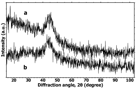
Table 1 shows the parameters of the X-ray amorphous halo approximated by a Gaussian line. For the samples irradiated with a high energy density the X-ray amorphous halo shifts toward smaller angles 2ϑ, from 44.5° for the initial to 44.0° for the irradiated. The full width at half maximum (FWHM) for the irradiated sample becomes slightly greater.
| Gaussian parameters | Initial sample | Irradiated sample |
|---|---|---|
| Center, 2ϑ° | 44.6 | 44 |
| Maximum, a.u. | 141.4 | 117.3 |
| Area, a.u. | 864 | 1061 |
| FWHM, 2ϑ° | 5.7 | 8.5 |
Mössbauer measurements also confirm the amorphous nature of the samples. The shape of the Mössbauer spectra is typical for disordered iron compounds with an almost uniform distribution of nonequivalent positions of Mössbauer nuclei in local atomic environment. There is a magnetically splitted sextet consisting of wide spectral lines in all spectra.
Figure 2 shows the Mössbauer spectra of the initial samples (a), the irradiated samples (b) and the initial and irradiated samples measured in a magnetic field (c). The difference between the irradiated and non-irradiated samples is a change in the magnetic texture, since the intensities of lines 2 and 5 of the wide sextet have changed. The spontaneous magnetization vector becomes closer to the normal to the foil plane in irradiated samples. This does not mean that the sample was so magnetized, since it is magnetically soft. It means that the induced magnetic anisotropy has become perpendicular. The shape of the Mössbauer spectra does not depend on the number of pulses of IPIB’s irradiation.

The mathematical treatment of the Mössbauer spectra was carried out with the restoration of the hyperfine field distribution function P(H). Table 2 shows the found ratio of line intensities and the average hyperfine field for the initial and irradiated samples.
| No external magnetic field | In external magnetic field | |||||
|---|---|---|---|---|---|---|
| , kOe | , kOe | |||||
| Initial sample | 3.94 | 0.71 | 215 | 2.59 | 1.25 | 212 |
| Irradiated sample | 5.68 | 0.29 | 221 | 2.74 | 1.25 | 212 |
It can be seen from this table that the average hyperfine field of the irradiated sample is slightly greater than for the initial one, which could be explained by a decrease in the free volume [8]. However, measurements of the Mössbauer spectra in a magnetic field showed that the effect of increasing the field is the influence of the magnetic texture. The spectra with the same direction of the magnetic moment of the nucleus are almost indistinguishable (figure 2c).
Despite weak changes in the structure of the alloy shown by XRD and Mössbauer studies, the magnetic properties and the eddy current magnetic impedance (figure 3a) vary significantly for the irradiated samples in comparison with the initial ones.
By integrating the field dependences of the differential magnetic susceptibility, we can obtain the dependences of the magnetizations on the magnetic field (hysteresis loops, figure 3b). A comparison of the hysteresis loops of the initial and irradiated samples showed that the magnetic anisotropy in the samples changes, the coercive field decreases, and the saturation magnetization increases as a result of irradiation. In the longitudinal orientation of the sample, when the lines of force of the magnetic field coincide with the plane of the sample and with the direction of the ribbon casting, the initial sample reaches saturation at lower fields (about 200 Oe) than the irradiated one (about 400 Oe).

The field dependence of the parameters of the oscillatory circuit was measured at frequencies of 1-2 MHz. Used technique of the alternating current magnetic susceptibility measurement (ACMS) caused certain increase of the observed coercive force. Therefore, the values obtained in present work should differ from the well-known ones measured in a constant magnetic field. However, this data can be used to compare values of the coercive field of different samples. Since the samples are metallic and have good electrical conductivity, the alternating field of the inductor creates eddy currents in them. These eddy currents in turn affect the complex resistance of the measuring oscillatory circuit. Eddy currents penetrate to the skin depth, which depends on the values of magnetic susceptibility. Thus, the value of the complex resistance of the oscillatory circuit, which contains the sample under study as a core of the inductance, depends on the strength of the external scanning magnetic field. The dependence of the complex resistance of the measuring circuit on the magnetic field represents the eddy current magnetic impedance. Typically, the magnetic impedance of amorphous alloys in the form of ribbons or wires is observed using a galvanic connection. In our case, the magnetic impedance is observed without galvanic contact, but by means of the eddy currents.
In the irradiated samples the relative values of the eddy current impedance decreases more than two times compared with the initial ones (figure 3a).
4 Conclusion
On the basis of the presented results, the amorphous FINEMET-type alloy remains amorphous after irradiation with intense pulsed ion beams (IPIBs). Internal mechanical stresses occur in the alloy after irradiation.
The value of the average hyperfine magnetic field for samples with applied external magnetic field 2400 Oe does not change within the measurement error. Magnetic properties are changed for modified samples. Magnetic anisotropy changes, the coercive field decreases (2–2.5 times), and the saturation magnetization slightly increases.
Acknowledgements
The device for measuring magnetic properties was developed and created
at the support of the subsidy allocated to Kazan Federal University
for the state assignment in the sphere of scientific activities (3.7352.2017/8.9).
This work was supported by the Russian Foundation for Basic Research (Grant No. 19-03-00847).
References
- [1] Remnev G E 2000 Bulletin of Tomsk Polytechnic Institute 303 59
- [2] Kovichak V S, Panova T V and Chernook T N 2017 Proc. 12th Int. Conf. on Interaction of Radiation with Solids 19-22 September 2017 (Minsk: Belarussian State University Press) p 146
- [3] Nazipov R A, Mitin A V, Vizhimov Yu M, Zuzin N A and Pyataev A V 2013 Physics and Chemistry of Materials Treatment 6 25
- [4] Nazipov R A, Zuzin N A, Pyataev A V, Batalov R I Bayazitov R M and Nurutdinov R M 2006 Proc. 10th Int. Youth Scientific School on ’Coherent Optics and Optical Spectroscopy’ (Kazan: Kazan State University Press) p 87
- [5] Nazipov R A, Pyataev A V, Ignatyev A A, Vizhimov Yu M, Batalov R I and Bayazitov R M 2015 Physics and Chemistry of Materials Treatment 6 5
- [6] Zhang Q, Mei X, Guan T, Zhang X, Remnev G E, Pavlov S K and Wang Y 2019 Fusion Engineering and Design 138 16
- [7] Zhong H, Zhang J, Shen J, Liang G, Zhang S, Huang W, Xu M, Yu X, Yan S, Remnev G E and Le X 2019 Nuclear Instruments and Methods in Physics Research B 461 226
- [8] Allia P, Ferro Milone A and Vinay F 1982 J. Appl. Phys. 53 7750