State-dependent regulation of cortical processing speed via gain modulation
Abstract
To thrive in dynamic environments, animals must be capable of rapidly and flexibly adapting behavioral responses to a changing context and internal state. Examples of behavioral flexibility include faster stimulus responses when attentive and slower responses when distracted. Contextual or state-dependent modulations may occur early in the cortical hierarchy and may be implemented via top-down projections from cortico-cortical or neuromodulatory pathways. However, the computational mechanisms mediating the effects of such projections are not known. Here, we introduce a theoretical framework to classify the effects of cell-type specific top-down perturbations on the information processing speed of cortical circuits. Our theory demonstrates that perturbation effects on stimulus processing can be predicted by intrinsic gain modulation, which controls the timescale of the circuit dynamics. Our theory leads to counter-intuitive effects such as improved performance with increased input variance. We tested the model predictions using large-scale electrophysiological recordings from the visual hierarchy in freely running mice, where we found that a decrease in single-cell intrinsic gain during locomotion led to an acceleration of visual processing. Our results establish a novel theory of cell-type specific perturbations, applicable to top-down modulation as well as optogenetic and pharmacological manipulations. Our theory links connectivity, dynamics, and information processing via gain modulation.
1 Introduction
Animals respond to the same stimulus with different reaction times depending on the context or the behavioral state. Faster responses may be elicited by expected stimuli or when the animal is aroused and attentive niemi1981foreperiod . Slower responses may occur in the presence of distractors or when the animal is disengaged from the task grueninger1969effects ; treisman1980feature ; desimone1995neural . Experimental evidence suggests that neural correlates of these contextual modulations occur early in the cortical hierarchy, already at the level of the primary sensory cortex jaramillo2010auditory ; samuelsen2012effects . During the waking state, levels of arousal, attention, and task engagement vary continuously and are associated with ongoing and large changes in the activity of neuromodulatory systems lee2014identification ; pinto2013fast ; fu2014cortical as well as cortico-cortical feedback pathways guo2014flow ; chen2017map ; nelson2013circuit ; leinweber2017sensorimotor ; zhang2014long . Activation of these pathways modulate the patterns of activity generated by cortical circuits and may affect their information-processing capabilities. However, the precise computational mechanism underlying these flexible reorganizations of cortical dynamics remains elusive.
Variations in behavioral and brain state, such as arousal, engagement and body movements may act on a variety of timescales, both slow (minutes, hours) and rapid (seconds or subsecond), and spatial scales, both global (pupil diameter, orofacial movements) and brain subregion-specific; and they can be recapitulated by artificial perturbations such as optogenetic, chemogenetic or electrical stimulation. These variations have been associated with a large variety of seemingly unrelated mechanisms operating both at the single cell and at the population level. At the population level, these mechanisms include modulations of low and high frequency rhythmic cortical activities mcginley2015waking ; changes in noise correlations cohen2009attention ; dadarlat2017locomotion ; and increased information flow between cortical and subcortical networks mcginley2015waking . On a cellular level, these variations have been associated with modulations of single-cell responsiveness and reliability dadarlat2017locomotion ; and cell-type specific gain modulation mcginley2015waking . These rapid, trial-by-trial modulations of neural activity may be mediated by neuromodulatory pathways, such as cholinergic and noradrenergic systems lee2014identification ; pinto2013fast ; fu2014cortical ; reimer2016pupil , or more precise cortico-cortical projections from prefrontal areas towards primary sensory areas guo2014flow ; chen2017map ; nelson2013circuit ; leinweber2017sensorimotor ; zhang2014long . The effects of these cortico-cortical projections can be recapitulated by optogenetic activation of glutamatergic feedback pathways zagha2015competing . In the face of this wide variety of physiological pathways, is there a common computational principle underlying the effects they elicit on sensory cortical circuits?
A natural way to model the effect of activating a specific pathway on a downstream circuit is in the form of a perturbation to the downstream circuit’s afferent inputs or recurrent couplings mazzucato2019expectation ; huang2019circuit . Here, we will present a theory explaining how these perturbations control the information-processing speed of a downstream cortical circuit. Our theory shows that the effects of perturbations that change the statistics of the afferents or the recurrent couplings can all be captured by a single mechanism of action: intrinsic gain modulation, where gain is defined as the rate of change of the intrinsic input/output transfer function of a neuron measured during periods of ongoing activity. Our theory is based on a biologically plausible model of cortical circuits using clustered spiking network Amit1997b . This class of models capture complex physiological properties of cortical dynamics such as state-dependent changes in neural activity, variability LitwinKumarDoiron2012 ; DecoHugues2012 ; mazzucato2015dynamics ; mazzucato2016stimuli ; rostami2020spiking and information-processing speed mazzucato2019expectation . Our theory predicts that gain modulation controls the intrinsic temporal dynamics of the cortical circuit and thus its information processing speed, such that decreasing the intrinsic single-cell gain leads to faster stimulus coding.
We tested our theory by examining the effect of locomotion on visual processing in the visual hierarchy. We found that locomotion decreased the intrinsic gain of visual cortical neurons in the absence of stimuli in freely running mice. The theory thus predicted a faster encoding of visual stimuli during running compared to rest, which we confirmed in the empirical data. Our theoretical framework links gain modulation to information-processing speed, providing guidance for the design and interpretation of future manipulation experiments by unifying the changes in brain state due to behavior, optogenetic, or pharmacological perturbations, under the same shared mechanism.
2 Methods
2.1 Spiking network model
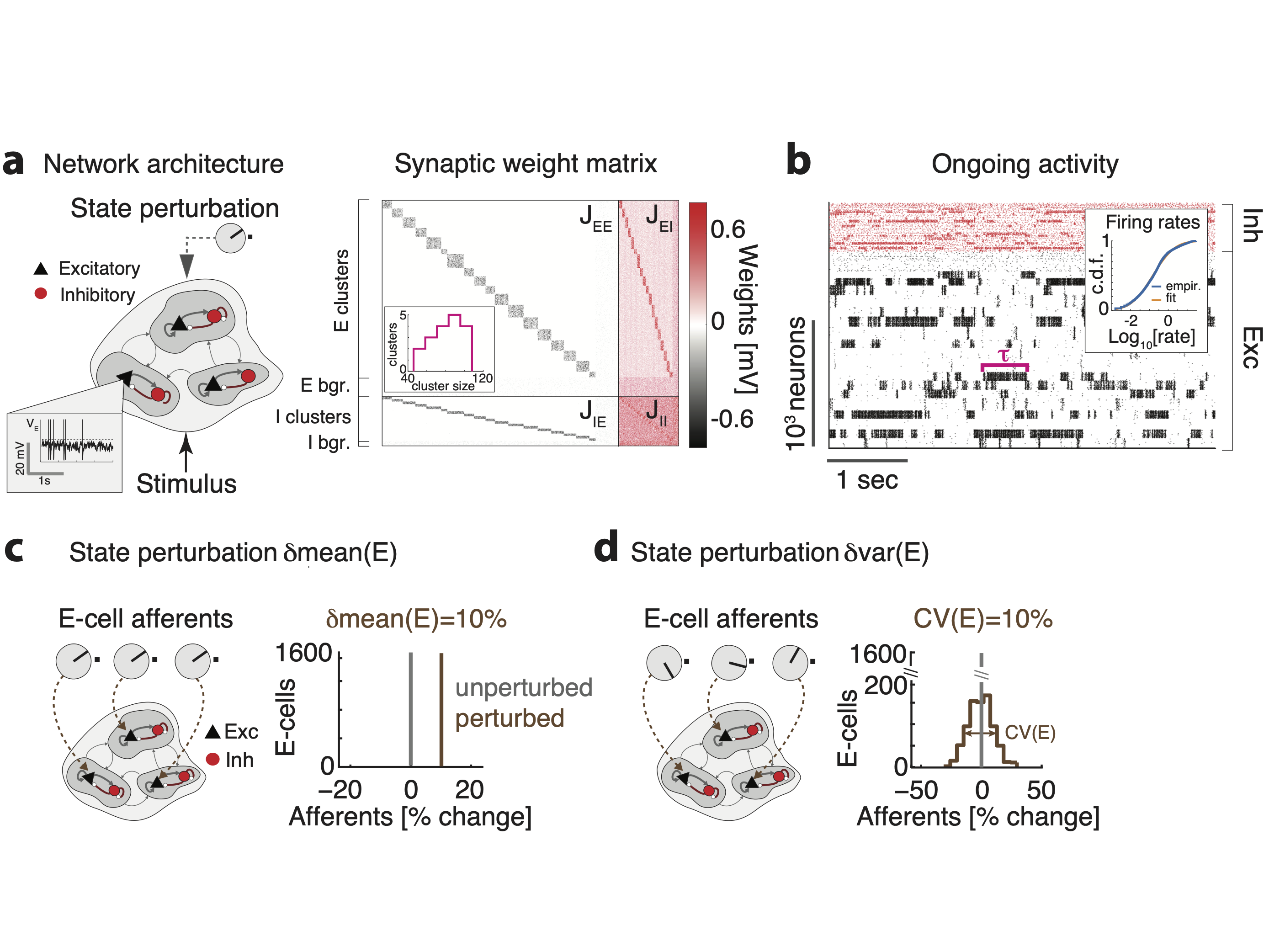
Architecture. We modeled the local cortical circuit as a network of excitatory (E) and inhibitory (I) neurons (with relative fraction and ) with random recurrent connectivity (Fig. 2). Connection probabilities were and . Nonzero synaptic weights from pre-synaptic neuron to post-synaptic neuron were , with sampled from a gaussian distribution with mean , for , and standard deviation . E and I neurons were arranged in clusters. E clusters had heterogeneous sizes drawn from a gaussian distribution with a mean of E-neurons and standard deviation. The number of clusters was then determined as , where is the fraction of background neurons in each population, i.e., not belonging to any cluster. I clusters had equal size . Clusters were defined by an increase in intra-cluster weights and a decrease in inter-cluster weights, under the constraint that the net input current to a neuron would remain unchanged compared to the case without clusters. Synaptic weights for within-cluster neurons where potentiated by a ratio factor . Synaptic weights between neurons belonging to different clusters were depressed by a factor . Specifically, we chose the following scaling: , , , and for , with , where is the fraction of E neurons in each cluster. Within-cluster E-to-E synaptic weights were further multiplied by cluster-specific factor equal to the ratio between the average cluster size and the size of each cluster, so that larger clusters had smaller within-cluster couplings. We chose network parameters so that the cluster timescale was 100 ms, as observed in cortical circuits Jones2007 ; mazzucato2015dynamics ; mazzucato2019expectation . Parameter values are in Table 1.
| Model parameters for clustered network simulations | ||
|---|---|---|
| Parameter | Description | Value |
| mean E-to-E synaptic weights | 0.6 mV | |
| mean E-to-I synaptic weights | 0.6 mV | |
| mean I-to-E synaptic weights | 1.9 mV | |
| mean I-to-I synaptic weights | 3.8 mV | |
| mean E-to-E synaptic weights | 2.6 mV | |
| mean I-to-I synaptic weights | 2.3 mV | |
| standard deviation of the synaptic weight distribution | ||
| Potentiated intra-cluster E-to-E weight factor | 14 | |
| Potentiated intra-cluster I-to-I weight factor | 5 | |
| Potentiation parameter for intra-cluster I-to-E weights | 10 | |
| Potentiation parameter for intra-cluster E-to-I weights | 8 | |
| Average baseline afferent rate to E and I neurons | 5 spks/s | |
| E-neuron threshold potential | 1.43 mV | |
| I-neuron threshold potential | 0.74 mV | |
| E- and I-neuron reset potential | 0 mV | |
| E- and I-neuron membrane time constant | 20 ms | |
| E- and I-neuron absolute refractory period | 5 ms | |
| E- and I-neuron synaptic time constant | 5 ms | |
Neuronal dynamics. We modeled spiking neurons as current-based leaky-integrate-and-fire (LIF) neurons whose membrane potential evolved according to the dynamical equation
where is the membrane time constant. Input currents included a contribution coming from the other recurrently connected neurons in the local circuit and an external current (units of mV s-1). The first term (for ) is a constant term representing input to the E or I neuron from other brain areas and ; while and represent the incoming sensory stimulus or the various types of perturbation (see Stimuli and perturbations below). When hits threshold (for ), a spike is emitted and is then held at the reset value for a refractory period . We chose the thresholds so that the homogeneous network (i.e.,where all ) was in a balanced state with average spiking activity at rates spks/s Amit1997b ; mazzucato2019expectation . Post-synaptic currents evolved according to the following equation
where is the synaptic time constant, are the recurrent couplings and is the time of the k-th spike from the j-th presynaptic neuron. Parameter values are in Table 1.
Sensory stimuli. We considered two classes of inputs: sensory stimuli and perturbations. In the “evoked” condition (Fig. 4a), we presented the network one of four sensory stimuli, modeled as changes in the afferent currents targeting of E-neurons in stimulus-selective clusters; each E-cluster had a probability of being selective to a sensory stimulus (mixed selectivity). In the first part of the paper (Fig. 1-6, I-clusters were not stimulus-selective. Moreover, in both the unperturbed and the perturbed stimulus-evoked conditions, stimulus onset occurred at time and each stimulus was represented by an afferent current , where is a linearly ramping increase reaching a value above baseline at . In the last part of the paper (Fig. 7), we introduced a new stimulation protocol where visual stimuli targeted both E and I clusters in pairs, corresponding to thalamic input onto both excitatory and inhibitory neurons in V1 zhuang2013layer ; miska2018sensory ; kloc2014target ; khan2018distinct .. Each E-I cluster pair had a 50% probability of being selective to each visual stimulus. If a E-I cluster pair was selective to a stimulus, then all E neurons and 50% of I neurons in that pair received the stimulus. The time course of visual stimuli was modeled as a double exponential profiles with rise and decay times of (0.05,0.5)s, and peak equal to a 20% increase compared to the baseline external current.
External perturbations. We considered several kinds of perturbations. In the perturbed stimulus-evoked condition (Fig. 4b, right panel), perturbation onset occurred at time and lasted until the end of the stimulus presentation at with a constant time course. We also presented perturbations in the absence of sensory stimuli (“ongoing” condition, Fig. 2-3); in that condition, the perturbation was constant and lasted for the whole duration of the trial (5s). Finally, when assessing single-cell responses to perturbations, we modeled the perturbation time course as a double exponential with rise and decay times s (Fig. 6). In all conditions, perturbations were defined as follows:
-
•
mean(E), mean(I): A constant offset in the mean afferent currents was added to all neurons in either E or I populations, respectively, expressed as a fraction of the baseline value (see Neuronal dynamics above), where for E neurons and for I neurons.
-
•
var(E), var(I): For each E or I neuron, respectively, the perturbation was a constant offset , where is a gaussian random variable with zero mean and standard deviation . We chose for E neurons and for I neurons. This perturbation did not change the mean afferent current but only its spatial variance across the E or I population, respectively. We measured the strength of these perturbations via their coefficient of variability , for , where and are the standard deviation and mean of the across-neuron distribution of afferent currents.
-
•
AMPA: A constant change in the mean synaptic couplings (for ), representing a modulation of glutamatergic synapses. We chose .
-
•
GABA: A constant change in the mean synaptic couplings (for ), representing a modulation of GABAergic synapses. We chose .
The range of the perturbations were chosen so that the network still produced metastable dynamics for all values.
Inhibition stabilization. We simulated a stimulation protocol used in experiments to test inhibition stabilization (Fig. 2-1b). This protocol is identical to the mean(I) perturbation during ongoing periods, where the perturbation targeted all I neurons with an external current applied for the whole length of 5s intervals, with and 40 trials per network and 10 networks for each value of the perturbation.
Simulations. All data analyses, model simulations, and mean field theory calculations were performed using custom software written in MATLAB, C and Python. Simulations in the stimulus-evoked conditions (both perturbed and unperturbed) comprised 10 realizations of each network (each network with different realization of synaptic weights), with 20 trials for each of the 4 stimuli. Simulations in the ongoing condition comprised 10 different realization of each network, with 40 trials per perturbation. Each network was initialized with random synaptic weights and simulated with random initial conditions in each trial. Sample sizes were similar to those reported in previous publications mazzucato2015dynamics ; mazzucato2016stimuli ; mazzucato2019expectation . Dynamical equations for the leaky-integrate-and-fire neurons were integrated with the Euler method with a 0.1ms step. MATLAB code to simulate the model with var(E) perturbation is located at https://github.com/mazzulab/perturb_spiking_net. Code to reproduce the full set of perturbations investigated in this paper are available upon request to the corresponding author.
| Model parameters for the reduced two-cluster network | ||
|---|---|---|
| Parameter | Description | Value |
| mean E-to-E synaptic weights | 0.8 mV | |
| mean I-to-E synaptic weights | 10.6 mV | |
| mean E-to-I synaptic weights | 2.5 mV | |
| mean I-to-I synaptic weights | 9.7 mV | |
| mean E-to-E synaptic weights | 14.5 mV | |
| mean I-to-I synaptic weights | 12.9 mV | |
| Potentiated intra-cluster E-to-E weight factor | 11.2 | |
| Average baseline afferent rate to E and I neurons | 7 spk/s | |
| E-neuron threshold potential | 4.6 mV | |
| I-neuron threshold potential | 8.7 mV | |
| E- and I-neuron synaptic time constant | 4 ms | |
| Fraction of background E neurons | 65 | |
2.2 Mean field theory
We performed a mean field analysis of a simplified two-cluster network for leaky-integrate-and-fire neurons with exponential synapses, comprising populations for Amit1997b ; mazzucato2019expectation : the first representing the two E clusters, the last two representing the background E and the I population. The infinitesimal mean and variance of the postsynaptic currents are:
| (1) |
| (2) |
where are the firing rates in the E-clusters; are the firing rates in the background E population, in the I population, and in the external current. Other parameters are described in Architecture and in Table 2. The network attractors satisfy the self-consistent fixed point equations:
| (3) |
where and , and is the current-to-rate transfer function for each population, which depend on the condition. In the absence of perturbations, all populations have the LIF transfer function
| (4) |
where and . and are terms accounting for the synaptic dynamics Fourcaud2002 . The perturbations var(E) and var(I) induced an effective population transfer function on the E and I populations, respectively, given by mazzucato2019expectation :
| (5) |
where and is a gaussian measure of zero mean and unit variance, is the external current and is the standard deviation of the perturbation with respect to baseline, denoted CV(E) and CV(I). Stability of the fixed point equation 3 was defined with respect to the approximate linearized dynamics of the instantaneous mean and variance of the input currents mazzucato2015dynamics ; mazzucato2019expectation :
| (6) |
where are defined in 2.2-2.2 and represents the appropriate transfer function 4 or 5. Fixed point stability required that the stability matrix
| (7) |
was negative definite. The full mean field theory described above was used for the comprehensive analysis of Fig. 3-2.For the schematic of Fig. 3c, we replaced the LIF transfer function 4 with the simpler function and the var(E) perturbation effect was then modeled as .
Effective mean field theory for a reduced network. To calculate the potential energy barrier separating the two network attractors in the reduced two-cluster network, we used the effective mean field theory developed in Mascaro1999 ; mattia2013heterogeneous ; mazzucato2019expectation . The idea is to first estimate the force acting on neural configurations with cluster firing rates outside the fixed points (3), then project the two-dimensional system onto a one-dimensional trajectory along which the force can be integrated to give an effective potential (Fig. 3-2). In the first step, we start from the full mean field equations for the populations in 3, and obtain an effective description of the dynamics for populations “in focus” describing E clusters ( in our case) by integrating out the remaining out-of-focus populations describing the background E neurons and the I neurons ( in our case). Given a fixed value for the in-focus populations, one obtains the stable fixed point firing rates of the out-of-focus populations by solving their mean field equations
| (8) |
for , as function of the in-focus populations , where stability is calculated with respect to the condition (7) for the reduced out-of-focus populations at fixed values of the in-focus rates . One then obtains a relation between the input and output values of the in-focus populations by inserting the fixed point rates of the out-of-focus populations calculated in (8):
| (9) |
for . The original fixed points are such that .
Potential energy barriers and transfer function gain. In a reduced network with two in-focus populations corresponding to the two E clusters, one can visualize Eq. (9) as a two-dimensional force vector at each point in the two-dimensional firing rate space . The force vanishes at the stable fixed points A and B and at the unstable fixed point C between them (Fig. 3-2). One can further reduce the system to one dimension by approximating its dynamics along the trajectory between A and B as Mascaro1999 :
| (10) |
where represents an effective transfer function and an effective force. We estimated the gain of the effective transfer function as , where and represent, respectively, the minimum and maximum of the force (see Fig. 3-2). From the one-dimensional dynamics (10) one can define a potential energy via . The energy minima represent the stable fixed points and and the saddle point between them represents the potential energy barrier separating the two attractors. The height of the potential energy barrier is then given by
| (11) |
which can be visualized as the area of the curve between the effective transfer function and the diagonal line (see Fig. 3).
2.3 Experimental data
We tested our model predictions using the open-source dataset of neuropixel recordings from the Allen Institute for Brain Science siegle2019survey . We focused our analysis on experiments where drifting gratings were presented at four directions (0°, 45°, 90°, 135°) and one temporal frequency (2 Hz). Out of the 54 sessions provided, only 7 sessions had enough trials per behavioral condition to perform our decoding analysis. Neural activity from the visual cortical hierarchy was collected and, specifically: primary visual cortex (V1) in 5 of these 7 sessions, with a median value of 75 neurons per session; lateral visual area (LM): 6 sessions, 47 neurons; anterolateral visual area (AL): 5 sessions, 61 neurons; posteromedial visual area (PM): 6 sessions, 55; anteromedial visual area (AM): 7 sessions, 48 neurons. We matched the number and duration of trials across condition and orientation and combined trials from the drifting gratings repeat stimulus set, and drifting grating contrast stimulus set. To do this, we combined trials with low-contrast gratings (0.08, 0.1, 0.13, 0.2; see Fig. 7-4) and trials with high-contrast gratings (0.6, 0.8, 1; see Fig. 7-3) into separate trial types to perform the decoding analysis, and analyzed the interval seconds aligned to stimulus onset.
For evoked activity, running trials were classified as those where the animal was running faster than 3 cm/s for the first 0.5 seconds of stimulus presentation. During ongoing activity, behavioral periods were broken up into windows of 1 second. Periods of running or rest were classified as such if 10 seconds had elapsed without a behavioral change. Blocks of ongoing activity were sorted and used based on the length of the behavior. Out of the 54 sessions provided, 14 sessions had enough time per behavioral condition (minimum of 2 minutes) to estimate single-cell transfer functions. Only neurons with a mean firing rate during ongoing activity greater than 5Hz were included in the gain analysis (2119 out of 4365 total neurons).
2.4 Stimulus decoding
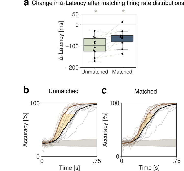
For both the simulations and data, a multi-class decoder was trained to discriminate between four stimuli from single-trial population activity vectors in a given time bin jezzini2013processing . To create a timecourse of decoding accuracy, we used a sliding window of 100ms (200ms) in the data (model), which was moved forward in 2ms (20ms) intervals in the data (model). Trials were split into training and test data-sets in a stratified 5-fold cross-validated manner, ensuring equal proportions of trials per orientation in both data-sets. In the model, a leave-2-out cross-validation was performed. To calculate the significance of the decoding accuracy, an iterative shuffle procedure was performed on each fold of the cross-validation. On each shuffle, the training labels were shuffled and the classifer accuracy was predicted on the unshuffled test data-set. This shuffle was performed 100 times to create a shuffle distribution to rank the actual decoding accuracy from the unshuffled decoder against and to determine when the mean decoding accuracy had increased above chance. This time point is what we referred to as the latency of stimulus decoding. To account for the speed of stimulus decoding (the slope of the decoding curve), we defined the -Latency between running and rest as the average time between the two averaged decoding curves from 40% up to 80% of the max decoding value at rest.
2.5 Firing rate distribution match
To control for increases of firing rate due to locomotion (Fig. 7b), we matched the distributions of population counts across the trials used for decoding in both behavioral conditions. This procedure was done independently for each sliding window of time along the decoding time course. Within each window, the spikes from all neurons were summed to get a population spike count per trial. A log-normal distribution was fit to the population counts across trials for rest and running before the distribution match (Fig 7-1a left). We sorted the distributions for rest and running in descending order, randomly removing spikes from trials in the running distribution to match the corresponding trials in the rest distribution (Fig 7-1a right). By doing this, we only removed the number of spikes necessary to match the running distribution to rest distribution. For example, trials where the rest distribution had a larger population count, no spikes were removed from either distribution. Given we performed this procedure at the population level rather than per neuron, we checked the change in PSTH between running and rest conditions before and after distribution matching (Fig 7-1b). This procedure was also performed on the simulated data (Fig. 7-5).
2.6 Single-cell gain
To infer the single-cell transfer function in simulations and data, we followed the method originally described in recanatesi2020metastable (see also lim2015inferring ; pereira2018attractor for a trial-averaged version). We estimated the transfer function on ongoing periods when no sensory stimulus was present. Briefly, the transfer function of a neuron was calculated by mapping the quantiles of a standard gaussian distribution of input currents to the quantiles of the empirical firing rate distribution during ongoing periods (Fig. 3d). We then fit this transfer function with a sigmoidal function. The max firing rate of the neuron in the sigmoidal fit was bounded to be no larger than 1.5 times that of the empirical max firing rate, to ensure realistic fits. We defined the gain as the slope at the inflection point of the sigmoid.
2.7 Single-cell response and selectivity
We estimated the proportion of neurons that were significantly excited or inhibited by cortical state perturbations in the model (Fig. 6) or locomotion in the data (Fig. 7) during periods of ongoing activity, in the absence of sensory stimuli. In the model, we simulated 40 trials per network, for 10 networks per each value of the perturbation; each trial in the interval s, with onset of the perturbation at (the perturbation was modeled as a double exponential with rise and decay times , Fig. 3a). In the data, we binned the spike counts in 500ms windows for each neuron after matching sample size between rest and running conditions, and significant difference between the conditions was assessed with a rank-sum test.
We estimated single neuron selectivity to sensory stimuli in each condition from the average firing rate responses of the -th neuron to stimulus in trial . For each pair of stimuli, selectivity was estimated as
where mean and var are estimated across trials. The d’ was then averaged across stimulus pairs.
3 Results
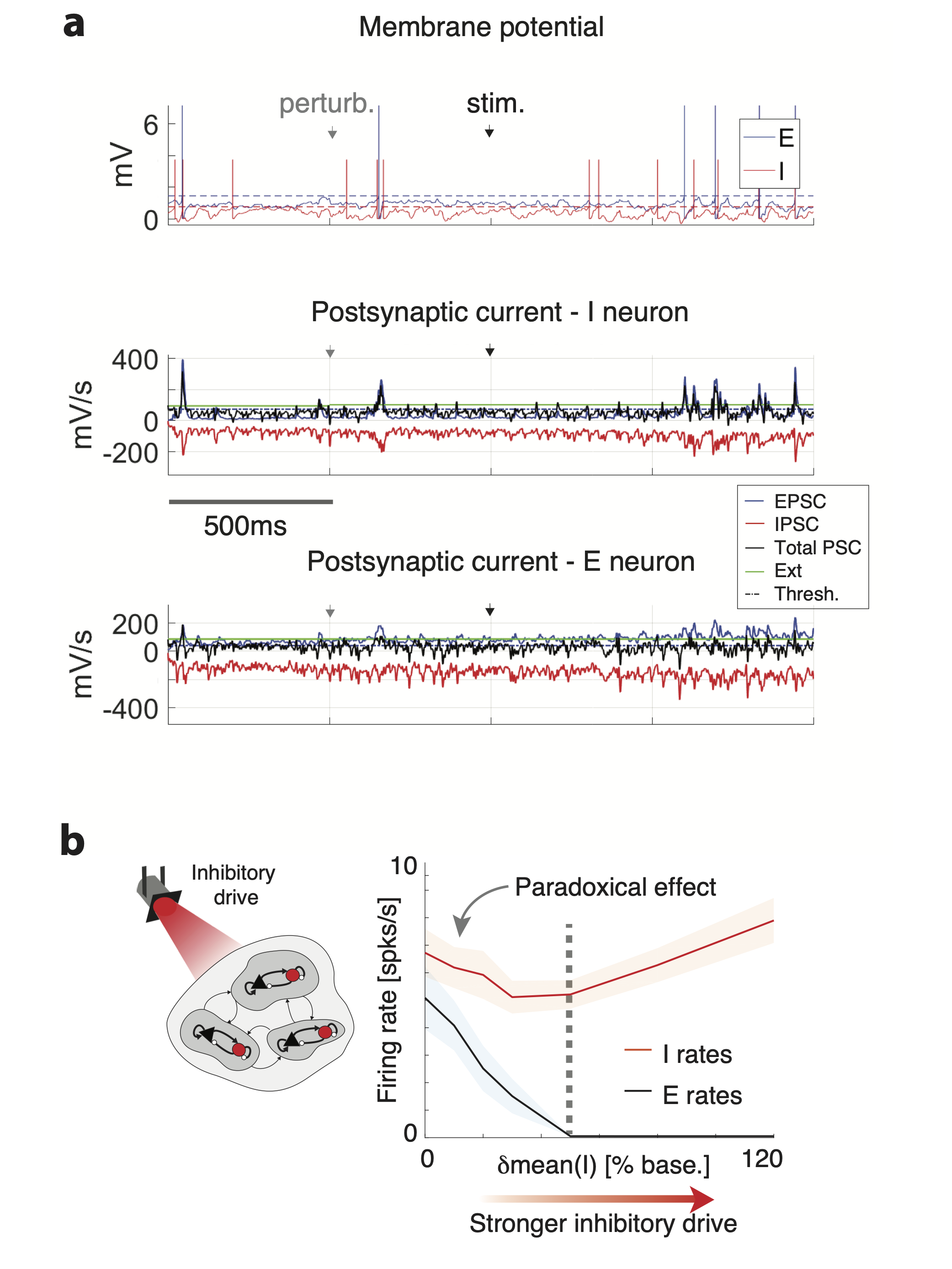
To elucidate the effect of state changes on cortical dynamics, we modeled the local circuit as a network of recurrently connected excitatory (E) and inhibitory (I) spiking neurons. Both E and I populations were arranged in clusters Amit1997b ; LitwinKumarDoiron2012 ; mazzucato2015dynamics ; schaub2015emergence ; mazzucato2019expectation , where synaptic couplings between neurons in the same cluster were potentiated compared to neurons in different clusters, reflecting the empirical observation of cortical assemblies of functionally correlated neurons (Fig. 2a; kiani2015natural, ; song2005highly, ; perin2011synaptic, ; lee2016anatomy, ). In the absence of external stimulation (ongoing activity), this E-I clustered network generates rich temporal dynamics characterized by metastable activity operating in inhibition stabilized regime, where E and I post-synaptic currents track each other achieving tight balance (Fig. 2-1). A heterogeneous distribution of cluster sizes leads to a lognormal distributions of firing rates (Fig. 2b). Network activity was characterized by the emergence of the slow timescale of cluster transient activation, with average activation lifetime of ms (hereby referred to as “cluster timescale," Fig. 2b), much larger than single neuron time constant (20ms; LitwinKumarDoiron2012, ; mazzucato2015dynamics, ).
To investigate how changes in cortical state may affect the network dynamics and information processing capabilities, we examined a vast range of state-changing perturbations (Fig. 2c-d, Table 3). State changes were implemented as perturbations of the afferent currents to cell-type specific populations, or as perturbations to the synaptic couplings. The first type of state perturbations mean(E) affected the mean of the afferent currents to E populations (Fig. 2c). E.g., a perturbation mean(E)=10% implemented an increase of all input currents to E neurons by 10% above their unperturbed levels. The perturbation mean(I) affected the mean of the afferent currents to I populations in an analogous way. The second type of state perturbations var(E) affected the across-neuron variance of afferents to E populations. Namely, in this perturbed state, the afferent current to each neuron in that population was sampled from a normal distribution with zero mean and fixed variance (Fig. 2d, measured by the coefficient of variability CV(E)=var(E)/mean(E) with respect to the unperturbed afferents). This perturbation thus introduced a spatial variance across neurons in the cell-type specific afferent currents, yet left the mean afferent current into the population unchanged. The state perturbation var(I) affected the variance of the afferent currents to I populations analogously. In the third type of state perturbations AMPA or GABA, we changed the average GABAergic or glutamatergic (AMPA) recurrent synaptic weights compared to their unperturbed values. We chose the range of state perturbations such that the network still retained non-trivial metastable dynamics within the whole range. We will refer to these state changes of the network as simply perturbations, and should not be confused with the presentation of the stimulus. We first established the effects of perturbations on ongoing network dynamics, and used those insight to explain their effects on stimulus-evoked activity.
3.1 State-dependent regulation of the network emergent timescale
A crucial feature of neural activity in clustered networks is metastable attractor dynamics, characterized by the emergence of a long timescale of cluster activation whereby network itinerant activity explores the large attractor landscape (Fig. 2b). We first examined whether perturbations modulated the network’s metastable dynamics and introduced a protocol where perturbations occurred in the absence of sensory stimuli (“ongoing activity”).
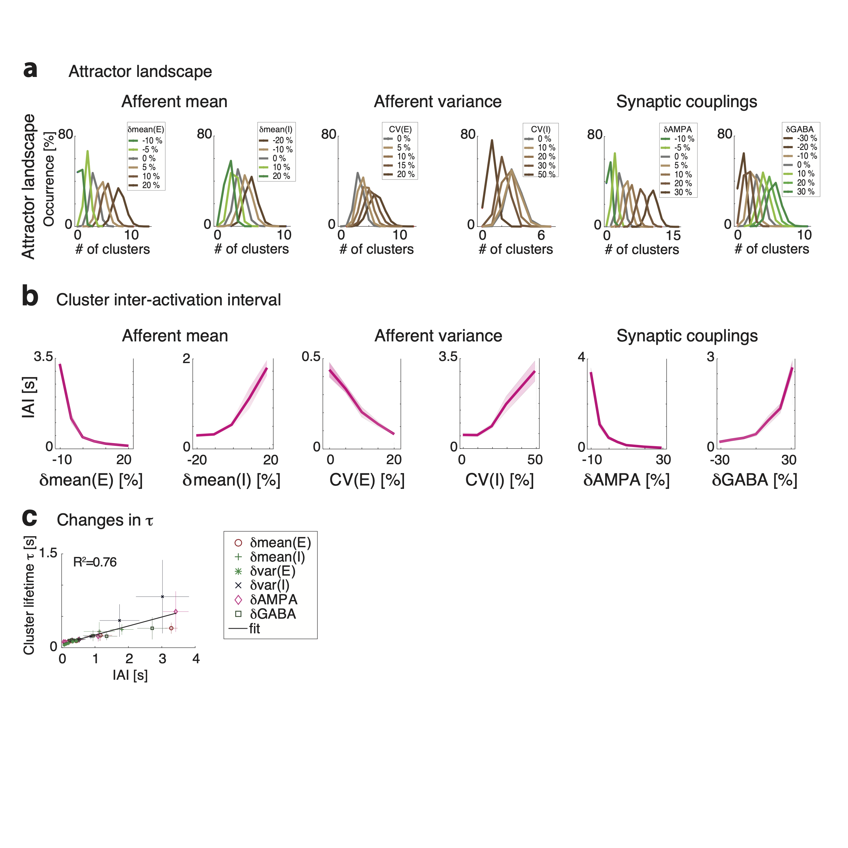
We found that perturbations strongly modulated the attractor landscape, changing the repertoire of attractors the network activity visited during its itinerant dynamics (Fig. 3a and 3-1a). Changes in attractor landscape were perturbation-specific. Perturbations increasing mean(E) (mean(I)) induced a consistent shift in the repertoire of attractors: larger perturbations led to larger (smaller) numbers of co-active clusters. Surprisingly, perturbations that increased var(E) (var(I)), led to network configurations with larger (smaller) sets of co-activated clusters. This effect occurred despite the fact that such perturbations did not change the mean afferent input to the network. Perturbations affecting AMPA and GABA had similar effects to mean(E) and mean(I), respectively.
We then examined whether perturbations affected the cluster activation timescale. We found that perturbations differentially modulated the average cluster activation timescale during ongoing periods, in the absence of stimuli (Fig. 3b). In particular, increasing mean(E), var(E), or AMPA led to a proportional acceleration of the network metastable activity and shorter ; while increasing mean(I), var(I) or GABA induced the opposite effect with longer . Changes in were congruent with changes in the duration of intervals between consecutive activations of the same cluster (cluster inter-activation intervals, Fig. 3-1).
3.2 Changes in cluster timescale are controlled by gain modulation
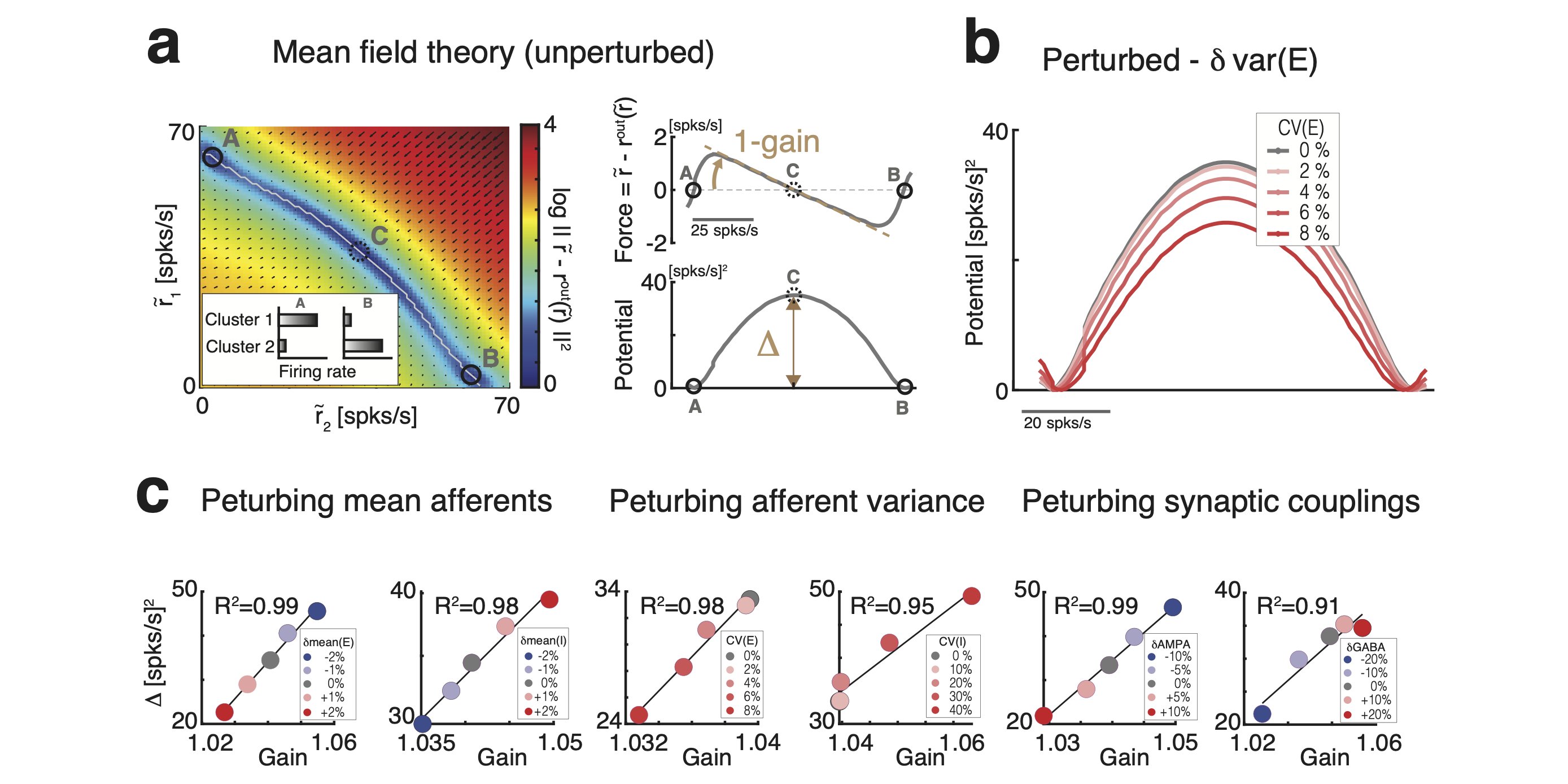
What is the computational mechanism mediating the changes in cluster timescale, induced by the perturbations? We investigated this question using mean field theory, where network attractors, defined by sets of co-activated clusters, are represented as potential wells in an attractor landscape Mascaro1999 ; LitwinKumarDoiron2012 ; mazzucato2015dynamics ; MattiaSanchezVives2012 ; mazzucato2019expectation . Let us illustrate this in a simplified network with two clusters (Fig. 3c and 3-2). Here, the attractor landscape consists of two potential wells, each well corresponding to a configuration where one cluster is active and the other is inactive. When the network activity dwells in the attractor represented by the left potential well, it may escape to the right potential well due to internally generated variability. This process will occur with a probability determined by the height of the barrier separating the two wells: the higher the barrier, the less likely the transition hanggi1990reaction ; LitwinKumarDoiron2012 ; MattiaSanchezVives2012 ; mazzucato2019expectation . Mean
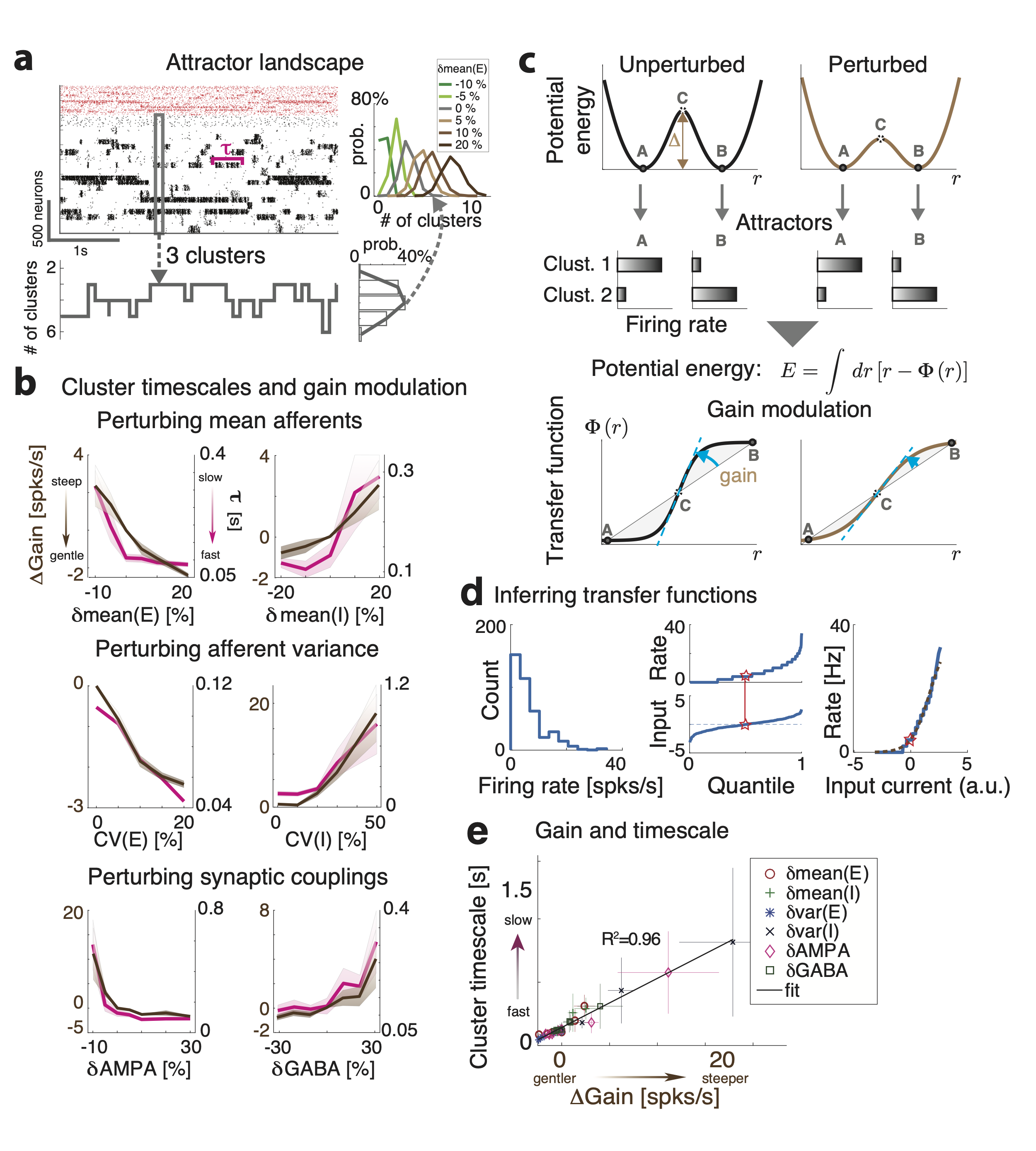
field theory thus established a relationship between the cluster timescale and the height of the barrier separating the two attractors. We found that perturbations differentially control the height of the barrier separating the two attractors (Fig. 3-2), explaining the changes in cluster timescale observed in the simulations (Fig. 3b).
Since reconstruction of the attractor landscape requires knowledge of the network’s structural connectivity, the direct test of the mean field relation between changes in attractor landscape and timescale modulations is challenging. We thus aimed at obtaining an alternative formulation of the underlying neural mechanism only involving quantities directly accessible to experimental observation. Using mean field theory, one can show that the double potential well representing the two attractors can be directly mapped to the effective transfer function of a neural population Mascaro1999 ; mazzucato2019expectation ; mattia2013heterogeneous . One can thus establish a direct relationship between changes in the slope (hereby referred to as “gain") of the intrinsic transfer function estimated during ongoing periods and changes in the barrier height separating metastable attractors (see Fig. 3c, 3-2 and Methods). In turn, this implies a direct relationship between gain modulation, induced by the perturbations, and changes in cluster activation timescale. In particular, perturbations inducing steeper gain will increase well depths and barrier heights, and thus increase the cluster timescale, and vice versa. Using mean field theory, we demonstrated a complete classification of the differential effect of all perturbations on barrier heights and gain (Fig. 3-2).
We then proceeded to verify these theoretical predictions, obtained in a simplified two-cluster network, in the high dimensional case of large networks with several clusters using simulations. While barrier heights and the network’s attractor landscape can be exactly calculated in the simplified two-cluster network, this task is infeasible in large networks with a large number of clusters where the number of attractors is exponential in the number of clusters. On the other hand, changes in barrier heights are equivalent to changes in gain, and the latter can be easily estimated from spiking activity (Fig. 3b and 3-2). We thus tested whether the relation between gain and timescale held in the high-dimensional case of a network with many clusters. We estimated single-cell transfer functions from their spiking activity during ongoing periods, in the absence of sensory stimuli but in the presence of different perturbations (Fig. 3d, lim2015inferring ; recanatesi2020metastable ). We found that network perturbations strongly modulated single-cell gain in the absence of stimuli, verifying mean field theory predictions in all cases (Fig. 3d). In particular, we confirmed the direct relationship between gain modulation and cluster timescale modulation: perturbations that decreased (increased) the gain also decreased (increased) cluster timescale (Fig. 3e, ). For all perturbations, gain modulations explained the observed changes in cluster timescale. ††Legend continued: Mean field theory provides a relation between potential energy and transfer function (bottom panel), thus linking cluster lifetime to neuronal gain in the absence of stimuli (dashed blue line, gain). d): A single-cell transfer function (bottom, empirical data in blue; sigmoidal fit in brown) can be estimated by matching a neuron’s firing rate distribution during ongoing periods (top) to a gaussian distribution of input currents (center, quantile plots; red stars denotes matched median values). e) Perturbation-induced changes in gain (x-axis: gain change in perturbed minus unperturbed condition, means.e.m. across 10 networks; color-coded markers represent different perturbations) explain changes in cluster lifetime (y-axis, linear regression, ) as predicted by mean field theory (same as in panel b).
3.3 Controlling information processing speed with perturbations
We found that changes in cortical state during ongoing activity, driven by external perturbations, control the circuit’s dynamical timescale. The neural mechanism mediating the effects of external perturbations is gain modulation, which controls the timescale of the network switching dynamics. How do such state changes affect the network information processing speed?
To investigate the effect of state perturbations on the network’s information-processing, we compared stimulus-evoked activity by presenting stimuli in an unperturbed and a perturbed condition. In unperturbed trials (Fig. 4a), we presented one of four sensory stimuli, modeled as depolarizing currents targeting a subset of stimulus-selective E neurons with linearly ramping time course. Stimulus selectivities were mixed and random, all clusters having equal probability of being stimulus-selective. In perturbed trials (Fig. 4b), in addition to the same sensory stimuli, we included a state perturbation, which was turned on before the stimulus and was active until the end of stimulus presentation. We investigated and classified the effect of several state changes implemented by perturbations affecting either the mean or variance of cell-type specific afferents to E or I populations, and the synaptic couplings. State perturbations were identical in all trials of the perturbed condition for each type; namely, they did not convey any information about the stimuli.
We assessed how much information about the stimuli was encoded in the population spike trains at each moment using a multiclass classifier (with four class labels corresponding to the four presented stimuli, Fig. 4c). In the unperturbed condition, the time course of the cross-validated decoding accuracy, averaged across stimuli, was significantly above chance after seconds (means.e.m. across 10 simulated networks, black curve in Fig. 4c) and reached perfect accuracy after a second. In the perturbed condition, stimulus identity was decoded at chance level in the period after the onset of the state perturbation but before stimulus presentation (Fig. 4c), consistent with the fact that the state perturbation did not convey information about the stimuli. We found that state perturbations significantly modulated the network information processing speed. We quantified this modulation as the average latency to reach a decoding accuracy between 40% and 80% (Fig. 4c, yellow area), and found that state perturbations differentially affected processing speed.
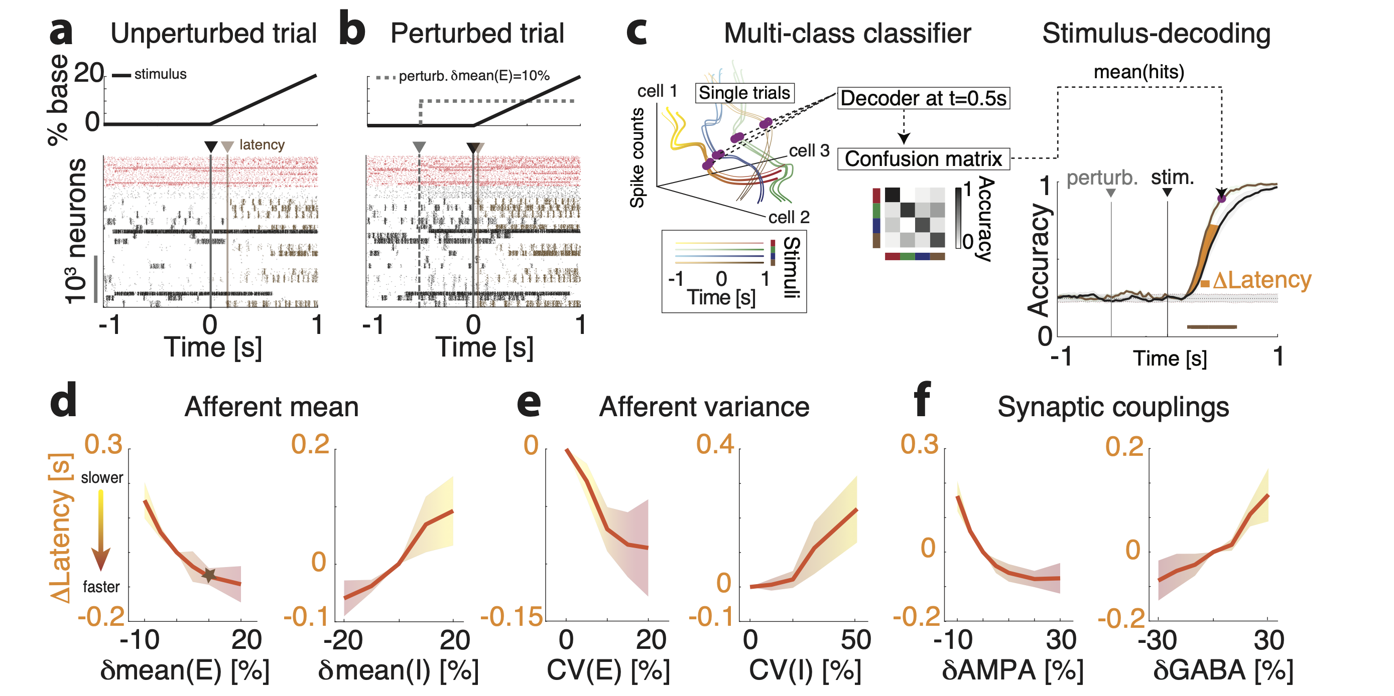
State perturbations had opposite effects depending on which cell-type specific populations they targeted. Increasing mean(E) monotonically improved network performance (Fig. 4d, left panel): in particular, positive perturbations induced an anticipation of stimulus-coding (shorter latency), while negative ones led to longer latency and slower coding. The opposite effect was achieved when increasing mean(I), which slowed down processing speed (Fig. 4d, right panel). State perturbations that changed the spatial variance of the afferent currents had counterintuitive effects (Fig. 4e). We measured the strength of these perturbations via their coefficient of variability , for , where and are the standard deviation and mean of the across-neuron distribution of afferent currents. Perturbations var(E) that increased led to faster processing speed. The opposite effect was achieved with perturbations var(I) inducing a spatial variance across afferents to I neurons, which slowed down stimulus-processing speed (Fig. 4e). Perturbations AMPA which increased the glutamatergic synaptic weights improved performance proportionally to the perturbation. The opposite effect was achieved by perturbations GABA that increased the GABAergic synaptic weights, which monotonically decreased network processing speed (Fig. 4f). We thus concluded that afferent current perturbations differentially modulated the speed at which network activity encoded information about incoming sensory inputs. Such modulations exhibited a rich dynamical repertoire (Table 3).
3.4 Gain modulation regulates the network information processing speed
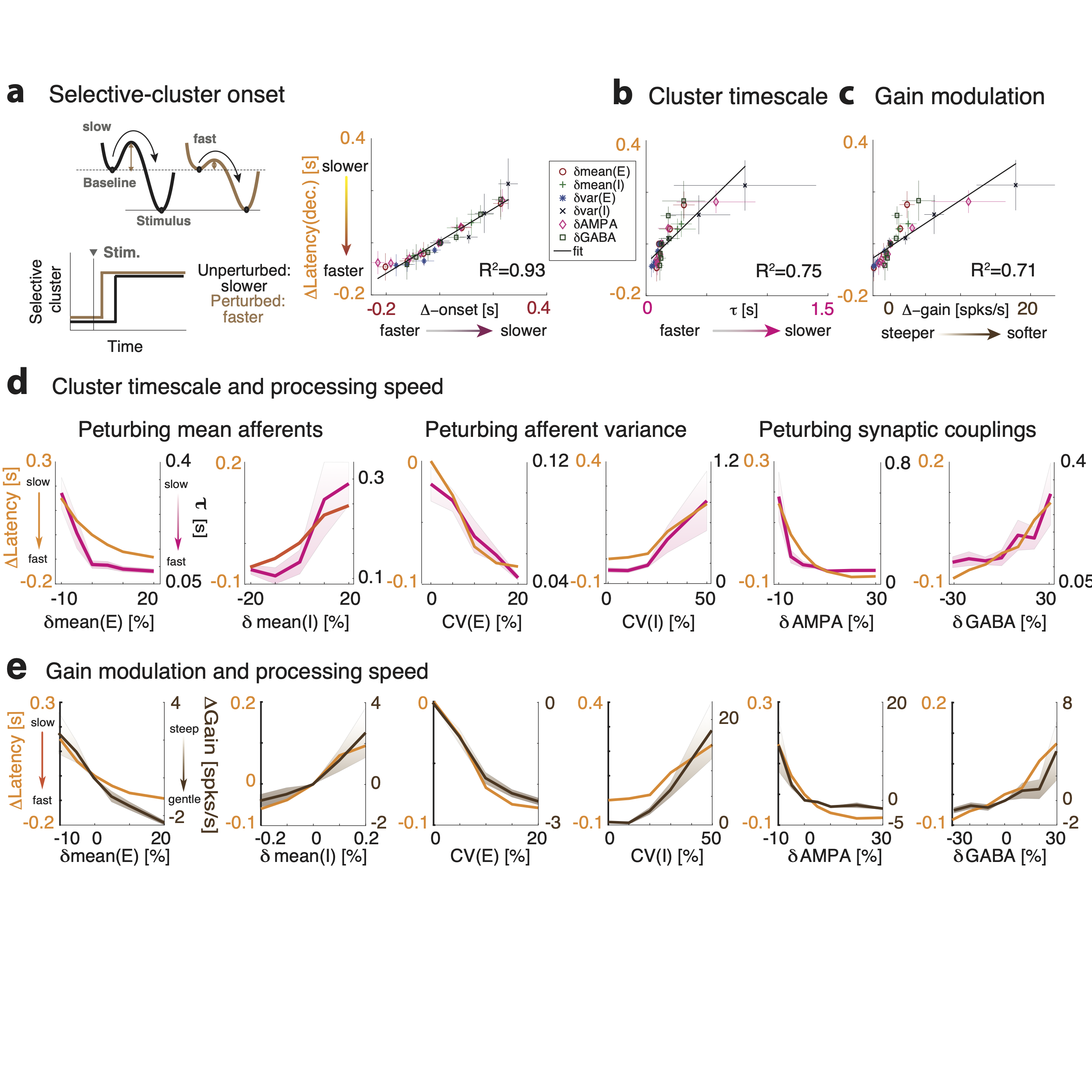
Our mean field framework demonstrates a direct relationship between the effects of perturbations on the network information processing speed and its effects on the cluster timescale (Fig. 3). In our simplified network with two clusters, stimulus presentation induces an asymmetry in the well depths, where the attractor B corresponding to the activated stimulus-selective cluster has a deeper well, compared to the attractor A where the stimulus-selective cluster is inactive. Upon stimulus presentation, the network ongoing state will be biased to transition towards the stimulus-selective attractor B with a transition rate determined by the barrier height separating A to B. Because external perturbations regulate the height of such barrier via gain modulation, they control in turn the latency of activation of the stimulus-selective cluster. We thus aimed at testing the prediction of our theory: that the perturbations modulate stimulus coding latency in the same way as they modulate cluster timescales during ongoing periods; and, as a consequence, that these changes in stimulus coding latency can be predicted by intrinsic gain modulation. Specifically, our theory predicts that perturbation driving a decrease (increase) in intrinsic gain during ongoing periods will induce a faster (slower) encoding of the stimulus.
We thus proceeded to test the relationship between perturbations effects on cluster timescales, gain modulation, and information processing speed. In the representative trial where the same stimulus was presented in the absence (Fig. 4a) or in the presence (Fig. 4b) of the perturbation mean(E), we found that stimulus-selective clusters (highlighted in brown) had a faster activation latency in response to the stimulus in the perturbed condition compared to the unperturbed one. A systematic analysis confirmed this mechanism showing that, for all perturbations, the activation latency of stimulus-selective clusters was a strong predictor of the change in decoding latency (Fig. 5a right panel, ). Moreover, we found that the perturbation-induced changes of the cluster timescale during ongoing periods predicted the effect of the perturbation on stimulus-processing latency during evoked periods (Fig. 5b,d). Specifically, perturbations inducing faster during ongoing periods, in turn accelerated stimulus coding; and vice versa for perturbations inducing slower .
We then tested whether perturbation-induced gain modulations during ongoing periods explained the changes in stimulus-processing speed during evoked periods, and found that the theoretical prediction was borne out in the simulations (Fig. 5c,e). Let us summarize the conclusion of our theoretical analyses. Motivated by mean field theory linking gain modulation to changes in transition rates between attractors, we found that gain modulation controls the cluster timescale during ongoing periods, and, in turn, regulates the onset latency of stimulus-selective clusters upon stimulus presentation. Changes in onset latency of stimulus-selective clusters explained changes in stimulus-coding latency. We thus linked gain modulation to changes in stimulus-processing speed (Fig. 5, Table 3).
| Latency | Activity | Response | Gain | ||
|---|---|---|---|---|---|
| mean(E) | E[], I[] | E[], I[] | |||
| mean(I) | E[], I[] | E[], I[mixed] | |||
| var(E) | E[], I[] | E[mixed], I[mixed] | |||
| var(I) | E[], I[] | E[], I[] | |||
| AMPA | E[], I[] | E[], I[] | |||
| GABA | E[], I[] | E[], I[mixed] | |||
| Locomotion | E[], I[] | E[mixed], I[mixed] |
3.5 Physiological responses to perturbations
Our results show that cortical processing speed can be accelerated or slowed down via external perturbations. We found that different types of perturbations may induce similar effects on processing speed: a dynamical acceleration may be obtained by either increasing the mean or the variance of the external input to E neurons, or either decreasing the mean or the variance of external inputs to I neurons. A dynamical deceleration may be obtained by the opposite perturbations. In order to devise an experimental test of our theory to dissect the specific effects of each type of perturbation, we then examined the single-cell responses to perturbations. By combining single-cell responses with dynamical effects, we will be able to isolate the effects of each perturbation.
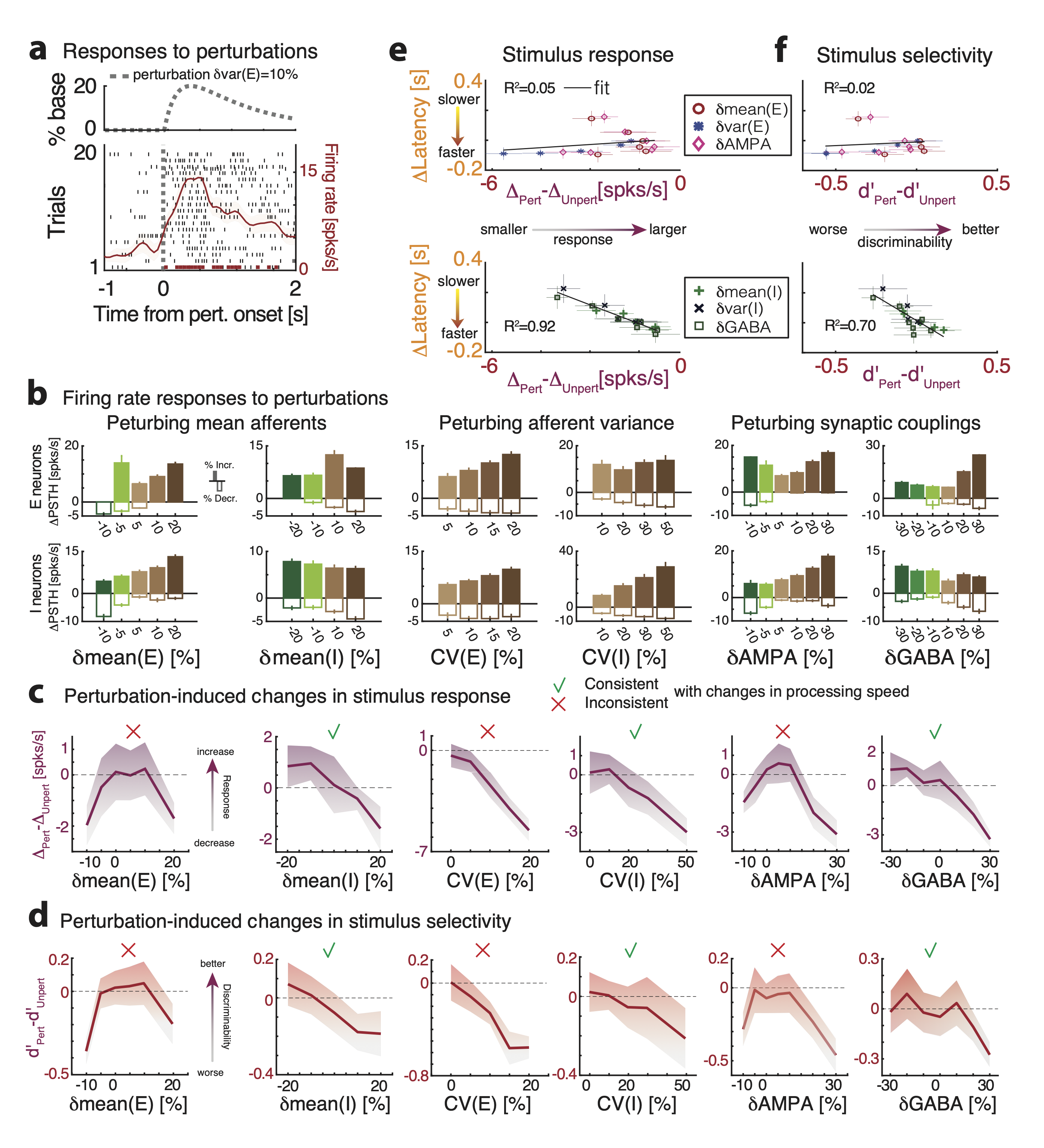
We characterized single-cell responses to perturbations during ongoing periods, in the absence of sensory stimuli (Fig. 6, 6-1). We found that perturbations differentially affected neuronal responses in a cell-type specific way. Perturbations changed the average population firing rates, and led to complex patterns of response across E and I populations (Fig. 6). Specifically, perturbations increasing mean(E) induced higher firing rates and induced proportionally excited responses in both E and I populations. On the other hand, perturbations that increased mean(I) led to a decrease in both E and I average firing rates (Fig. 6). This paradoxical effect tsodyks1997paradoxical revealed that the network operates in the inhibition stabilized regime (Fig. 2-1). When increasing the inhibitory current beyond mean(I)=, the network reached a reversal point where the E population activity became silent and the I population rebounded, starting to increase their firing rates again (Fig. 2-1).
Perturbations increasing the variance var(E) and var(I) led to surprising effects (Fig. 6, 6-1). Increasing var(E) induced higher firing rates in both E and I populations, despite leaving the mean afferents unchanged; moreover, it led to mixed responses at the single cell level, with a prevalence of excited responses in both E and I populations. We will see below that this set of responses is consistent with locomotion-induced effects in the visual cortical hierarchy. Increasing var(I) left firing rates of I populations unchanged but led to a decrease of E population firing rates. This perturbation also induced mixed responses at the single cell level, with a prevalence of inhibited responses in both populations. Finally, perturbations AMPA and GABA led to responses similar to those found when driving the mean E- or I-afferents, respectively.
Our theory thus suggests that it is possible to identify a specific perturbation by combining all its effects, including gain modulation, changes in stimulus-processing speed and single-cell physiological responses (Table 3) .
3.6 Changes in single-cell responses cannot explain the effects of perturbations on evoked activity
Although we found that gain modulation captures the effects of perturbations on network activity, we investigate whether alternative explanations were also possible, in terms of traditional measures of stimulus responsiveness and selectivity.
We found that perturbations strongly affected the peak of single-cell responses to stimuli compared to baseline (PSTH, Fig. 6-1b), as well as single-cell selectivity to stimuli with significant changes in their d’ (Fig. 6-1c). We then tested whether perturbation-induced changes in stimulus responses or selectivity could explain the observed changes in stimulus-processing speed. We first hypothesized that, if the response increase induced by the perturbation were larger for stimulus-selective compared to nonselective neurons (i.e., if PSTH(sel)>PSTH(nonsel)), then a perturbation increasing stimulus-responses could lead to faster stimulus-processing speed. Likewise, we hypothesized that faster stimulus-processing speed may be induced by perturbations improving single-cell selectivity (d’) to stimuli. Surprisingly, we found a complex relation between changes in single-cell responsiveness and selectivity to stimuli, induced by the perturbations, and modulation of stimulus-processing speed (Fig. 6-1). For perturbations targeting I populations (mean(I), var(I), and GABA) changes in responsiveness and selectivity were consistent with changes in processing speed ( for responsiveness and selectivity, respectively). However, for perturbations targeting E populations (mean(E), var(E), and AMPA) changes in responsiveness and selectivity were not consistent with changes in processing speed ( for responsiveness and selectivity, respectively). Strikingly, in the case of the perturbation var(E), processing speed increased with larger perturbations even though responses and selectivity increasingly degraded. In the case of the perturbation mean(E) and AMPA, network performance likewise increased but single-cell metrics where non-monotonic in the value of the perturbation (Fig. 6-1e, f). Because changes in single-cell stimulus properties were only consistent with changes in processing speed for some perturbations (mean(I), var(I), and GABA), but inconsistent for other perturbations, we thus conclude that they could not represent an alternative mechanism underlying the observed effects of perturbations.

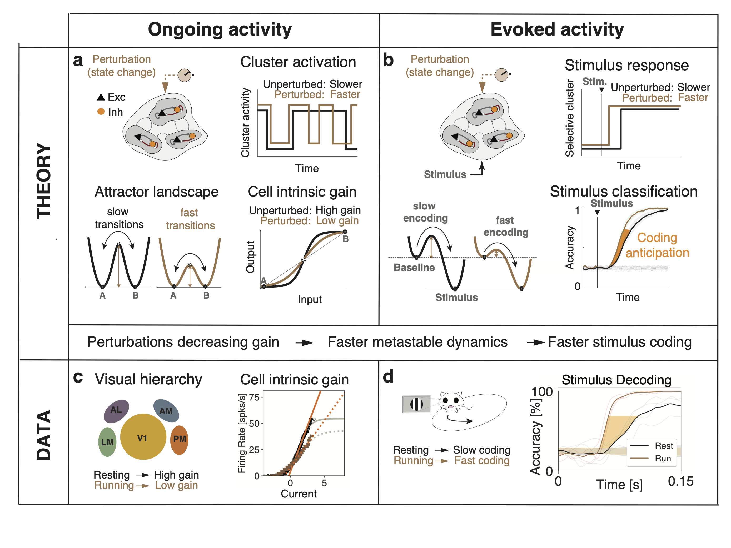
3.7 Locomotion decreases single-cell gain and accelerates visual processing speed
Our theory predicts a link between gain modulations measured during ongoing periods and changes in stimulus-processing speed during evoked periods. We sought to experimentally test this prediction in freely running mice using electrophysiological recordings from the visual hierarchy including the primary visual cortex (V1) and 4 higher cortical visual areas LM, AL, PM, AM (open-source neuropixels dataset available from the Allen Institute, siegle2019survey, ). We interpreted periods where the animal was resting as akin to the “unperturbed” condition in our model, and periods where the animal was running as the “perturbed” condition (Fig. 7a in the data). We thus set out to test our theory in the following three steps: i) in each area, we estimated the effect of locomotion on single-cell responses and on intrinsic gain during ongoing periods, ii) based on these changes, we built a biologically plausible model of cortical circuits processing visual stimuli and predicted whether locomotion would accelerate or slow down visually-evoked responses; iii) we tested the prediction in each area with a decoding analysis of visually-evoked population activity.
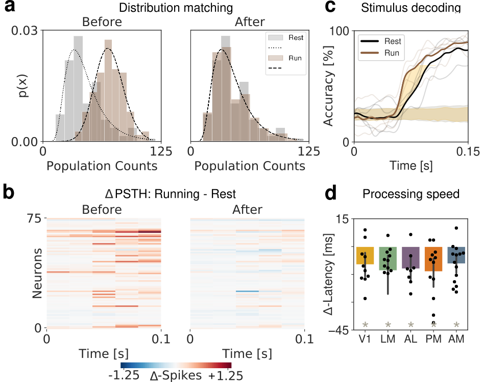
During periods of ongoing activity (in the absence of visual stimuli), we found that locomotion induced an overall increase in firing rate across all visual cortical areas (Fig. 7b left), in agreement with previous studies niell2010modulation ; stringer2019high ; dipoppa2018vision . Although, we found that locomotion led to complex responses inducing mixed excited and inhibited responses across neurons (Fig. 7b right), as previously reported dipoppa2018vision . We then estimated the single-cell transfer functions from spiking activity during ongoing periods both when the animal was at rest and in motion (Fig. 7c). We found that locomotion strongly modulated the single-cell gain in the absence of stimuli in all visual cortical areas (Fig. 7d). Specifically, we found that locomotion on average decreased the single-cell gain.
Our theory predicts that, in all visual cortical areas, the locomotion-induced increase in firing rates, the mixed excited and inhibited neural responses and the decrease in intrinsic gain are consistent with a state-changing perturbation mediated by an increase in the variance of the input currents to E neurons (var(E), Table 3). According to our theory, the decrease in gain leads to an acceleration of stimulus-processing speed in all visual cortical areas.
We aimed at refining the model predictions on the locomotion effects on V1 and the visual cortical hierarchy by introducing a biologically plausible stimulation protocol in our spiking network. Following experimental evidence on anatomical connectivity in the visual pathway zhuang2013layer ; miska2018sensory ; kloc2014target ; khan2018distinct , we then modeled incoming visual stimuli as a transient increase in the input currents to both E and I neurons (Fig. 7e). We then modeled the effect of locomotion as an external perturbation inducing an increase in the variance of the inputs to E neurons var(E), capturing the observed empirical effects of locomotion on ongoing periods in terms of gain decrease and mixed single-cell responses. In this model of visual processing, we found that locomotion accelerated visual processing speed during evoked period by ms on average (meanS.D. across 10 sessions, Fig. 7f). We thus set out to test this prediction in the empirical data.
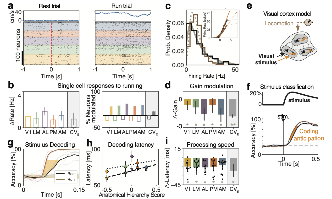
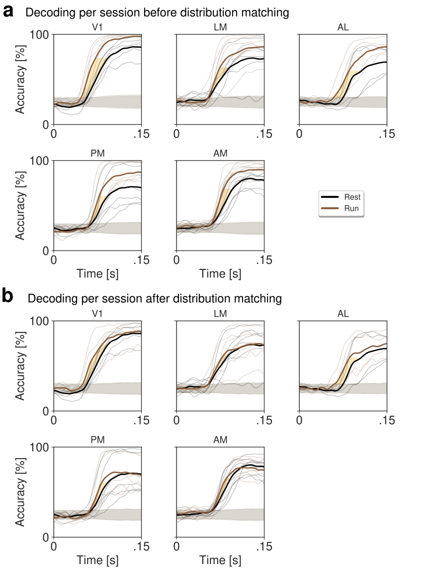
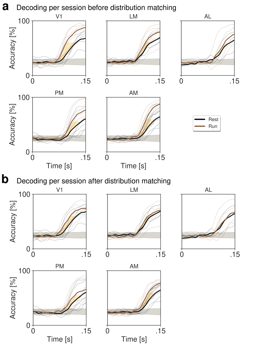
Previous studies have observed an improvement in peak decoding performance during locomotion dadarlat2017locomotion , but changes in decoding latency have not been investigated. To probe the speed and accuracy of visual responses in perturbed and unperturbed conditions, we performed a cross-validated classification analysis to assess the amount of information regarding the orientation of drifting grating stimuli present in population spiking activity along the visual cortical hierarchy. Crucially, because decoding accuracy depends on sample size, we equalized number of trials between resting and running conditions. We found that trials in which the animal was running revealed both an increase in peak decoding accuracy and an anticipation of stimulus coding (shorter latency) as compared to trials where the animal was stationary (Fig. 7g), consistently across the whole visual hierarchy (Fig. 7i). Furthermore, the time to reach significant decoding for each cortical area followed the anatomical hierarchy score in both unperturbed and perturbed conditions, consistent with the idea that information about the visual stimulus travels up a visual hierarchy in a feed-forward fashion (Fig. 7h, siegle2019survey, ).
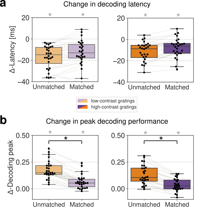
Given that locomotion induced an increase in firing rates in all cortical areas (Fig. 7b), we then examined the extent to which the observed effects of locomotion (increased peak accuracy and anticipation) were merely due to the increase in firing rates. We thus matched the distribution of firing rates between running and resting (see methods and Fig. 7-1). We found that after rate matching the change in peak decoding accuracy decreased significantly (Fig. 7-2-7-4). Crucially, the anticipation of stimulus processing speed induced by locomotion was still present in the rate-matched condition (Fig. 7-2), confirming that it was independent of changes in firing rates. The same effect was preserved in the rate-matched model simulations as well (Fig. 7-5). We thus concluded that the anticipation of visual processing speed induced by locomotion is consistent with a mechanism whereby locomotion decreases single-cell gain via an increase in the afferent variance var(E) as predicted by our theory (Table 3).
4 Discussion
Cortical circuits flexibly adapt their information processing capabilities to changes in environmental demands and internal state. Empirical evidence suggests that these state-dependent modulations may occur already in the sensory cortex where they may be induced by top-down pathways or neuromodulation. Here, we presented a mechanistic theory explaining how stimulus-processing speed can be regulated in a state-dependent manner via gain modulation, induced by transient changes in the afferent currents or in the strength of synaptic transmission.
Our theory entails a recurrent spiking network where excitatory and inhibitory neurons are arranged in clusters, generating metastable activity in the form of transient activation of subsets of clusters. We showed that gain modulation controls the timescale of metastable activity and thus the network’s information-processing speed and reaction times upon stimulus presentation. Specifically, our theory predicted that perturbations that decrease (increase) the intrinsic single-cell gain during ongoing periods accelerate (slow down) the latency of stimulus responses.
We tested this prediction by examining the effect of locomotion on visual processing in freely running mice. We found that locomotion reduced the intrinsic single-cell gain during ongoing periods, thus accelerating stimulus-coding speed across the visual cortical hierarchy. Our theory suggests that the observed effects of locomotion are consistent with a perturbation that increases the spatial variance of the afferent currents to the local excitatory population. These results establish a new theory of state-dependent adaptation of cortical responses via gain modulation, unifying the effect of different pathways under a shared computational mechanism.
4.1 Metastable activity in cortical circuits
The crucial dynamical feature of our model is its metastable activity, whereby single-trial ensemble spike trains unfold through sequences of metastable states. State are long-lasting, with abrupt transitions between consecutive states. Metastable activity has been ubiquitously observed in a variety of cortical and subcortical areas, across species and tasks Abeles1995a ; Jones2007 ; engel2016selective ; PonceAlvarez2012 ; maboudi2018uncovering ; rich2016decoding ; sadacca2016behavioral ; taghia2018uncovering ; deco2019awakening . Metastable activity can be used to predict behavior and was implicated as a neural substrate of cognitive function, such as attention engel2016selective , expectation mazzucato2019expectation , and decision making rich2016decoding ; taghia2018uncovering ; recanatesi2020metastable . Metastable activity was observed also during ongoing periods, in the absence of sensory stimulation, suggesting that it may be an intrinsic dynamical regime of cortical circuits mazzucato2015dynamics ; engel2016selective . Here, we showed how cortical circuits can flexibly adjust their performance and information-processing speed via modulations of their metastable dynamics.
Metastable activity may naturally arise in circuits where multiple stable states, or attractors, are destabilized by external perturbations MillerKatz2010 or intrinsically generated variability DecoHugues2012 ; LitwinKumarDoiron2012 ; mazzucato2015dynamics ; schaub2015emergence ; mazzucato2019expectation ; rostami2020spiking ; recanatesi2020metastable . Biologically plausible models of metastable dynamics have been proposed in terms of recurrent spiking networks where neurons are arranged in clusters, reflecting the empirically observed assemblies of functionally correlated neurons kiani2015natural ; song2005highly ; perin2011synaptic ; lee2016anatomy . Clustered network models of metastable dynamics provide a parsimonious explanation of several physiological observations such as stimulus-induced reductions of trial-to-trial variability DecoHugues2012 ; LitwinKumarDoiron2014 ; mazzucato2015dynamics ; rostami2020spiking ; Churchland2010 , of firing rate multistability mazzucato2015dynamics , and of neural dimensionality mazzucato2016stimuli . Compared to previous models of metastable dynamics, our results extend the biological plausibility of clustered networks in several aspects. The introduction of pairs of E and I clusters induces a tight balance where the E and I contributions to the postsynaptic currents of each neuron closely track each other with opposite signs (Fig. 2-1), as observed experimentally in cortical circuits okun2008instantaneous . Moreover, we showed that these networks operate in the inhibition stabilized regime (Fig. 2-1), which is believed to be the operational regime of cortical circuits ozeki2009inhibitory ; sanzeni2019inhibition ; moore2018rapid . We showed that a heterogeneous distribution of cluster sizes naturally give rise to lognormal distribution of firing rates (Fig. 2b inset), as observed in cortical circuits shafi2007variability ; hromadka2008sparse ; o2010neural ; roxin2011distribution . We then generalize the results in mazzucato2019expectation to establish gain modulation as the general mechanism controlling that state-dependent changes in processing speed in recurrent circuits with metastable dynamics. This class of models thus provide a biologically plausible, mechanistic link between connectivity, dynamics, and information-processing.
4.2 Linking metastable activity to flexible cognitive function via gain modulation
Recent studies have shown that cortical circuits may implement a variety of flexible cognitive computations by modulating the timescale of their intrinsic metastable dynamics mattia2013heterogeneous ; mazzucato2019expectation ; engel2016selective ; rich2016decoding ; deco2019awakening . Our results establish a comprehensive framework to investigate the extent of this hypothesis. We propose that gain modulation is the neural mechanism underlying flexible state-dependent cortical computation. Specifically, we showed that gain modulation controls the timescale of metastable dynamics, which, in turn, determines the network’s information-processing speed.
Our theoretical framework to link gain modulation to changes in potential barrier heights is based on the effective mean field theory following Mascaro1999 ; mattia2013heterogeneous ; mazzucato2019expectation , which we used to reduce a multidimensional system to obtain an effective potential describing a single population. Although this approach is exact in the case of networks with symmetric connectivity, it represents only an approximation to the full network dynamics in the case of networks with asymmetric couplings such as the ones considered in this study rodriguez2020climbing . It would be interesting to extend our results to an exact framework by estimating the network Lyapunov function yan2013nonequilibrium .
4.3 Alternative models of gain modulation
Previous studies have suggested gain modulation as a mechanism to sharpen single-cell tuning curves without affecting selectivity cardin2008cellular ; haider2009rapid , potentially mediating attention mcadams1999effects ; treue1999feature ; rabinowitz2015attention . In those studies, gain modulation was defined as change in the single-neuron response function to stimuli of increasing contrast. Here, we have taken a different approach and defined gain as the slope of the intrinsic neuronal current-to-rate function during ongoing periods (i.e., in the absence of stimuli, see also chance2002gain, ; haider2009rapid, ; mazzucato2019expectation, ), as opposed to the contrast response function. We have classified mechanisms of gain modulation which act by changing the mean or spatial variance across neurons of the cell-type specific afferent currents to the local cortical circuit, where we modeled afferent currents as constant biases; or by changing the recurrent couplings. The rationale for our choice was to investigate the effects on internally generated variability in a network whose dynamics were entirely deterministic. Alternatively, one could model external currents as time-dependent inputs with fast noise, such as Poisson processes or colored noise. In that case, changes in background noise due to barrages of synaptic inputs are capable of inducing gain modulation as well chance2002gain ; haider2009rapid . Previous work compared these different kinds of perturbations (Poisson noise or afferent spatial variance) in the case of the perturbation var(E) mazzucato2019expectation , showing they may lead to similar outcomes.
4.4 Physiological mechanisms of gain modulation
Several different physiological pathways can modulate the gain of the intrinsic neuronal transfer function, including neuromodulation, top-down and cortico-cortical interactions. Gain modulation can also be induced artificially by means of optogenetic or pharmacological manipulations. The perturbations investigated in our model may be related to different pathways and implicated in various types of cognitive function.
Neuromodulation
Neuromodulatory pathways strongly affect sensory processing in cortical circuits by changing cell-type specific afferent currents to the circuit, in some cases controlling their dynamical regime mcginley2015waking . Our theory may be applicable to explain the effects of cholinergic and serotonergic activation on sensory cortex.
Cholinergic pathways, modulating ionic currents in pyramidal cells mccormick1992neurotransmitter , can control cortical states and mediate the effects of arousal and locomotion. Artificial stimulation of cholinergic pathways was shown to improve sensory coding in visual goard2009basal ; pinto2013fast and barrel cortex eggermann2014cholinergic . Cholinergic stimulation alone in the absence of sensory stimuli was shown to induce mixed responses with different neural populations increasing or decreasing their spiking activity goard2009basal . Our theory shows that these combined experimental observations (coding improvement and mixed firing rate changes) are consistent with a mechanism whereby cholinergic activation induces an increase in var(E) afferents to sensory cortex, inducing an acceleration of sensory processing (Fig. 2e and S1a).
Activation of serotonergic pathways by stimulation of dorsal raphe serotonergic neurons or local iontophoresis was shown to transiently degrade stimulus coding in sensory cortex, decreasing responses to mechanosensory stimuli dugue2014optogenetic and increasing the latency of the first spike evoked by auditory stimuli hurley2002serotonin . Serotonergic stimulation was shown to decrease firing rates in the olfactory cortex lottem2016optogenetic , inferior colliculus hurley2002serotonin , and primary visual cortex michaiel2019hallucinogenic ; seillier2017serotonin . Our theory shows that these experimental observations (coding degradation and decreased firing rates) are consistent with two alternative mechanisms (Fig. 2d and S1a): either an increase in the afferent currents to I populations (i.e., mean(I)) implementing the paradoxical inhibition effect tsodyks1997paradoxical ; or a decrease in the afferents to E populations (i.e., mean(E)). Future experiments could test between these two alternatives.
Top-down projections
A prominent feature of sensory cortex is the integration of feedforward and cortico-cortical feedback pathways at each stage of sensory processing felleman1991distributed . In particular, top-down projections from higher cortical areas to sensory cortex are known to modulate the speed and accuracy of sensory processing mazzucato2019expectation . Our theory may explain the effects of activating several cortico-cortical pathways.
Activation of feedback axons from motor cortex (M1) to somatosensory cortex (S1) was shown to increase activity in S1 during whisking petreanu2012activity and led to faster and more accurate responses to whisker stimulation zagha2013motor . Suppression of the same pathway induced slower S1 responses to whisking in awake mice. Our theory shows that the effect of these cortico-cortical perturbations is consistent with an increase in the mean afferent currents to E populations in S1 (i.e., the mean(E) perturbation in Fig. 2d), leading to higher firing rates and faster processing speed.
Expectation and arousal are known to strongly modulate neural activity in sensory cortices salkoff2020movement . Expected stimuli are processed faster and more accurately than unexpected stimuli both in auditory jaramillo2010auditory and gustatory cortex samuelsen2012effects . Experimental evidence shows that the anticipation of sensory processing induced by expectation is mediated by top-down projections from the amygdala to the gustatory cortex samuelsen2012effects , whose activation elicits mixed excited and inhibited responses in both pyramidal and inhibitory cells in the gustatory cortex samuelsen2012effects ; vincis2016associative . Our model shows that, while an acceleration of stimulus processing speed may in principle be mediated by different state-changing perturbations, only the var(E) perturbation is consistent with the empirically observed mixed responses. Indeed, our theory suggests that these top-down projections may operate by inducing an increase in the spatial variance of the afferent currents to the E population (var(E) in Fig. 2d, extending previous results in mazzucato2019expectation, , to networks including inhibitory clusters) .
In attentional tasks, distractors slow down reaction times grueninger1969effects ; treisman1980feature , a behavioral effect that may be mediated by changes in the speed and accuracy of sensory processing in cortical circuits desimone1995neural . The presence of distracting stimuli within a neurons receptive field suppresses its responses to the preferred stimulus knierim1992neuronal . The underlying mechanism may recruit lateral inhibition onto the local cortical circuit reynolds2009normalization ; gilbert2013top . Our theory shows that this mechanism is consistent with a modulation of the afferents to local I populations, mediated by either an increase in mean(I) or var(I) in Fig. 2d. It would be interesting to discriminate between these two perturbations with future experiments.
Optogenetic and pharmacological manipulations
Our theory may shed light on the effects of manipulation experiments. Optogenetic activation (inactivation) of specific E or I cells arenkiel2007vivo ; li2019spatiotemporal has been modeled as an increase (decrease) of the afferent currents to those cells ebsch2018imbalanced ; mahrach2020mechanisms ; sanzeni2019inhibition . However, protein expression may not be complete across all cells of the targeted population, and even in the case of complete expression across the targeted population, different cells may be more or less sensitive to laser stimulation. Thus the effect of optogenetic stimulation on the targeted population may then be more accurately modeled by a concurrent change in both mean and variance of the targeted cell-type specific afferents (e.g., mean(E) and var(E) for E populations; mean(I) and var(I) for I populations). Recent studies showed that, while a homogeneous stimulation of all I cell types simultaneously can be captured by a model of E-I recurrently coupled neurons (as in our model), partial activation of specific inhibitory cell-types may induced more complex responses mahrach2020mechanisms ; sanzeni2019inhibition ; li2019spatiotemporal ; otchy2015acute ; zur2008direct ; phillips2016asymmetric . We plan to revisit this issue in the future.
Our theory may also be applicable to the effects of pharmacological manipulations of different synaptic receptors. In particular, the effects of combined local injection of AMPA/kainate and NMDA receptor antagonists (agonists) may be recapitulated by a decrease (increase) in AMPA, which correspondingly perturb the value of couplings (Fig. 2d). Similarly, the effects of local injection of GABA receptor antagonists (agonists) may be recapitulated by a decrease (increase) in GABA, which correspondingly perturb the value of couplings.
4.5 Locomotion and gain modulation
Locomotion has been shown to modulate visually evoked activity niell2010modulation and is sufficient in driving activity in mouse V1 leinweber2017sensorimotor ; saleem2013integration . Our results were consistent with previous studies in showing that locomotion affects the activity of neurons in the visual cortical hierarchy during both ongoing and stimulus-evoked activity. We found that locomotion in the absence of sensory stimuli induces an average increase in firing rates. At the single-cell level we reported a complex mix of excited and inhibited responses in both E and I cells, also consistent with previous results fu2014cortical ; dipoppa2018vision . Crucially, we uncovered that locomotion decreased the single-cell gain during ongoing activity across the board in the visual cortical hierarchy (Fig. 7d). Our theory predicted that the observed decrease in gain would lead to an acceleration of visual processing during locomotion in cortex. This prediction was confirmed in the data (Fig. 7i). The acceleration of processing speed observed in cortex did not depend on the locomotion-induced changes in firing rates and was still present even after matching the firing rate distributions between running and rest conditions (Fig. 7-1). Our model of the perturbation effects induced by locomotion (increased firing rates with mixed excited and inhibited responses, and faster visual processing) suggests that the effect of locomotion may be mediated by a increase in the spatial variance of the afferent current to the E populations (var(E) perturbation) ayaz2013locomotion ; niell2010modulation ; fu2014cortical . Concretely, gain modulation may be implemented via the combined effect of activating neuromodulatory pathways such as cholinergic fu2014cortical and noradrenergic polack2013cellular inputs.
5 Acknowledgements
This work was supported by a National Institute of Deafness and Other Communication Disorders Grant K25-DC013557 and a National Institute of Neurological Disorders and Stroke Grant NS118461 (BRAIN Initiative). We would like to thank G. La Camera, Y. Ahmadian, and M. Wehr for useful discussions and suggestions.
References
- [1] Pekka Niemi and Risto Näätänen. Foreperiod and simple reaction time. Psychological bulletin, 89(1):133, 1981.
- [2] Walter E Grueninger and Karl H Pribram. Effects of spatial and nonspatial distractors on performance latency of monkeys with frontal lesions. Journal of comparative and physiological psychology, 68(2p1):203, 1969.
- [3] Anne M Treisman and Garry Gelade. A feature-integration theory of attention. Cognitive psychology, 12(1):97–136, 1980.
- [4] Robert Desimone and John Duncan. Neural mechanisms of selective visual attention. Annual review of neuroscience, 18(1):193–222, 1995.
- [5] Santiago Jaramillo and Anthony Zador. Auditory cortex mediates the perceptual effects of acoustic temporal expectation. Nature Precedings, pages 1–1, 2010.
- [6] Chad L Samuelsen, Matthew PH Gardner, and Alfredo Fontanini. Effects of cue-triggered expectation on cortical processing of taste. Neuron, 74(2):410–422, 2012.
- [7] A Moses Lee, Jennifer L Hoy, Antonello Bonci, Linda Wilbrecht, Michael P Stryker, and Cristopher M Niell. Identification of a brainstem circuit regulating visual cortical state in parallel with locomotion. Neuron, 83(2):455–466, 2014.
- [8] Lucas Pinto, Michael J Goard, Daniel Estandian, Min Xu, Alex C Kwan, Seung-Hee Lee, Thomas C Harrison, Guoping Feng, and Yang Dan. Fast modulation of visual perception by basal forebrain cholinergic neurons. Nature neuroscience, 16(12):1857–1863, 2013.
- [9] Yu Fu, Jason M Tucciarone, J Sebastian Espinosa, Nengyin Sheng, Daniel P Darcy, Roger A Nicoll, Z Josh Huang, and Michael P Stryker. A cortical circuit for gain control by behavioral state. Cell, 156(6):1139–1152, 2014.
- [10] Zengcai V Guo, Nuo Li, Daniel Huber, Eran Ophir, Diego Gutnisky, Jonathan T Ting, Guoping Feng, and Karel Svoboda. Flow of cortical activity underlying a tactile decision in mice. Neuron, 81(1):179–194, 2014.
- [11] Tsai-Wen Chen, Nuo Li, Kayvon Daie, and Karel Svoboda. A map of anticipatory activity in mouse motor cortex. Neuron, 94(4):866–879, 2017.
- [12] Anders Nelson, David M Schneider, Jun Takatoh, Katsuyasu Sakurai, Fan Wang, and Richard Mooney. A circuit for motor cortical modulation of auditory cortical activity. Journal of Neuroscience, 33(36):14342–14353, 2013.
- [13] Marcus Leinweber, Daniel R Ward, Jan M Sobczak, Alexander Attinger, and Georg B Keller. A sensorimotor circuit in mouse cortex for visual flow predictions. Neuron, 95(6):1420–1432, 2017.
- [14] Siyu Zhang, Min Xu, Tsukasa Kamigaki, Johnny Phong Hoang Do, Wei-Cheng Chang, Sean Jenvay, Kazunari Miyamichi, Liqun Luo, and Yang Dan. Long-range and local circuits for top-down modulation of visual cortex processing. Science, 345(6197):660–665, 2014.
- [15] Matthew J McGinley, Martin Vinck, Jacob Reimer, Renata Batista-Brito, Edward Zagha, Cathryn R Cadwell, Andreas S Tolias, Jessica A Cardin, and David A McCormick. Waking state: rapid variations modulate neural and behavioral responses. Neuron, 87(6):1143–1161, 2015.
- [16] Marlene R Cohen and John HR Maunsell. Attention improves performance primarily by reducing interneuronal correlations. Nature neuroscience, 12(12):1594, 2009.
- [17] Maria C Dadarlat and Michael P Stryker. Locomotion enhances neural encoding of visual stimuli in mouse v1. Journal of Neuroscience, 37(14):3764–3775, 2017.
- [18] Jacob Reimer, Matthew J McGinley, Yang Liu, Charles Rodenkirch, Qi Wang, David A McCormick, and Andreas S Tolias. Pupil fluctuations track rapid changes in adrenergic and cholinergic activity in cortex. Nature communications, 7(1):1–7, 2016.
- [19] Edward Zagha, Xinxin Ge, and David A McCormick. Competing neural ensembles in motor cortex gate goal-directed motor output. Neuron, 88(3):565–577, 2015.
- [20] Luca Mazzucato, Giancarlo La Camera, and Alfredo Fontanini. Expectation-induced modulation of metastable activity underlies faster coding of sensory stimuli. Nature neuroscience, page 1, 2019.
- [21] Chengcheng Huang, Douglas A Ruff, Ryan Pyle, Robert Rosenbaum, Marlene R Cohen, and Brent Doiron. Circuit models of low-dimensional shared variability in cortical networks. Neuron, 101(2):337–348, 2019.
- [22] D. J. Amit and N. Brunel. Model of global spontaneous activity and local structured activity during delay periods in the cerebral cortex. Cereb Cortex, 7(3):237–52, 1997.
- [23] A. Litwin-Kumar and B. Doiron. Slow dynamics and high variability in balanced cortical networks with clustered connections. Nat Neurosci, 15(11):1498–505, 2012.
- [24] G. Deco and E. Hugues. Neural network mechanisms underlying stimulus driven variability reduction. PLoS Comput Biol, 8(3):e1002395, 2012.
- [25] Luca Mazzucato, Alfredo Fontanini, and Giancarlo La Camera. Dynamics of multistable states during ongoing and evoked cortical activity. The Journal of Neuroscience, 35(21):8214–8231, 2015.
- [26] Luca Mazzucato, Alfredo Fontanini, and Giancarlo La Camera. Stimuli reduce the dimensionality of cortical activity. Frontiers in systems neuroscience, 10:11, 2016.
- [27] Vahid Rostami, Thomas Rost, Alexa Riehle, Sacha J van Albada, and Martin P Nawrot. Spiking neural network model of motor cortex with joint excitatory and inhibitory clusters reflects task uncertainty, reaction times, and variability dynamics. bioRxiv, 2020.
- [28] L. M. Jones, A. Fontanini, B. F. Sadacca, P. Miller, and D. B. Katz. Natural stimuli evoke dynamic sequences of states in sensory cortical ensembles. Proc Natl Acad Sci U S A, 104(47):18772–7, 2007.
- [29] Jun Zhuang, Carl R Stoelzel, Yulia Bereshpolova, Joseph M Huff, Xiaojuan Hei, Jose-Manuel Alonso, and Harvey A Swadlow. Layer 4 in primary visual cortex of the awake rabbit: contrasting properties of simple cells and putative feedforward inhibitory interneurons. Journal of Neuroscience, 33(28):11372–11389, 2013.
- [30] Nathaniel J Miska, Leonidas MA Richter, Brian A Cary, Julijana Gjorgjieva, and Gina G Turrigiano. Sensory experience inversely regulates feedforward and feedback excitation-inhibition ratio in rodent visual cortex. Elife, 7:e38846, 2018.
- [31] Michelle Kloc and Arianna Maffei. Target-specific properties of thalamocortical synapses onto layer 4 of mouse primary visual cortex. Journal of Neuroscience, 34(46):15455–15465, 2014.
- [32] Adil G Khan, Jasper Poort, Angus Chadwick, Antonin Blot, Maneesh Sahani, Thomas D Mrsic-Flogel, and Sonja B Hofer. Distinct learning-induced changes in stimulus selectivity and interactions of gabaergic interneuron classes in visual cortex. Nature neuroscience, 21(6):851–859, 2018.
- [33] N. Fourcaud and N. Brunel. Dynamics of the firing probability of noisy integrate-and-fire neurons. Neural Comput, 14(9):2057–110, 2002.
- [34] M. Mascaro and D. J. Amit. Effective neural response function for collective population states. Network, 10(4):351–73, 1999.
- [35] Maurizio Mattia, Pierpaolo Pani, Giovanni Mirabella, Stefania Costa, Paolo Del Giudice, and Stefano Ferraina. Heterogeneous attractor cell assemblies for motor planning in premotor cortex. Journal of Neuroscience, 33(27):11155–11168, 2013.
- [36] Joshua H Siegle, Xiaoxuan Jia, Séverine Durand, Sam Gale, Corbett Bennett, Nile Graddis, Greggory Heller, Tamina K Ramirez, Hannah Choi, Jennifer A Luviano, et al. A survey of spiking activity reveals a functional hierarchy of mouse corticothalamic visual areas. bioRxiv, page 805010, 2019.
- [37] Ahmad Jezzini, Luca Mazzucato, Giancarlo La Camera, and Alfredo Fontanini. Processing of hedonic and chemosensory features of taste in medial prefrontal and insular networks. The Journal of Neuroscience, 33(48):18966–18978, 2013.
- [38] Stefano Recanatesi, Ulises Pereira, Masayoshi Murakami, Zachary Mainen, and Luca Mazzucato. Metastable attractors explain the variable timing of stable behavioral action sequences. bioRxiv, 2020.
- [39] Sukbin Lim, Jillian L McKee, Luke Woloszyn, Yali Amit, David J Freedman, David L Sheinberg, and Nicolas Brunel. Inferring learning rules from distributions of firing rates in cortical neurons. Nature neuroscience, 18(12):1804, 2015.
- [40] Ulises Pereira and Nicolas Brunel. Attractor dynamics in networks with learning rules inferred from in vivo data. Neuron, 99(1):227–238, 2018.
- [41] Michael T Schaub, Yazan N Billeh, Costas A Anastassiou, Christof Koch, and Mauricio Barahona. Emergence of slow-switching assemblies in structured neuronal networks. PLoS computational biology, 11(7), 2015.
- [42] Roozbeh Kiani, Christopher J Cueva, John B Reppas, Diogo Peixoto, Stephen I Ryu, and William T Newsome. Natural grouping of neural responses reveals spatially segregated clusters in prearcuate cortex. Neuron, 85(6):1359–1373, 2015.
- [43] Sen Song, Per Jesper Sjöström, Markus Reigl, Sacha Nelson, and Dmitri B Chklovskii. Highly nonrandom features of synaptic connectivity in local cortical circuits. PLoS biology, 3(3), 2005.
- [44] Rodrigo Perin, Thomas K Berger, and Henry Markram. A synaptic organizing principle for cortical neuronal groups. Proceedings of the National Academy of Sciences, 108(13):5419–5424, 2011.
- [45] Wei-Chung Allen Lee, Vincent Bonin, Michael Reed, Brett J Graham, Greg Hood, Katie Glattfelder, and R Clay Reid. Anatomy and function of an excitatory network in the visual cortex. Nature, 532(7599):370–374, 2016.
- [46] M. Mattia and M. V. Sanchez-Vives. Exploring the spectrum of dynamical regimes and timescales in spontaneous cortical activity. Cogn Neurodyn, 6(3):239–50, 2012.
- [47] Peter Hänggi, Peter Talkner, and Michal Borkovec. Reaction-rate theory: fifty years after kramers. Reviews of modern physics, 62(2):251, 1990.
- [48] Misha V Tsodyks, William E Skaggs, Terrence J Sejnowski, and Bruce L McNaughton. Paradoxical effects of external modulation of inhibitory interneurons. Journal of neuroscience, 17(11):4382–4388, 1997.
- [49] Cristopher M Niell and Michael P Stryker. Modulation of visual responses by behavioral state in mouse visual cortex. Neuron, 65(4):472–479, 2010.
- [50] Carsen Stringer, Marius Pachitariu, Nicholas Steinmetz, Matteo Carandini, and Kenneth D Harris. High-dimensional geometry of population responses in visual cortex. Nature, page 1, 2019.
- [51] Mario Dipoppa, Adam Ranson, Michael Krumin, Marius Pachitariu, Matteo Carandini, and Kenneth D Harris. Vision and locomotion shape the interactions between neuron types in mouse visual cortex. Neuron, 98(3):602–615, 2018.
- [52] M. Abeles, H. Bergman, I. Gat, I. Meilijson, E. Seidemann, N. Tishby, and E. Vaadia. Cortical activity flips among quasi-stationary states. Proc Natl Acad Sci USA, 92:8616–8620, 1995.
- [53] Tatiana A Engel, Nicholas A Steinmetz, Marc A Gieselmann, Alexander Thiele, Tirin Moore, and Kwabena Boahen. Selective modulation of cortical state during spatial attention. Science, 354(6316):1140–1144, 2016.
- [54] A. Ponce-Alvarez, V. Nacher, R. Luna, A. Riehle, and R. Romo. Dynamics of cortical neuronal ensembles transit from decision making to storage for later report. J Neurosci, 32(35):11956–69, 2012.
- [55] Kourosh Maboudi, Etienne Ackermann, Laurel Watkins de Jong, Brad E Pfeiffer, David Foster, Kamran Diba, and Caleb Kemere. Uncovering temporal structure in hippocampal output patterns. eLife, 7:e34467, 2018.
- [56] Erin L Rich and Jonathan D Wallis. Decoding subjective decisions from orbitofrontal cortex. Nature neuroscience, 19(7):973, 2016.
- [57] Brian F Sadacca, Narendra Mukherjee, Tony Vladusich, Jennifer X Li, Donald B Katz, and Paul Miller. The behavioral relevance of cortical neural ensemble responses emerges suddenly. Journal of Neuroscience, 36(3):655–669, 2016.
- [58] Jalil Taghia, Weidong Cai, Srikanth Ryali, John Kochalka, Jonathan Nicholas, Tianwen Chen, and Vinod Menon. Uncovering hidden brain state dynamics that regulate performance and decision-making during cognition. Nature communications, 9(1):2505, 2018.
- [59] Gustavo Deco, Josephine Cruzat, Joana Cabral, Enzo Tagliazucchi, Helmut Laufs, Nikos K Logothetis, and Morten L Kringelbach. Awakening: Predicting external stimulation to force transitions between different brain states. Proceedings of the National Academy of Sciences, 116(36):18088–18097, 2019.
- [60] P. Miller and D. B. Katz. Stochastic transitions between neural states in taste processing and decision-making. J Neurosci, 30(7):2559–70, 2010.
- [61] A. Litwin-Kumar and B. Doiron. Formation and maintenance of neuronal assemblies through synaptic plasticity. Nat Commun, 5:5319, 2014.
- [62] M. M. Churchland, B. M. Yu, J. P. Cunningham, L. P. Sugrue, M. R. Cohen, G. S. Corrado, W. T. Newsome, A. M. Clark, P. Hosseini, B. B. Scott, D. C. Bradley, M. A. Smith, A. Kohn, J. A. Movshon, K. M. Armstrong, T. Moore, S. W. Chang, L. H. Snyder, S. G. Lisberger, N. J. Priebe, I. M. Finn, D. Ferster, S. I. Ryu, G. Santhanam, M. Sahani, and K. V. Shenoy. Stimulus onset quenches neural variability: a widespread cortical phenomenon. Nat Neurosci, 13(3):369–78, 2010.
- [63] Michael Okun and Ilan Lampl. Instantaneous correlation of excitation and inhibition during ongoing and sensory-evoked activities. Nature neuroscience, 11(5):535–537, 2008.
- [64] Hirofumi Ozeki, Ian M Finn, Evan S Schaffer, Kenneth D Miller, and David Ferster. Inhibitory stabilization of the cortical network underlies visual surround suppression. Neuron, 62(4):578–592, 2009.
- [65] A Sanzeni, Bradley Akitake, Hannah C Goldbach, Caitlin E Leedy, Nicolas Brunel, and Mark H Histed. Inhibition stabilization is a widespread property of cortical networks. bioRxiv, page 656710, 2019.
- [66] Alexandra K Moore, Aldis P Weible, Timothy S Balmer, Laurence O Trussell, and Michael Wehr. Rapid rebalancing of excitation and inhibition by cortical circuitry. Neuron, 97(6):1341–1355, 2018.
- [67] M Shafi, Y Zhou, J Quintana, C Chow, J Fuster, and M Bodner. Variability in neuronal activity in primate cortex during working memory tasks. Neuroscience, 146(3):1082–1108, 2007.
- [68] Tomáš Hromádka, Michael R DeWeese, and Anthony M Zador. Sparse representation of sounds in the unanesthetized auditory cortex. PLoS biology, 6(1), 2008.
- [69] Daniel H O’Connor, Simon P Peron, Daniel Huber, and Karel Svoboda. Neural activity in barrel cortex underlying vibrissa-based object localization in mice. Neuron, 67(6):1048–1061, 2010.
- [70] Alex Roxin, Nicolas Brunel, David Hansel, Gianluigi Mongillo, and Carl van Vreeswijk. On the distribution of firing rates in networks of cortical neurons. Journal of Neuroscience, 31(45):16217–16226, 2011.
- [71] Pablo Rodríguez-Sánchez, Egbert H Van Nes, and Marten Scheffer. Climbing escher’s stairs: A way to approximate stability landscapes in multidimensional systems. PLOS Computational Biology, 16(4):e1007788, 2020.
- [72] Han Yan, Lei Zhao, Liang Hu, Xidi Wang, Erkang Wang, and Jin Wang. Nonequilibrium landscape theory of neural networks. Proceedings of the National Academy of Sciences, 110(45):E4185–E4194, 2013.
- [73] Jessica A Cardin, Larry A Palmer, and Diego Contreras. Cellular mechanisms underlying stimulus-dependent gain modulation in primary visual cortex neurons in vivo. Neuron, 59(1):150–160, 2008.
- [74] Bilal Haider and David A McCormick. Rapid neocortical dynamics: cellular and network mechanisms. Neuron, 62(2):171–189, 2009.
- [75] Carrie J McAdams and John HR Maunsell. Effects of attention on orientation-tuning functions of single neurons in macaque cortical area v4. Journal of Neuroscience, 19(1):431–441, 1999.
- [76] Stefan Treue and Julio C Martinez Trujillo. Feature-based attention influences motion processing gain in macaque visual cortex. Nature, 399(6736):575–579, 1999.
- [77] Neil C Rabinowitz, Robbe L Goris, Marlene Cohen, and Eero P Simoncelli. Attention stabilizes the shared gain of v4 populations. Elife, 4:e08998, 2015.
- [78] Frances S Chance, Larry F Abbott, and Alex D Reyes. Gain modulation from background synaptic input. Neuron, 35(4):773–782, 2002.
- [79] David A McCormick. Neurotransmitter actions in the thalamus and cerebral cortex and their role in neuromodulation of thalamocortical activity. Progress in neurobiology, 39(4):337–388, 1992.
- [80] Michael Goard and Yang Dan. Basal forebrain activation enhances cortical coding of natural scenes. Nature neuroscience, 12(11):1444, 2009.
- [81] Emmanuel Eggermann, Yves Kremer, Sylvain Crochet, and Carl CH Petersen. Cholinergic signals in mouse barrel cortex during active whisker sensing. Cell reports, 9(5):1654–1660, 2014.
- [82] Guillaume P Dugué, Magor L Lörincz, Eran Lottem, Enrica Audero, Sara Matias, Patricia A Correia, Clément Léna, and Zachary F Mainen. Optogenetic recruitment of dorsal raphe serotonergic neurons acutely decreases mechanosensory responsivity in behaving mice. PloS one, 9(8), 2014.
- [83] Laura M Hurley, Ann M Thompson, and George D Pollak. Serotonin in the inferior colliculus. Hearing research, 168(1-2):1–11, 2002.
- [84] Eran Lottem, Magor L Lörincz, and Zachary F Mainen. Optogenetic activation of dorsal raphe serotonin neurons rapidly inhibits spontaneous but not odor-evoked activity in olfactory cortex. Journal of Neuroscience, 36(1):7–18, 2016.
- [85] Angie M Michaiel, Philip RL Parker, and Cristopher M Niell. A hallucinogenic serotonin-2a receptor agonist reduces visual response gain and alters temporal dynamics in mouse v1. Cell reports, 26(13):3475–3483, 2019.
- [86] Lenka Seillier, Corinna Lorenz, Katsuhisa Kawaguchi, Torben Ott, Andreas Nieder, Paria Pourriahi, and Hendrikje Nienborg. Serotonin decreases the gain of visual responses in awake macaque v1. Journal of Neuroscience, 37(47):11390–11405, 2017.
- [87] Daniel J Felleman and DC Essen Van. Distributed hierarchical processing in the primate cerebral cortex. Cerebral cortex (New York, NY: 1991), 1(1):1–47, 1991.
- [88] Leopoldo Petreanu, Diego A Gutnisky, Daniel Huber, Ning-long Xu, Dan H O’Connor, Lin Tian, Loren Looger, and Karel Svoboda. Activity in motor–sensory projections reveals distributed coding in somatosensation. Nature, 489(7415):299, 2012.
- [89] Edward Zagha, Amanda E Casale, Robert NS Sachdev, Matthew J McGinley, and David A McCormick. Motor cortex feedback influences sensory processing by modulating network state. Neuron, 79(3):567–578, 2013.
- [90] David B Salkoff, Edward Zagha, Erin McCarthy, and David A McCormick. Movement and performance explain widespread cortical activity in a visual detection task. Cerebral Cortex, 30(1):421–437, 2020.
- [91] Roberto Vincis and Alfredo Fontanini. Associative learning changes cross-modal representations in the gustatory cortex. Elife, 5:e16420, 2016.
- [92] James J Knierim and David C Van Essen. Neuronal responses to static texture patterns in area v1 of the alert macaque monkey. Journal of neurophysiology, 67(4):961–980, 1992.
- [93] John H Reynolds and David J Heeger. The normalization model of attention. Neuron, 61(2):168–185, 2009.
- [94] Charles D Gilbert and Wu Li. Top-down influences on visual processing. Nature Reviews Neuroscience, 14(5):350–363, 2013.
- [95] Benjamin R Arenkiel, Joao Peca, Ian G Davison, Catia Feliciano, Karl Deisseroth, George J Augustine, Michael D Ehlers, and Guoping Feng. In vivo light-induced activation of neural circuitry in transgenic mice expressing channelrhodopsin-2. Neuron, 54(2):205–218, 2007.
- [96] Nuo Li, Susu Chen, Zengcai V Guo, Han Chen, Yan Huo, Hidehiko K Inagaki, Guang Chen, Courtney Davis, David Hansel, Caiying Guo, et al. Spatiotemporal constraints on optogenetic inactivation in cortical circuits. Elife, 8, 2019.
- [97] Christopher Ebsch and Robert Rosenbaum. Imbalanced amplification: A mechanism of amplification and suppression from local imbalance of excitation and inhibition in cortical circuits. PLoS computational biology, 14(3):e1006048, 2018.
- [98] Alexandre Mahrach, Guang Chen, Nuo Li, Carl van Vreeswijk, and David Hansel. Mechanisms underlying the response of mouse cortical networks to optogenetic manipulation. eLife, 9, 2020.
- [99] Timothy M Otchy, Steffen BE Wolff, Juliana Y Rhee, Cengiz Pehlevan, Risa Kawai, Alexandre Kempf, Sharon MH Gobes, and Bence P Ölveczky. Acute off-target effects of neural circuit manipulations. Nature, 528(7582):358–363, 2015.
- [100] Boaz Keren-Zur, Luca Mazzucato, and Yaron Oz. Direct mediation and a visible metastable supersymmetry breaking sector. Journal of High Energy Physics, 2008(10):099, 2008.
- [101] Elizabeth AK Phillips and Andrea R Hasenstaub. Asymmetric effects of activating and inactivating cortical interneurons. Elife, 5:e18383, 2016.
- [102] Aman B Saleem, Aslı Ayaz, Kathryn J Jeffery, Kenneth D Harris, and Matteo Carandini. Integration of visual motion and locomotion in mouse visual cortex. Nature neuroscience, 16(12):1864–1869, 2013.
- [103] Aslı Ayaz, Aman B Saleem, Marieke L Schölvinck, and Matteo Carandini. Locomotion controls spatial integration in mouse visual cortex. Current Biology, 23(10):890–894, 2013.
- [104] Pierre-Olivier Polack, Jonathan Friedman, and Peyman Golshani. Cellular mechanisms of brain state–dependent gain modulation in visual cortex. Nature neuroscience, 16(9):1331, 2013.