Automated Smartphone based System for
Diagnosis of Diabetic Retinopathy
Abstract
Early diagnosis of diabetic retinopathy for treatment of the disease has been failing to reach diabetic people living in rural areas. Shortage of trained ophthalmologists, limited availability of healthcare centers, and expensiveness of diagnostic equipment are among the reasons. Although many deep learning-based automatic diagnosis of diabetic retinopathy techniques have been implemented in the literature, these methods still fail to provide a point-of-care diagnosis. This raises the need for an independent diagnostic of diabetic retinopathy that can be used by a non-expert. Recently the usage of smartphones has been increasing across the world. Automated diagnoses of diabetic retinopathy can be deployed on smartphones in order to provide an instant diagnosis to diabetic people residing in remote areas. In this paper, inception based convolutional neural network and binary decision tree-based ensemble of classifiers have been proposed and implemented to detect and classify diabetic retinopathy. The proposed method was further imported into a smartphone application for mobile-based classification, which provides an offline and automatic system for diagnosis of diabetic retinopathy.
1 Introduction
1.1 Diabetic Retinopathy Diagnosis
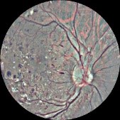
Diabetic Retinopathy (DR) is a retinal complication caused when retinal blood vessels are damaged by diabetes. Signs of DR start with microaneurysms, which are small red spots that appear when there is a blood escape from retinal blood vessels. If microaneurysms are not treated early walls of capillaries may get broken which form hemorrhages. Exudates may appear on the retina if treatment of the disease is delayed, and this can lead to permanent vision loss or vision impairment.
DR diagnosis is clinically performed by ophthalmologists with the help of high-end fundus images capturing devices. In order to capture retinal images, different imaging techniques, such as optical coherence, tomography, and fundus photography, have been used (Salz & Witkin, 2015). All of these techniques come with the challenge of expensive design, deployment, and usage costs. Trained professionals are needed to use these techniques. In addition to the trained professional need of these techniques, an ophthalmologist or more are required to study and diagnose a fundus image that is captured by the imaging methodology. An ophthalmologist usually requires two to seven days for retinal image diagnosis. Diabetes patients that reside in rural and remote areas usually suffer from delayed diagnosis and treatment of DR because of the expensive deployment of diagnosing equipment and shortage of ophthalmologists and health care centers.
Limited access to point-of-care diagnostic services was identified as one of the barriers of medical diagnosis in rural areas (Huaynate et al., 2015). Foster & Resnikoff (2005) put four strategies to fight the challenges the diagnoses process of DR faces in order to implement treatment for preventable blindness; (1) Creating academic, public and governmental awareness of the effects of blindness and visual loss, and the fact that 75% of diseases that cause blindness are preventable; (2) Automating and mobilizing existing techniques and methods; (3) Implementing district-specific and country-specific prioritizing strategies of diagnosing and treatment resources for a productive process; (4) Providing comprehensive, maintainable and fair diagnosis services of visual diseases at district level, which includes staff training, distributing diagnosis and treatment resources, and infrastructure, such as health care center buildings.
A timely and accurate automatic diagnosis of DR could enable diabetic patients to get treatment for preventable visual diseases, thereby avoiding permanent vision loss or impairment. Point-of-care DR diagnostic service, which aims to diagnose DR instantly at a patient’s place, is achieved in this paper using a smartphone for data collection and trained model for detection.
1.2 Annotated Training Data
One of the main issues with incorporating deep learning in medical image analyses is the shortage of available annotated training dataset (Miotto et al., 2017)(Razzak et al., 2018). Transfer learning techniques have gained wider acceptance because of the unavailability of enough annotated training data in the design and training of deep convolutional neural network models (Erhan et al., 2010)(Litjens et al., 2017). In Altaf et al. (2019), annotated training data insufficiency was identified as the main challenge of applying deep learning models in the healthcare automation industry. Furthermore, Altaf et al. (2019) recommended that methods that exploit deep learning using reduced data need to be devised and implemented. We have combined inception module based convolutional neural networks with a binary tree-based ensemble of classifiers to increase the performance of our proposed neural network model with limited training data.
2 Related Work
Although the literature of automated DR detection and classification contains various works, the methods employed can be generally classified into two, which are feature extraction based and image-level based classifications. In order to provide an onsite diagnosis of DR, mobile detection works have also been proposed and implemented. Deep learning models such as Convolutional Neural Networks (CNN) are usually used to classify fundus images. A fundus images dataset that is uploaded to the Kaggle website for DR detection competition (California-Healthcare-Foundation, 2015) has been extensively used in the image level DR classification literature. End-to-end approach has been used to train a deep learning model from scratch. In order to avoid the cost of training, transfer learning has also been used.
In multi-class classification, DR has been classified into one of five stages, which are normal, mild, moderate, severe, and proliferative stages, as proposed by Wilkinson et al. (2003). In the following subsections, different works that have been performed in detecting DR will be reviewed.
2.1 Feature Extraction Based Classification of Diabetic Retinopathy
Lesions of DR such as macular edema, exudates, microaneurysms, and hemorrhages have been automatically identified, segmented and detected in order to classify DR. End-to-end and transfer learning of Convolutional Neural Networks (CNN) and Fully Convolved Residual Networks (FCRN) have been employed for classifying the extracted lesions. Table A.1 (See Appendix A) presents a summary of lesion detection based approaches for detecting DR.
2.2 Image Level Classification of Diabetic Retinopathy
In image level-based classification of DR from fundus images, there is no need to segment and extract lesions. The detection is performed on the whole fundus image. Table A.2 (See Appendix A) summarizes the image level DR detection literature.
2.3 Mobile Classification of Diabetic Retinopathy
One of the strategies suggested by Foster & Resnikoff (2005) for prevention of treatable blindness was the distribution of maintainable and unbiased eye care services at the district level. Tele-retina has been implemented to capture retinal images by non-experts from remote areas and transferring to the ophthalmic center for DR diagnosis (Cavallerano et al., 2004)(Bursell et al., 2012)(Coronado, 2014). Tele retina needs a strong network connection between patients and health care centers. Teleophthalmology, which includes analysis of collected images, can be implemented to provide DR diagnosis remotely (Mohammadpour et al., 2017).
As is put by Foster & Resnikoff (2005), in order to automate and mobilize existing techniques and methods, a smartphone device with mobile retinal image capturing cameras could be used to diagnose DR in remote and rural areas. This can solve the cost and time challenges of the traditional diagnosis of DR.
One of the main challenges of deploying DR automatic diagnosis on a smartphone is the quality of the retinal images captured and the limited processing power and memory inside. A speedy, easy to use, and cheap retinal imaging device could be used as an add-on with a smartphone to collect fundus images of patients as in Kim et al. (2018).
Smartphone based automatic diagnosis of DR can be implemented in two ways. The first way is an internet-based diagnosis, and the second one is a stand-alone independent smartphone application software-based diagnosis. In an internet-based diagnosis, the smartphone is only used to capture a fundus image of a patient with the help of an add-on retinal camera. After a fundus image is captured, it is sent to a professional ophthalmologist for diagnosis, and results would be sent back over the internet. This would require the user to have a stable internet connection for sending the fundus images and receiving back the results of the diagnosis. The challenge of low internet coverage can be solved using an independent smartphone application that is able to process a captured fundus image and automatically diagnose the stage of DR without any network connection.
3 Proposed Methodology
In order to provide an easy to use and independent smartphone based diagnoses of DR, a deep learning model needs to be trained first. After training, the model needs to be deployed in a smartphone application. For clarification, Figure 1(a) presents the flow of the proposed development process.


3.1 Deep Learning Model Training
For training a deep learning model, a dataset of fundus images that are uploaded to Kaggle website (California-Healthcare-Foundation, 2015) has been used. The dataset contains fundus images labeled into five stages of DR. Inspired by the inception module presented in Szegedy et al. (2015) inception based convolutional neural network was designed and implemented. We further discuss image pre- processing, inception based network, and binary decision tree-based ensemble of classifiers.
3.1.1 Image Pre-processing
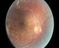
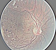
For model training, 200 images were selected from each stage of DR. In order to exploit high performance from a deep learning model and smaller training dataset; image pre-processing was performed by adopting Graham (2015)’s method. The fundus images were cropped to remove unnecessary black background pixels. The local average was subtracted from the cropped fundus image. Cropping of the images’ 10% was repeated to remove white rounding noise. Figure 2(a), 2(b) and 2(c) show the implemented preprocessing on a sample fundus image.
3.1.2 Inception based Network
After the automatic diagnosis is performed, the designed deep learning model should be smartphone adaptable in order to be imported into a mobile application and fit to the memory limit of smartphones. For this reason, an inception module based convolutional neural network was designed. The inception module’s architecture is presented in Figure 3(b). It was designed based on the inception module presented in Szegedy et al. (2015).
The framework of our proposed inception based convolutional neural network is illustrated in Figure 3(a). The network was trained using Stochastic Gradient Decent (SGD) with an ascending learning rate of 0.0001.


3.1.3 Binary Decision Tree-based Ensemble of Classifiers
In order to increase the performance of our proposed inception based convolutional neural network, a binary tree-based ensemble of four binary classifiers were used. Four binary classifiers were trained to classify between normal and mild, normal and moderate, normal and severe, and normal and proliferative stages of DR. The mild, moderate, severe and proliferative stages of the disease are considered to be unhealthy states of the retina. Figure 1(b) presents our proposed binary decision tree-based ensemble of classifiers.
3.2 Smartphone Application Development
A standalone smartphone application software was designed to import the trained binary deep learning networks and implement a binary decision tree-based ensemble to classify fundus images into healthy and unhealthy. The smartphone device’s operating system used was ColorOS version 6.0, which is a variety of Android. Size of the trained binary classifying models was 5757 kilobytes.
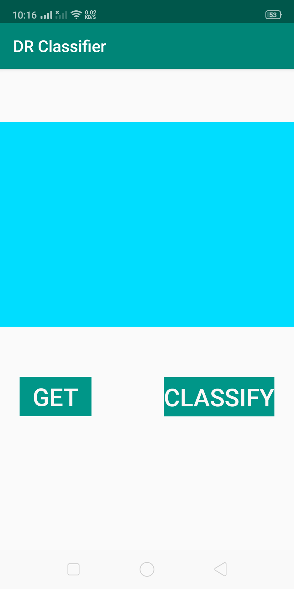
A retinal image is proposed to be captured using add on camera or from the smartphone’s gallery by tapping on the “GET” button as is shown in Figure A.1(a). And the label of the imported retinal image is predicted by tapping on the “CLASSIFY” button.
4 Results and Discussion
In this section, we compare the performance of our trained model with similar works, which have classified DR into binary classes using the Kaggle dataset (California-Healthcare-Foundation, 2015), as shown in Table 4.1.
| POINTS OF COMPARISON | Mohammadian et al. (2017) | Hagos & Kant (2019) | PROPOSED METHOD | |
| Training parameters | Training dataset size | 35126 | 2500 | 1000 |
| Optimizer | ADAM | SGD | SGD | |
| Learning rate | - | Ascending 0.0001 | Ascending 0.0001 | |
| Loss function | - | Cosine loss | Cosine loss | |
| Data augmentation used? | Yes | No | No | |
| Results | Accuracy | 87.12% | 90.90% | 99.86 |
| Sensitivity(%) | - | - | 99.25 | |
| Specificity(%) | - | - | 99.60 | |
| Loss(%) | - | 3.94 | - | |
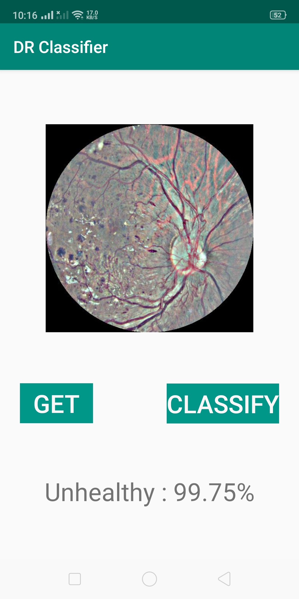
From Table 4.1, our proposed model achieved the best performance with 99.86% accuracy, 99.25% sensitivity and 99.60% specificity followed by the work of Hagos & Kant (2019) and Mohammadian et al. (2017). From this analysis, it shows that our proposed method with limited training data, and without data augmentation performs better than other previous methods. Figure A.1(b) shows our developed smartphone application’s classification output on a sample fundus image.
5 Conclusion
In this paper, inception based convolutional neural network, binary decision tree-based ensemble of classifiers, and android smartphone application development have been combined in order to provide a standalone smartphone based automatic diagnosis of DR with an accuracy of 99.86%. Using our implemented system, diabetic people residing in remote and rural areas with less coverage of healthcare centers and shortage of professional ophthalmologists only need an Android smartphone, and a portable fundus camera add on for an instant DR diagnosis. The methods followed in this paper can lead the way in providing a non-expert usable point-of-care diagnostic services for DR in specific and other medical image classification tasks in general. At this stage, the developed application only diagnoses images from a phone’s gallery. For future work, we plan to perform a multi-class classification of DR, and incorporate a handheld fundus camera to start deployment.
References
- Abràmoff et al. (2016) Michael David Abràmoff, Yiyue Lou, Ali Erginay, Warren Clarida, Ryan Amelon, James C Folk, and Meindert Niemeijer. Improved automated detection of diabetic retinopathy on a publicly available dataset through integration of deep learning. Investigative ophthalmology & visual science, 57(13):5200–5206, 2016.
- Adly et al. (2019) Mohamed M. Adly, Amr S. Ghoneim, and A. Aliaa Youssif. On the grading of diabetic retinopathies using a binary-tree-based multiclass classifier of cnns. International Journal of Computer Science and Information Security (IJCSIS), 17(1), 2019.
- Altaf et al. (2019) Fouzia Altaf, Syed Islam, Naveed Akhtar, and Naeem K Janjua. Going deep in medical image analysis: Concepts, methods, challenges and future directions. arXiv preprint arXiv:1902.05655, 2019.
- Bursell et al. (2012) Sven-Erik Bursell, Laima Brazionis, and Alicia Jenkins. Telemedicine and ocular health in diabetes mellitus. Clinical and Experimental Optometry, 95(3):311–327, 2012.
- California-Healthcare-Foundation (2015) California-Healthcare-Foundation. Diabetic retinopathy detection, 2015. URL https://www.kaggle.com/c/diabetic-retinopathy-detection.
- Cavallerano et al. (2004) J Cavallerano, MG Lawrence, I Zimmer-Galler, W Bauman, S Bursell, WK Gardner, M Horton, L Hildebrand, J Federman, L Carnahan, et al. Telehealth practice recommendations for diabetic retinopathy. Telemedicine journal and e-health: the official journal of the American Telemedicine Association, 10(4):469–482, 2004.
- Colas et al. (2016) E Colas, A Besse, A Orgogozo, B Schmauch, N Meric, and E Besse. Deep learning approach for diabetic retinopathy screening. Acta Ophthalmologica, 94, 2016.
- Coronado (2014) Andrea C Coronado. Diagnostic accuracy of tele-ophthalmology for diabetic retinopathy assessment: A meta-analysis and economic analysis. 2014.
- Costa & Campilho (2017) Pedro Costa and Aurélio Campilho. Convolutional bag of words for diabetic retinopathy detection from eye fundus images. IPSJ Transactions on Computer Vision and Applications, 9(1):10, 2017.
- Dutta et al. (2018) Suvajit Dutta, Bonthala CS Manideep, Syed Muzamil Basha, Ronnie D. Caytiles, and N. Ch. S. N. Iyengar. Classification of diabetic retinopathy images by using deep learning models. International Journal of Grid and Distributed Computing, 11(1):99–106, January 2018. doi: 10.14257/ijgdc.2018.11.1.09. URL https://doi.org/10.14257/ijgdc.2018.11.1.09.
- Erhan et al. (2010) Dumitru Erhan, Yoshua Bengio, Aaron Courville, Pierre-Antoine Manzagol, Pascal Vincent, and Samy Bengio. Why does unsupervised pre-training help deep learning? Journal of Machine Learning Research, 11(Feb):625–660, 2010.
- Foster & Resnikoff (2005) Allen Foster and Serge Resnikoff. The impact of vision 2020 on global blindness. Eye, 19(10):1133, 2005.
- Gargeya & Leng (2017) Rishab Gargeya and Theodore Leng. Automated identification of diabetic retinopathy using deep learning. Ophthalmology, 124(7):962–969, 2017.
- Gondal et al. (2017) Waleed M Gondal, Jan M Köhler, René Grzeszick, Gernot A Fink, and Michael Hirsch. Weakly-supervised localization of diabetic retinopathy lesions in retinal fundus images. In 2017 IEEE International Conference on Image Processing (ICIP), pp. 2069–2073. IEEE, 2017.
- González-Gonzalo et al. (2019) Cristina González-Gonzalo, Verónica Sánchez-Gutiérrez, Paula Hernández-Martínez, Inés Contreras, Yara T Lechanteur, Artin Domanian, Bram van Ginneken, and Clara I Sánchez. Evaluation of a deep learning system for the joint automated detection of diabetic retinopathy and age-related macular degeneration. arXiv preprint arXiv:1903.09555, 2019.
- Graham (2015) Ben Graham. Kaggle diabetic retinopathy detection competition report. University of Warwick, 2015.
- Gulshan et al. (2016) Varun Gulshan, Lily Peng, Marc Coram, Martin C Stumpe, Derek Wu, Arunachalam Narayanaswamy, Subhashini Venugopalan, Kasumi Widner, Tom Madams, Jorge Cuadros, et al. Development and validation of a deep learning algorithm for detection of diabetic retinopathy in retinal fundus photographs. Jama, 316(22):2402–2410, 2016.
- Hagos & Kant (2019) Misgina Tsighe Hagos and Shri Kant. Transfer learning based detection of diabetic retinopathy from small dataset. arXiv preprint arXiv:1905.07203, 2019.
- Huaynate et al. (2015) Cynthia Fiorella Anticona Huaynate, Monica Jehnny Pajuelo Travezaño, Malena Correa, Holger Mayta Malpartida, Richard Oberhelman, Laura L Murphy, and Valerie A Paz-Soldan. Diagnostics barriers and innovations in rural areas: insights from junior medical doctors on the frontlines of rural care in peru. BMC health services research, 15(1):454, 2015.
- Jamil et al. (2018) Ahmad Zeeshan Jamil, Luqman Ali, Muhammad Younis Tahir, and Fazal Shah Shiraz. Smart phone: A smart technology for fundus photography in diabetic retinopathy screening. Pakistan Journal of Ophthalmology, 34(4), 2018.
- Kashyap et al. (2017) Nikita Kashyap, Dharmendra Kumar Singh, and Girish Kumar Singh. Mobile phone based diabetic retinopathy detection system using ann-dwt. In 2017 4th IEEE Uttar Pradesh Section International Conference on Electrical, Computer and Electronics (UPCON), pp. 463–467. IEEE, 2017.
- Kashyap et al. (2019) Nikita Kashyap, Dharmendra Kumar Singh, and Girish Kumar Singh. Color histogram-and smartphone-based diabetic retinopathy detection system. In Engineering Vibration, Communication and Information Processing, pp. 669–678. Springer, 2019.
- Kim et al. (2018) Tyson N Kim, Frank Myers, Clay Reber, PJ Loury, Panagiota Loumou, Doug Webster, Chris Echanique, Patrick Li, Jose R Davila, Robi N Maamari, et al. A smartphone-based tool for rapid, portable, and automated wide-field retinal imaging. Translational vision science & technology, 7(5):21–21, 2018.
- Litjens et al. (2017) Geert Litjens, Thijs Kooi, Babak Ehteshami Bejnordi, Arnaud Arindra Adiyoso Setio, Francesco Ciompi, Mohsen Ghafoorian, Jeroen Awm Van Der Laak, Bram Van Ginneken, and Clara I Sánchez. A survey on deep learning in medical image analysis. Medical image analysis, 42:60–88, 2017.
- Mansour (2018) Romany F. Mansour. Deep-learning-based automatic computer-aided diagnosis system for diabetic retinopathy. Biomedical Engineering Letters, 8(1):41–57, Feb 2018. ISSN 2093-985X. doi: 10.1007/s13534-017-0047-y. URL https://doi.org/10.1007/s13534-017-0047-y.
- Mateen et al. (2018) Muhammad Mateen, Junhao Wen, Nasrullah, Sun Song, and Zhouping Huang. Fundus image classification using VGG-19 architecture with PCA and SVD. Symmetry, 11(1):1, December 2018. doi: 10.3390/sym11010001. URL https://doi.org/10.3390/sym11010001.
- Miotto et al. (2017) Riccardo Miotto, Fei Wang, Shuang Wang, Xiaoqian Jiang, and Joel T Dudley. Deep learning for healthcare: review, opportunities and challenges. Briefings in bioinformatics, 19(6):1236–1246, 2017.
- Mo et al. (2018) Juan Mo, Lei Zhang, and Yangqin Feng. Exudate-based diabetic macular edema recognition in retinal images using cascaded deep residual networks. Neurocomputing, 290:161–171, 2018.
- Mohammadian et al. (2017) Saboora Mohammadian, Ali Karsaz, and Yaser M Roshan. Comparative study of fine-tuning of pre-trained convolutional neural networks for diabetic retinopathy screening. In 2017 24th National and 2nd International Iranian Conference on Biomedical Engineering (ICBME), pp. 1–6. IEEE, 2017.
- Mohammadpour et al. (2017) Mehrdad Mohammadpour, Zahra Heidari, Masoud Mirghorbani, and Hassan Hashemi. Smartphones, tele-ophthalmology, and vision 2020. International journal of ophthalmology, 10(12):1909, 2017.
- Nagasawa et al. (2019) Toshihiko Nagasawa, Hitoshi Tabuchi, Hiroki Masumoto, Hiroki Enno, Masanori Niki, Zaigen Ohara, Yuki Yoshizumi, Hideharu Ohsugi, and Yoshinori Mitamura. Accuracy of ultrawide-field fundus ophthalmoscopy-assisted deep learning for detecting treatment-naïve proliferative diabetic retinopathy. International ophthalmology, pp. 1–7, 2019.
- Orlando et al. (2018) Jose Ignacio Orlando, Elena Prokofyeva, Mariana del Fresno, and Matthew B Blaschko. An ensemble deep learning based approach for red lesion detection in fundus images. Computer methods and programs in biomedicine, 153:115–127, 2018.
- Perdomo et al. (2016) Oscar Perdomo, Sebastian Otalora, Francisco Rodríguez, John Arevalo, and Fabio A González. A novel machine learning model based on exudate localization to detect diabetic macular edema. 2016.
- Perdomo et al. (2017) Oscar Perdomo, John Arevalo, and Fabio A González. Convolutional network to detect exudates in eye fundus images of diabetic subjects. In 12th International Symposium on Medical Information Processing and Analysis, volume 10160, pp. 101600T. International Society for Optics and Photonics, 2017.
- Prasanna et al. (2013) Prateek Prasanna, Shubham Jain, Neelakshi Bhagat, and Anant Madabhushi. Decision support system for detection of diabetic retinopathy using smartphones. In 2013 7th International Conference on Pervasive Computing Technologies for Healthcare and Workshops, pp. 176–179. IEEE, 2013.
- Pratt et al. (2016) Harry Pratt, Frans Coenen, Deborah M Broadbent, Simon P Harding, and Yalin Zheng. Convolutional neural networks for diabetic retinopathy. Procedia Computer Science, 90:200–205, 2016.
- Prentašić & Lončarić (2016) Pavle Prentašić and Sven Lončarić. Detection of exudates in fundus photographs using deep neural networks and anatomical landmark detection fusion. Computer methods and programs in biomedicine, 137:281–292, 2016.
- Quellec et al. (2017) Gwenolé Quellec, Katia Charrière, Yassine Boudi, Béatrice Cochener, and Mathieu Lamard. Deep image mining for diabetic retinopathy screening. Medical image analysis, 39:178–193, 2017.
- Rajalakshmi et al. (2018) Ramachandran Rajalakshmi, Radhakrishnan Subashini, Ranjit Mohan Anjana, and Viswanathan Mohan. Automated diabetic retinopathy detection in smartphone-based fundus photography using artificial intelligence. Eye, 32(6):1138, 2018.
- Razzak et al. (2018) Muhammad Imran Razzak, Saeeda Naz, and Ahmad Zaib. Deep learning for medical image processing: Overview, challenges and the future. In Classification in BioApps, pp. 323–350. Springer, 2018.
- Salz & Witkin (2015) David A Salz and Andre J Witkin. Imaging in diabetic retinopathy. Middle East African journal of ophthalmology, 22(2):145, 2015.
- Szegedy et al. (2015) Christian Szegedy, Wei Liu, Yangqing Jia, Pierre Sermanet, Scott Reed, Dragomir Anguelov, Dumitru Erhan, Vincent Vanhoucke, and Andrew Rabinovich. Going deeper with convolutions. In Proceedings of the IEEE conference on computer vision and pattern recognition, pp. 1–9, 2015.
- Ting et al. (2017) Daniel Shu Wei Ting, Carol Yim-Lui Cheung, Gilbert Lim, Gavin Siew Wei Tan, Nguyen D Quang, Alfred Gan, Haslina Hamzah, Renata Garcia-Franco, Ian Yew San Yeo, Shu Yen Lee, et al. Development and validation of a deep learning system for diabetic retinopathy and related eye diseases using retinal images from multiethnic populations with diabetes. Jama, 318(22):2211–2223, 2017.
- Verbraak et al. (2019) Frank D Verbraak, Michael D Abramoff, Gonny CF Bausch, Caroline Klaver, Giel Nijpels, Reinier O Schlingemann, and Amber A van der Heijden. Diagnostic accuracy of a device for the automated detection of diabetic retinopathy in a primary care setting. Diabetes care, 42(4):651–656, 2019.
- Wilkinson et al. (2003) CP Wilkinson, Frederick L Ferris III, Ronald E Klein, Paul P Lee, Carl David Agardh, Matthew Davis, Diana Dills, Anselm Kampik, R Pararajasegaram, Juan T Verdaguer, et al. Proposed international clinical diabetic retinopathy and diabetic macular edema disease severity scales. Ophthalmology, 110(9):1677–1682, 2003.
- Xu et al. (2016) Xiayu Xu, Wenxiang Ding, Xuemin Wang, Ruofan Cao, Maiye Zhang, Peilin Lv, and Feng Xu. Smartphone-based accurate analysis of retinal vasculature towards point-of-care diagnostics. Scientific reports, 6:34603, 2016.
Appendix A Appendix
| AUTHORS | METHODS | TRAINING | LESIONS | PERFORMANCE | ||
|---|---|---|---|---|---|---|
| SN(%) | SP(%) | ACC(%) | ||||
| Perdomo et al. (2016) | CNN | Transfer learning | Macula edema | 56.50 | 92.80 | 77.00 |
| Prentašić & Lončarić (2016) | CNN with 11 layers | End-to-end | Exudates | 78.00 | - | - |
| Abràmoff et al. (2016) | CNN | End-to-end | Macula edema | 100.00 | - | - |
| Perdomo et al. (2017) | LeNet CNN | Transfer learning | Exudates | 99.80 | 99.60 | 99.60 |
| Mo et al. (2018) | FCRN | End-to-end | Macula edema | 92.55 | - | - |
| Gondal et al. (2017) | Octree based CNN and Transfer learning | Transfer learning | Microaneurysms | 52.00 | - | - |
| Haemorrhages | 91.00 | - | - | |||
| AUTHORS | METHODS | TRAINING | PERFORMANCE | |||
|---|---|---|---|---|---|---|
| SN(%) | SP(%) | ACC(%) | AUC | |||
| Gulshan et al. (2016) | Inception-V3 CNN | Transfer learning | 90.30 | 90.00 | - | 0.99 |
| Colas et al. (2016) | CNN | End-to-end | 96.20 | 66.60 | - | 0.95 |
| Pratt et al. (2016) | CNN with 13 layers | End-to-end | 95.00 | 30.00 | 75.00 | - |
| Mohammadian et al. (2017) | Inception-V3 | Transfer learning | - | - | 87.12 | - |
| Quellec et al. (2017) | AlexNet CNN | Transfer learning | - | - | - | 0.95 |
| Costa & Campilho (2017) | CNN | End-to-end | 78.00 | - | - | 0.97 |
| Abràmoff et al. (2016) | CNN | End-to-end | 96.80 | 87.00 | - | 0.98 |
| Gargeya & Leng (2017) | ResNet | End-to-end | 94.00 | 98.00 | - | - |
| Ting et al. (2017) | CNN | End-to-end | 90.5 | 91.60 | - | 0.94 |
| Dutta et al. (2018) | Deep neural network | End-to-end | - | - | 86.30 | - |
| Mateen et al. (2018) | VGGNet-19 with SVM | End-to-end | - | - | 98.34 | - |
| Mansour (2018) | AlexNet with SVM classifier | Transfer learning | 100.00 | 93.00 | 97.93 | - |
| Orlando et al. (2018) | CNN | End-to-end | 97.21 | 50.00 | - | 0.93 |
| Adly et al. (2019) | VggNet | End-to-end | 81.80 | 89.30 | 83.20 | - |
| Nagasawa et al. (2019) | CNN | End-to-end | 94.70 | 97.20 | - | 0.97 |
| Verbraak et al. (2019) | Detection Validation of a device | End-to-end | 100.00 | - | - | - |
| González-Gonzalo et al. (2019) | Performance validation | End-to-end | 92.00 | 92.10 | - | 0.97 |
| Hagos & Kant (2019) | Inception-V3 network | Transfer learning with fine-tuning | - | - | 90.90 | - |
| AUTHORS | TECHNIQUES | CAMERA | PERFORMANCE(%) | |||
|---|---|---|---|---|---|---|
| SN | SP | PRECISION | ACC | |||
| Prasanna et al. (2013) | Feature extraction based classification | ophthalmoscope | - | - | - | - |
| Xu et al. (2016) | Vessel segmentation | - | - | - | - | 93.30 |
| Kashyap et al. (2017) | DWT based classification | LED with a condensing lens | 57.00 | - | 63.00 | - |
| Rajalakshmi et al. (2018) | Cloud-based diagnostic | Smartphone | 99.10 | 80.40 | - | - |
| Jamil et al. (2018) | Performance validation of mobile diagnosis | 20 dioptre condensing lens with a phone camera | - | - | - | - |
| Kashyap et al. (2019) | Histogram comparison | LED with a condensing lens | 53.00 | - | 62.00 | - |