Realization of the orbital-selective Mott state at the molecular level in \chBa3LaRu2O9
Abstract
Molecular magnets based on heavy transition metals have recently attracted significant interest in the quest for novel magnetic properties. For systems with an odd number of valence electrons per molecule, high or low molecular spin states are typically expected in the double exchange or quasi-molecular orbital limits respectively. In this work, we use bulk characterization, muon spin relaxation, neutron diffraction, and inelastic neutron scattering to identify a rare intermediate spin-3/2 per dimer state in the 6H-perovskite Ba3LaRu2O9 that cannot be understood in a double exchange or quasi-molecular orbital picture and instead arises from orbital-selective Mott insulating behavior at the molecular level. Our measurements are also indicative of collinear stripe magnetic order below 26(1) K for these molecular spin-3/2 degrees-of-freedom, which is consistent with expectations for an ideal triangular lattice with significant next nearest neighbor in-plane exchange. Finally, we present neutron diffraction and Raman scattering data under applied pressure that reveal low-lying structural and spin state transitions at modest pressures P 1 GPa, which highlights the delicate balance between competing energy scales in this system.
I Introduction
The interplay between charge, spin, lattice and orbital degrees of freedom leads to a tremendous variety of exotic phenomena in strongly-correlated electron systems. One particularly famous example is the metal-Mott insulator transition, where a Coulomb repulsion that is strong relative to orbital hopping leads to a significant modification of the band structure and promotes complete electron localization and hence local moment physics. More recently, the intriguing concept of an orbital-selective Mott phase was proposed in order to explain the coexistence of itinerant and localized electron character in the ruthenate system SrCaRuO4 Anisimov et al. (2002). This state can be achieved in multi-orbital systems with disparate orbital hoppings, leading to a situation where some valence electrons are localized while others are itinerant. Although the realization of the orbital-selective Mott phase is still under debate for SrCaRuO4 Wang et al. (2004); Balicas et al. (2005); Liebsch and Ishida (2007); Neupane et al. (2009); de’ Medici et al. (2009), this state has been the subject of several theoretical investigations Koga et al. (2004); de’Medici et al. (2005); Ferrero et al. (2005); Liebsch (2005); Biermann et al. (2005); Rincón et al. (2014a, b) and has been discussed in the context of iron-based superconductivity Yu and Si (2012, 2013), high- cuprates Venturini et al. (2002), the metal-insulator transition in V2O3 Laad et al. (2006), and the magnetic properties of double perovskites Chen (2018) and BaFe2Se3 Mourigal et al. (2015); Herbrych et al. (2018).
Orbital-selective Mott physics may also play an important role in heavy transition metal (i.e. 4 and 5) molecular magnets Streltsov and Khomskii (2016, 2017). The large spatial extent of the orbitals and the reduction of Hund’s coupling generate competing energy scales that can invalidate the local moment / double exchange picture generally expected in their 3 transition metal counterparts and therefore have significant consequences on their electronic ground states and magnetic properties. For a system with two types of orbitals (c and d), three regimes are possible depending on the relative strengths of , , , and : (i) the quasi-molecular orbital limit (, , ), (ii) the local moment / double exchange limit (, , ), and (iii) the orbital-selective limit ( , , 0) Streltsov and Khomskii (2014). An accurate determination of the electronic ground state for these molecular magnets is the first step towards developing a detailed understanding of their collective magnetic properties, which may be quite interesting if the molecules are strongly-interacting.
Heavy transition metal molecular magnets with an odd number of electrons per dimer are an excellent playground for identifying systems and establishing trends where the double exchange picture breaks down at the molecular level. The competition between Hund’s coupling and orbital hopping is particularly striking in this case, as the electronic ground state evolves from high to low spin with increasing orbital hopping Streltsov and Khomskii (2016). In principle, for particular electron configurations an intermediate spin ground state can also be realized in the orbital-selective regime but experimental examples are lacking. This class of materials may therefore enable detailed investigations of low-lying spin state transitions, which are not common in magnetic materials based on single ion building blocks. Furthermore, in the quasi-molecular orbital limit they can also generate new 1/2 quantum magnets with ideal frustrated lattice geometries that are less susceptible to Jahn-Teller distortions Ziat et al. (2017) and in some cases are under active consideration as quantum spin liquid candidates Sheckelton et al. (2012); Mourigal et al. (2014); Akbari-Sharbaf et al. (2018).
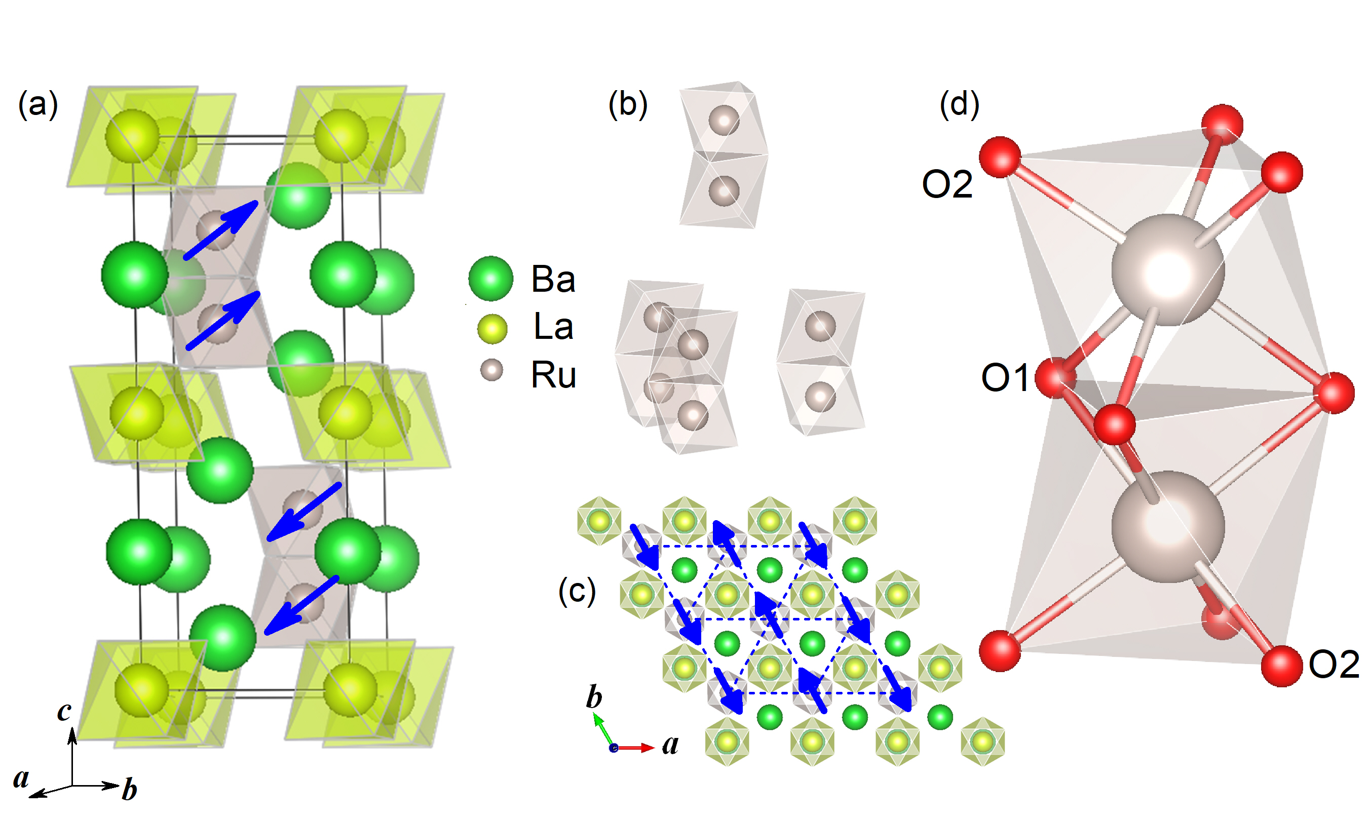
The 6H-perovskites, with the general chemical formula Ba3MR2O9, consist of transition metal dimers decorating a triangular lattice as illustrated in Fig. 1 and they have already been shown to host a variety of interesting electronic ground states at the molecular level. For example, Ba3NaRu2O9 exhibits interdimer charge order below 210 K Kimber et al. (2012), Ba3(Y,In,Lu)Ru2O9 are quantum magnets with the 1/2 degree of freedom delocalized over the Ru dimers Ziat et al. (2017), and Ba3CeRu2O9 has a non-magnetic ground state that arises from quasi-molecular orbital formation combined with a large zero field splitting Chen et al. (2019). The rich molecular behavior in this family likely arises from the ability of this structure to accommodate heavy transition metal dimers based on face-sharing octahedra, which feature metal-metal distances shorter than the nearest neighbor distance in the corresponding elemental metal Chen et al. (2019) in some cases. In fact, quasi-molecular orbital formation has been argued to give rise to the quantum magnetism in Ba3(Y,In,Lu)Ru2O9 Ziat et al. (2017) rather than the large spin state of 5/2 per dimer that would be realized by a double exchange mechanism Bechlars et al. (2010).
Interestingly, despite the same valence electron count of seven per Ru dimer, the magnetic properties of the isostructural system Ba3LaRu2O9 have been shown to be drastically different. The effective and ordered moments per dimer, extracted via magnetic susceptibility Doi et al. (2002) and neutron diffraction Senn et al. (2013) measurements respectively, are much larger and cannot be explained by an 1/2 electronic ground state for the dimers. No satisfactory explanation for this different behavior has been proposed to-date and the true electronic ground state of the Ru dimers in Ba3LaRu2O9 has remained an open question. In this work, we combine magnetometry, heat capacity, muon spin relaxation, neutron diffraction, and inelastic neutron scattering to identify an 3/2 electronic ground state for the dimers that we argue arises from an orbital-selective mechanism at the molecular level. We also establish an ordering temperature of 26(1) K for the stripe spin configuration that is expected for a triangular lattice with a significant next-nearest neighbor in-plane exchange interaction. Finally, we use neutron powder diffraction and Raman scattering under applied pressure to show that moderate pressures of 1 GPa generate both structural and spin state transitions in Ba3LaRu2O9.
II Experimental methods
Polycrystalline samples of \chBa3LaRu2O9 were synthesized by a solid-state reaction using a stoichiometric amount of the starting materials \chBaCO3, Ru, and \chLa2O3 (fine powder predried at 950°C overnight) with purities of 99.9% or higher. The starting materials were mixed in agate mortars, pressed into pellets, annealed in air at 900°C for 12 hours, and then annealed at 1200°C for 20 hours with intermediate grinding and pelletizing.
The room temperature X-ray powder diffraction (XRD) patterns were collected using a HUBER Image Plate Guinier Camera 670 with Ge monochromatized Cu radiation ( Å) to check the quality of the powder samples. No obvious impurity peaks were observed.
The dc magnetic susceptibility and magnetization measurements were performed in the temperature range of 2-320 K using a Quantum Design superconducting interference device (SQUID) magnetometer. The high-temperature magnetic susceptibility was measured with a Quantum Design Magnetic Property Measurement System (MPMS) in the temperature range of 300-800 K. The specific heat measurements were performed using the relaxation method with a commercial Physical Property Measurement System (PPMS) from Quantum Design.
Neutron powder diffraction (NPD) was performed on 6.5 g of polycrystalline \chBa3LaRu2O9 using the HB-2A powder diffractometer of the High Flux Isotope Reactor (HFIR) at Oak Ridge National Laboratory (ORNL) Calder et al. (2018). The sample was loaded in a cylindrical vanadium can, and the data were collected at different temperatures ranging from 1.5 K to 300 K with neutron wavelengths of 1.54 Å and 2.41 Å and a collimation of open-21′-12′. The ambient pressure HB-2A data was refined using the FullProf software suite Rodríguez-Carvajal (1993) and the magnetic structure symmetry analysis was performed using SARAh Wills (2000). Further NPD studies were also carried out on HB-2A with 4.5 g of polycrystalline \chBa3LaRu2O9 and a Fluorinert pressure medium first loaded in a teflon tube and then placed in a Cu-Be clamp cell capable of applying hydrostatic pressures up to 1 GPa. Elastic neutron scattering measurements, complementary to the ambient pressure NPD experiment described above, were performed on the 14.6 meV fixed-incident-energy triple-axis spectrometer HB-1A of the HFIR at ORNL using the same polycrystalline sample of \chBa3LaRu2O9 measured in ambient pressure at HB-2A over a temperature range 1.5 K to 40 K. For this experiment, the sample was loaded in a cylindrical Al can to minimize incoherent nuclear scattering that could prevent the detection of weak magnetic Bragg peaks not observed in the initial HB-2A experiment. The overall background was minimized by using a double-bounce monochromator system, mounting two-highly oriented pyrolytic graphite (PG) filters in the incident beam to remove higher-order wavelength contamination, and placing an analyzer of PG crystals before the single He-3 detector for energy discrimination. A collimation of 40′-40′-40′-80′ resulted in an energy resolution at the elastic line just over 1 meV (FWHM).
Inelastic neutron-scattering (INS) measurements were performed on the direct-geometry time-of-flight chopper spectrometer SEQUOIA Granroth et al. (2010) of the Spallation Neutron Source (SNS) at ORNL, using the same \chBa3LaRu2O9 polycrystalline measured in the ambient pressure HB-2A experiment. The sample was loaded in a cylindrical Al can and spectra were collected with incident energies 25 and 100 meV at temperatures of 4 K (both incident energies), 30 K (25 meV only), and 300 K (100 meV only). An empty aluminum can was measured in identical experimental conditions for a similar counting time. The resulting background spectra were subtracted from the corresponding sample spectra after normalization with a vanadium standard to account for variations of the detector response and the solid angle coverage.
Muon spin relaxation (SR) measurements were performed on the M20 surface muon beamline at TRIUMF. A low-background “veto” set-up was employed with the sample mounted inside a mylar packet placed in the path of the muon beam within a helium flow cryostat. Measurements were performed in zero field and longitudinal field geometries over a temperature range of 1.5 to 60 K. A good review of the SR technique can be found in Ref. Yaouanc and De Reotier (2011).
Raman scattering was performed under compression between 0 to 10.36 GPa using diamond anvil cell techniques. Polycrystalline material was loaded into a symmetric diamond anvil cell along with an annealed ruby ball, and KBr was used as the pressure medium for the measurement. This assured a quasi-hydrostatic environment for the sample. Fluorescence from the ruby ball was used to determine pressure Mao et al. (1986). These experiments were carried out using the COMPRES beamline facility at the National Synchrotron Light Source II at Brookhaven National Laboratory. We employed = 532 nm; 1 mW power; 30 sec integration, averaged three times. All data were collected at room temperature.
III Results and Discussion
III.1 (I) Electronic ground state of the Ru dimers
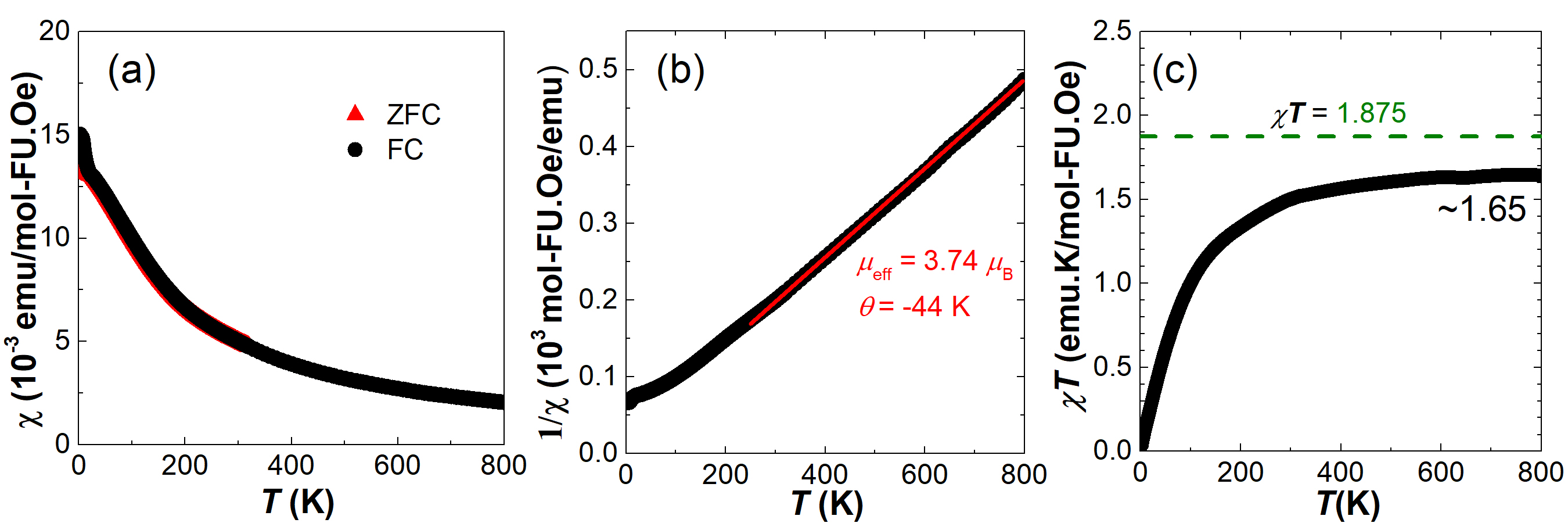
Figure 2(a) shows the dc magnetic susceptibility (plotted as ) vs temperature for \chBa3LaRu2O9 measured from 2 to 800 K in an applied magnetic field of 1 kOe under both zero-field cooled (ZFC) and field-cooled (FC) conditions. The low-temperature data below 25 K is indicative of magnetic order and will be discussed more in the next section. The Curie-Weiss fit of the high-temperature inverse susceptibility data (above 250 K) is shown in Fig. 2(b). This fit results in a Curie-Weiss temperature K and an effective moment /FU (\chBa3LaRu2O9 formula unit), which is close to the expected value of 3.87/dimer for a molecular spin-3/2 state. The versus plot shown in Fig. 2(c) reaches a saturation value of 1.65 that is slightly below for spin-3/2, which could be due to the thermal population of spin-1/2 excited states in the high-temperature region (up to 800 K). This suggests that the Ru-dimers in \chBa3LaRu2O9 adopt an unusual spin-3/2 ground state, in sharp contrast to the isostructural analogs Ba3(Y,In,Lu)Ru2O9 that are known to host spin-1/2 dimer ground states Ziat et al. (2017).
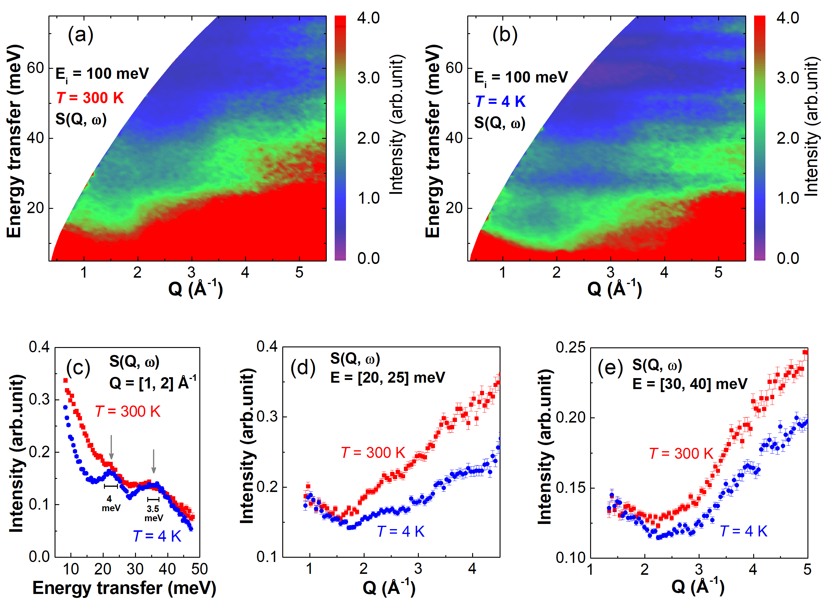
To obtain additional evidence for the exotic intermediate spin state of the Ru dimers in \chBa3LaRu2O9, INS measurements were performed on the SEQUOIA spectrometer with an incident energy meV. Figure 3(a) and (b) show the color contour plots of the dynamical structure factor multiplied by the magnetic form factor squared at room temperature (300 K) and base temperature (4 K), respectively. The spectra are dominated by the phonon modes in the high momentum transfer () regions, which makes it challenging to identify any weak magnetic modes. For this reason, constant- cuts of for both the 4 K and 300 K data sets are plotted in Fig. 3(c) with a integration range of 1 to 2 Å-1. We can now clearly observe two peaks centered at energy transfers 22 and 35 meV, indicated by gray arrows, corresponding to magnetic excitation candidates. The -dependence of these two peaks is shown with constant energy cuts plotted in Fig. 3(d) and (e) respectively. The intensity of both peaks decreases with increasing in the low- ( 2 Å-1) region and therefore they have a magnetic origin. The persistence of these two modes up to 300 K suggests that they correspond to d-d excitations and not collective magnetic excitations (i.e. spin waves). On the other hand, we identified a third magnetic mode just above the elastic line that is dispersive in nature with the expected temperature-dependence for a spin wave origin. This mode is most clearly observed in lower 25 meV data and will be discussed in more detail later.
| B’ | In (3.5 K) | Y (3.5 K) | Lu (1.5 K) | La (1.5 K) |
|---|---|---|---|---|
| a(Å) | 5.7947(1) | 5.8565(1) | 5.8436(1) | 5.95103(14) |
| c(Å) | 14.2738(2) | 14.4589(1) | 14.3978(2) | 15.0087(4) |
| Ba2-z | 0.9116(2) | 0.9075(1) | 0.9084(2) | 0.8910(2) |
| Ru-z | 0.1611(1) | 0.1632(1) | 0.1620(1) | 0.16458(16) |
| O1-x | 0.4874(5) | 0.4879(4) | 0.4887(5) | 0.4866(5) |
| O2-x | 0.1712(4) | 0.1758(2) | 0.1741(3) | 0.1787(4) |
| O2-z | 0.4150(1) | 0.4124(1) | 0.4138(1) | 0.40457(8) |
| 8.82 | 6.27 | 6.18 | 5.83 | |
| Ru-Ru(Å) | 2.538(3) | 2.511(2) | 2.533(3) | 2.564(3) |
| Ru-O1(Å) | 2.001(3) | 2.009(2) | 2.019(2) | 2.034(3) |
| Ru-O2(Å) | 1.956(2) | 1.936(1) | 1.947(2) | 1.902(3) |
| Ru-O1-Ru(∘) | 78.8(1) | 77.4(1) | 77.7(1) | 78.16(15) |
Our previous magnetic susceptibility and INS work on Ba3(Y,In,Lu)Ru2O9 Ziat et al. (2017) revealed a single d-d excitation in all three cases with an energy transfer ranging between 31.5 and 34 meV, which we identified as a molecular transition from the 1/2 ground state to the 3/2 excited state within a quasi-molecular orbital picture. This interpretation is consistent with the neutron scattering selection rule 0,1 Furrer et al. (2010). Although these excited modes were not resolution-limited, the broadening may arise from a finite amount of zero field splitting. The current INS data on Ba3LaRu2O9 provides an interesting contrast, as two d-d excitations are observed in a similar energy regime. This observation is consistent with an 3/2 electronic ground state for the Ru dimers if these two modes represent transitions to the 1/2 and 5/2 manifolds, as these excitations are both allowed by selection rules. We assign the lower and upper modes to the 1/2 and 5/2 transitions, respectively, as the former is nearly resolution-limited while the latter exhibits increased broadening expected to arise from significant zero field splitting of an 5/2 state. We also note that this assignment is consistent with the high-temperature magnetic susceptibility data described above and the previous determination that the nearly isostructural systems Ba3(Y,In,Lu)Ru2O9 host 1/2 electronic ground states.
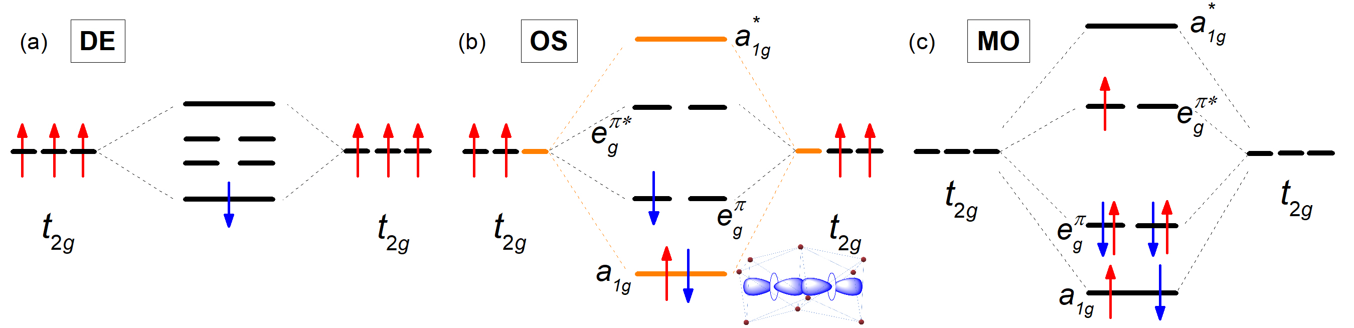
The orbital diagram for Ba3(Y,In,Lu)Ru2O9, with seven valence electrons per Ru dimer, has been discussed previously and consists of a lower-energy bonding level and a higher-energy bonding level Ziat et al. (2017) that are made up of linear combinations of the atomic -orbitals Kugel et al. (2015). In these cases, the extremely short Ru-Ru distances arising from the face-sharing octahedral geometry of the Ru dimers and the spatially-extended 4 orbitals lead to the low-spin (i.e. 1/2) molecular orbital diagram illustrated in Fig. 4(c), rather than the high-spin (i.e. 5/2) double exchange scenario shown in Fig. 4(a) typically expected for molecular magnets based on 3 transition metals. To gain some insight into why the Ru dimers realize a different electronic ground state in \chBa3LaRu2O9, we revisited the low-temperature crystal structure of this system using the HB-2A powder diffractometer with a neutron wavelength of 1.54 Å. Our new refinement results for \chBa3LaRu2O9 are presented in Table I and compared to our previous work on Ba3(Y,In,Lu)Ru2O9 Ziat et al. (2017). While we find broad agreement with earlier diffraction work Doi et al. (2002); Senn et al. (2013), we note that our low-temperature Ru-Ru and Ru-O1 distances are slightly larger for \chBa3LaRu2O9 compared to Ba3(Y,In,Lu)Ru2O9. Since the orbitals are aligned and overlap directly, as shown in the inset of Fig. 4(b), the orbital hopping is most effectively tuned with the Ru-Ru distance. The situation is quite different for the orbitals, where the reduced direct overlap ensures that the orbital hopping is smaller and determined by both the Ru-Ru and Ru-O1 distances. It appears that the seemingly subtle differences in the Ru-Ru and Ru-O1 distances in \chBa3LaRu2O9 and Ba3(Y,In,Lu)Ru2O9 are still large enough to effectively tune and generate a spin state transition in this family of materials. This scenario provides a natural explanation for the intermediate spin state of the Ru dimers in \chBa3LaRu2O9, as it can arise from the orbital diagram presented in Fig. 4(b). Due to the large and the comparatively smaller , only the manifold participates in molecular bonding. Presumably, the other five electrons engage in a double exchange process to generate the 3/2 spin degree of freedom.
III.2 (II) Collective static magnetic properties
With the electronic ground state of the Ru dimers in \chBa3LaRu2O9 firmly established as 3/2 due to orbital-selective behavior, we now examine the collective static magnetic properties of this molecular magnet. There are two previous reports on this topic Doi et al. (2002); Senn et al. (2013), but several open questions remain. The initial X-ray diffraction and bulk characterization study reveals two possible magnetic transitions in the specific heat at 6 K and 22 K, while the magnetic susceptibility shows a clear peak at and a very subtle bump at . Follow-up neutron powder diffraction work using the WISH spectrometer at ISIS revealed magnetic Bragg peaks for 10 K, but no precise magnetic transition temperature was reported so the origin of and remains unknown. Furthermore, the magnetic Bragg peaks observed in the NPD data were modeled within a local moment picture with intradimer ferromagnetic exchange that was noted to be unusual and the magnetic structure could only be explained by a model consisting of two irreducible representations with a confounding moment direction.
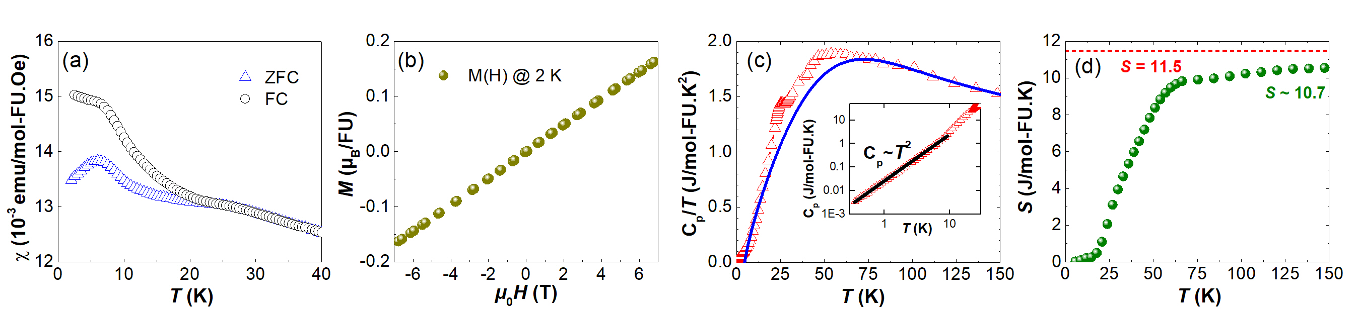
We performed a series of measurements to explore these issues. First, we present dc magnetic susceptibility data up to 40 K on our \chBa3LaRu2O9 polycrystalline samples in Fig. 5(a). We find a zero-field-cooled (ZFC) / field-cooled (FC) divergence that onsets below 25 K and a broad peak at 6 K in both the ZFC and FC data. These findings are in broad agreement with previous work Doi et al. (2002), although our value is slightly higher. We also plot the magnetization as a function of field at 2 K in Fig. 5(b) and we find a linear response which is indicative of a collinear antiferromagnetic ground state. Next, we show heat capacity data over a much wider temperature range than published previously Doi et al. (2002) in Fig. 5(c). Interestingly, this data shows a clear anomaly at 25 K and a Schottky anomaly centered around 50 K, but no obvious feature around . We also note that the lowest temperature data measured between 300 mK and 10 K exhibits a -dependence as shown in the Fig. 5(c) inset. This -dependence can arise from different origins, including a spin wave contribution from a gapless quasi-two-dimensional antiferromagnet, and will be discussed in more detail later. The magnetic entropy was extracted from the data after subtracting the lattice contribution that was approximated by a Thirring model Thirring (1913); Gordon et al. (1989); Cheng et al. (2005) and the result is presented in Fig. 5(d). We find that the entropy recovered up to 150 K ( 10.7 J/mol FU-K) is only slightly lower than expectations for a 3/2 molecular degree of freedom ( 11.5 J/mol FU-K), which is consistent with the Ru dimer electronic ground state that we described above. This result also suggests that the Schottky anomaly arises from a small zero field splitting of the 3/2 state.
Since we were not able to identify a definitive origin for the magnetic transition in \chBa3LaRu2O9 from our bulk characterization measurements, we performed muon spin relaxation (SR) on our samples. This technique is extremely sensitive to local magnetic fields and magnetic volume fractions, so it can be used to readily differentiate between scenarios where the transition arises from a small magnetic impurity phase or a magnetic structure change intrinsic to \chBa3LaRu2O9. Muon spin polarization (i.e. polarization) plotted as a function of time at various temperatures is shown in Fig. 6(a) and reveals clear evidence of static magnetism appearing below a temperature of roughly 25 K, which is in excellent agreement with our bulk characterization data. Closer inspection of the lowest temperature data reveals highly-damped oscillations, as shown in Fig. 6(b), indicating long-range order with appreciable decoherence (short ). The polarization could be successfully fit with the following function:
| (1) |
where is the dephasing or decoherence rate and is the spin-lattice relaxation rate. Two different frequencies were used, which likely correspond to muon stopping sites near the two inequivalent oxygen sites in the structure since 1Å muon-oxygen bonds are commonly found in oxides Holzschuh et al. (1983). It was not possible to successfully fit the data with the Koptev-Tarasov function Yaouanc and De Reotier (2011), which accounts for damping of oscillations through significant inhomogeneity of the internal fields, rather than decoherence. Similar results were obtained for two samples studied (A and B). Slight discrepancies are observed, but the oscillation frequencies are the same within the uncertainty on the fitting parameters. More precisely, in sample A we obtained s-1 and s-1. In sample B, s-1 and s-1. Since this data for the two samples is in excellent agreement, we focus on the sample A measurements in the rest of this section.
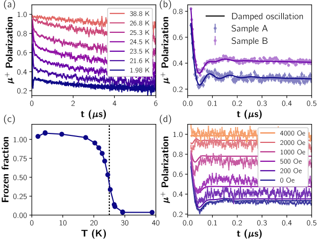
To identify the onset of the magnetic order and hence the transition temperature, we have fit the slowly-relaxing part of the zero-field polarization at s for sample A to a simple exponential function, , where represents the frozen fraction. Here the amplitude of the slowly-relaxing component contains both the non-frozen fraction of the sample and the 1/3 tail from the static fraction. The temperature-dependence of the ordered volume fraction extracted from this analysis is shown in Fig. 6(c) and essentially consistent with a fully-ordered sample.
In Fig. 6(d), a longitudinal field scan of the muon spin polarization is shown and demonstrates that Oe is sufficient to completely decouple the muon spins from internal magnetic fields (which are at most around 850 Oe). A simulation of the expected longitudinal field behavior for each muon stopping site was performed using the following equation:
| (2) |
with the magnitude of the magnetic field given by
| (3) |
and with and . Here, represents the internal field for the th muon stopping site in the absence of applied field. While this simulation [see Fig. 6(d)] is not entirely successful, it is also quite an oversimplification. Here we only consider the effects of the addition of the longitudinal field onto a unique (but randomly-oriented) internal field with linewidth and relaxation effects added afterwards in an ad hoc fashion. The main deviations from theory are at low fields where the linewidth is comparable to the applied field and our model is particularly crude. The fields at which complete decoupling is achieved are well-captured by the model and overall this analysis is fully consistent with the notion of static magnetism in \chBa3LaRu2O9.
These SR measurements provide support for two important conclusions. Firstly, the highest frequency observed in these samples (72 s-1 for sample A and 79 s-1 for sample B) is roughly six times higher than the highest frequency observed in the isostructural material \chBa3LuRu2O9 Ziat et al. (2017). This finding can be easily rationalized with the higher local magnetic fields expected for the 3/2 Ru dimer ground state discussed above. Naively, one would expect a factor of three increase in internal field, but this expectation completely neglects the rather different orbital configurations that will result in these two distinct situations. Secondly, there is no abrupt change in the magnetic volume fraction of the data at . This suggests that the 6 K magnetic transition arises from a small magnetic impurity in the sample, and in fact the 12-L perovskite \chBa4LaRu3O12 is known to order at this temperature Shimoda et al. (2010). We note that the true ordering temperature of \chBa3LuRu2O9 is significantly higher than its isostructural counterparts Ba3(Y,In,Lu)Ru2O9 Doi et al. (2002); Ziat et al. (2017) and this is fully consistent with expectations for an electronic ground state with a larger spin.
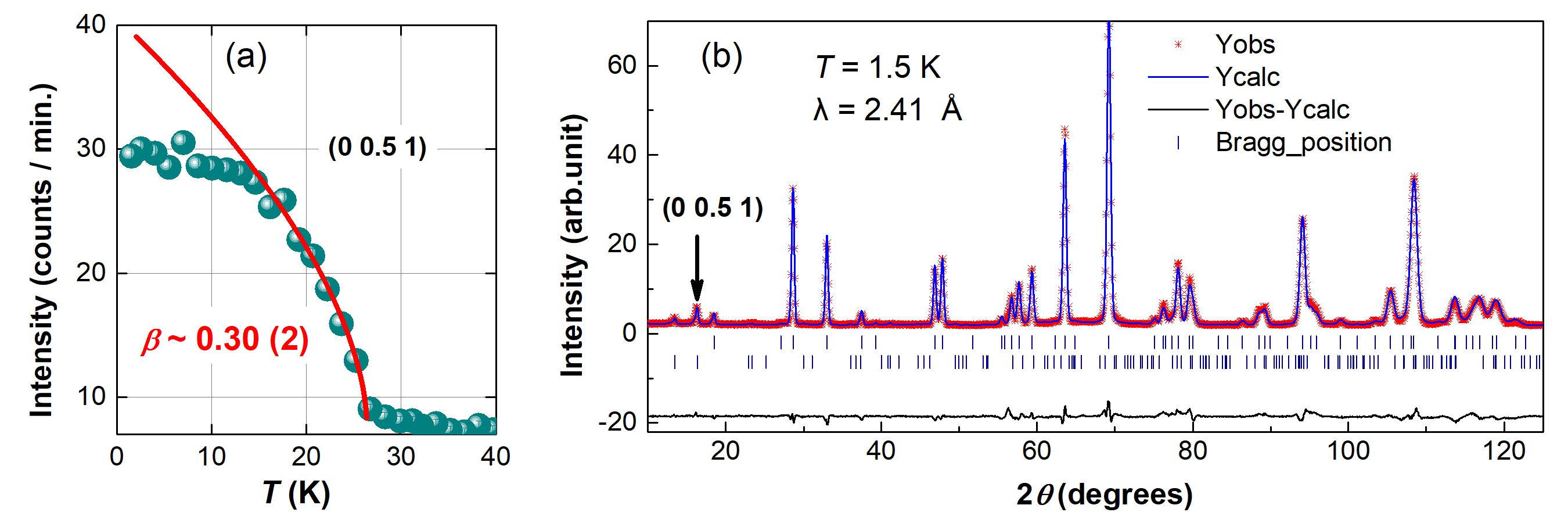
In order to obtain confirmation of the true magnetic transition temperature and to better understand the critical behavior at this transition, complementary elastic neutron scattering measurements were performed on a polycrystalline sample of \chBa3LaRu2O9 using the HB-1A triple axis spectrometer at HFIR. Fig. 7(a) shows the temperature dependence of the intensity for the strongest magnetic peak, which can be indexed by (0 0.5 1) as explained below. A simple power law was applied to fit the peak intensity near the transition temperature:
| (4) |
where is the Neél temperature and is the critical exponent of the order parameter (OP). The fitting result yields 26(1) K and no sharp change in the intensity is detected around 6 K, which is consistent with our other measurements described above. We also find that 0.30(2), which is close to the values expected for a three-dimensional universality class ( 0.326 and 0.345) and much larger than expected for a quasi-2D Ising model ( 0.125). This result is surprising since a -dependence for the low-temperature specific heat often arises from spin wave contributions of a gapless, quasi-2D antiferromagnet.
With the true magnetic transition temperature of \chBa3LuRu2O9 now established, we return to the open questions surrounding the magnetic structure measured previously Senn et al. (2013). We collected neutron powder diffraction data on polycrystalline \chBa3LaRu2O9 at 1.5 and 40 K using the HB-2A powder diffractometer with a neutron wavelength of 2.41 Å. The 1.5 K diffraction pattern is similar to the previous measurements of Senn et al Senn et al. (2013) and consists of both nuclear and magnetic Bragg peaks, as shown in Fig. 7(b). The 1.5 K data can be refined in the space group, as noted above, and the magnetic peaks observed can be indexed with the same propagation vector (0 0.5 0) identified previously. To model the magnetic structure we first performed a symmetry analysis using SARAh Wills (2000). Assuming a second order phase transition at , the most likely magnetic models should correspond to one of the eight irreducible representations described in Ref. Senn et al. (2013). However, we found that none of these models could fully explain our data, and therefore we also tried linear combinations of them. Ultimately, we find that the best magnetic refinement of the 1.5 K diffraction pattern is achieved by using the same model as before Senn et al. (2013). The component is required to explain the magnetic intensity at the (-1 0.5 0) position, which is not captured by the model. To estimate the ordered moment size, we used a local moment model with the Ru5+ magnetic form factor that has been reported elsewhere Parkinson et al. (2003). The refined moment sizes per Ru ion at 1.5 K are , , and . These values are consistent with the reported ordered moment sizes of , , and reported previously Senn et al. (2013) and close to the expectation of for a 3/2 Ru dimer electronic ground state. We also note that the orbital-selective Mott state for the Ru dimers naturally describes the ferromagnetic intradimer coupling revealed from the analysis of the NPD data, but the refined moment direction is difficult to understand. A schematic of the refined magnetic structure is presented in Figs. 1(a) and (c); this is the collinear stripe spin configuration that is predicted for a triangular lattice with a significant in-plane next nearest neighbor exchange interaction (i.e. Seabra and Shannon (2011)).
III.3 (III) Collective spin dynamics
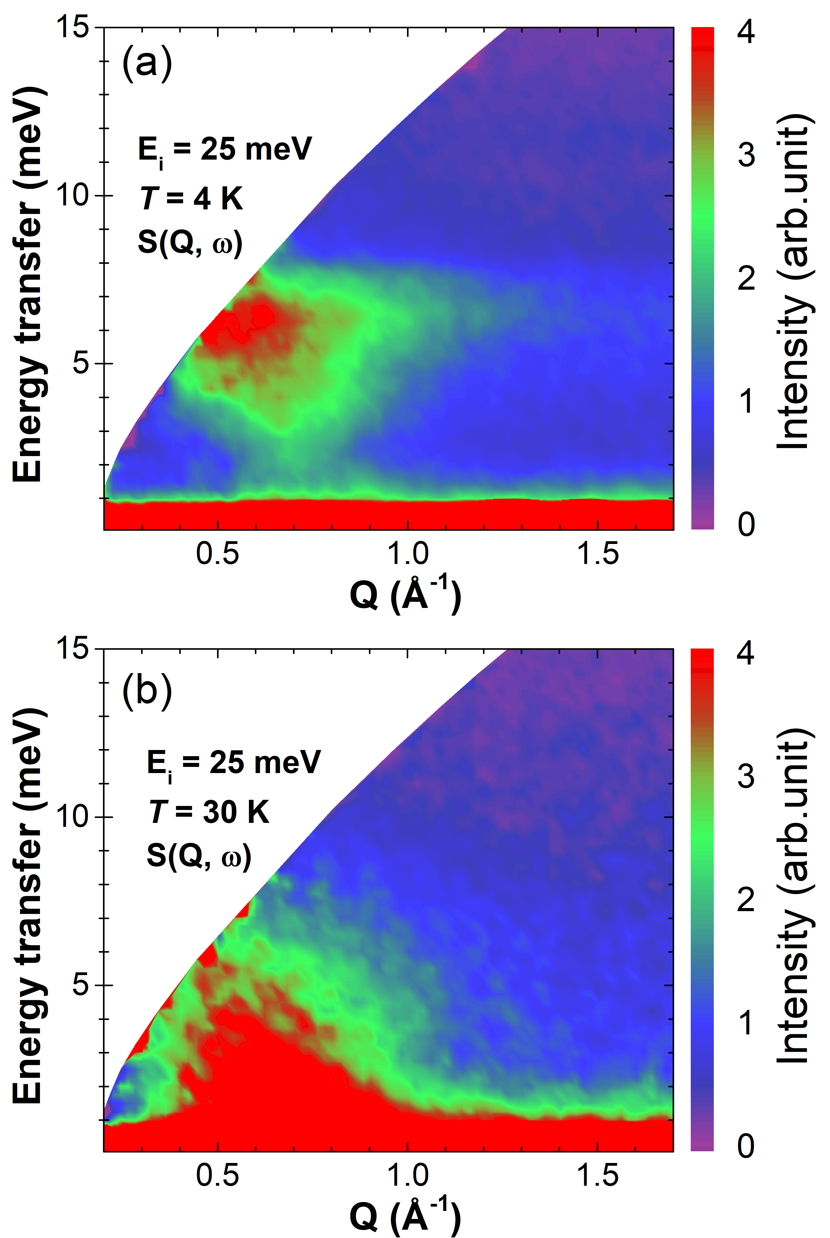
We now return to the spin wave mode measured with inelastic neutron scattering using the SEQUOIA spectrometer. This excitation was best observed by collecting data with an incident energy 25 meV, which is presented in Fig. 8(a) and (b) at 4 and 30 K respectively. Notably, there is a strong band of inelastic scattering at 30 K centered above the (0 0.5 0) and (0 0.5 1) magnetic Bragg peaks that shows a significant shift in spectral weight up to higher energy transfers at 4 K. This phenomenon has been observed in many other ordered systems when cooling below Carlo et al. (2013); Aczel et al. (2014); Taylor et al. (2016) and may suggest that the spin wave spectrum of Ba3LaRu2O9 is gapped. However, the spin wave contribution to the low-temperature specific heat would then show an activated behavior rather than a dependence. This discrepancy, combined with the inconsistent universality class conclusions obtained from the specific heat and the HB-1A neutron scattering data, suggest that the -dependence of the low-temperature specific heat does not arise from a spin wave contribution.
We also considered modeling the spin wave data with a magnetic Hamiltonian, but we failed to find a simple model that could explain the moment direction obtained from neutron powder diffraction. For this hexagonal crystal structure, Heisenberg models with zero field splitting (i.e. single ion anisotropy) can only produce moments in the ab-plane ( 0) or along the c-axis ( 0), but not somewhere in between these two extremes. To obtain the correct ground state found experimentally, it appears that the addition of exchange anisotropy is essential, but this consideration goes beyond the scope of our work on a powder sample. Single crystal inelastic neutron scattering measurements on \chBa3LaRu2O9 will be invaluable for elucidating the magnetic Hamiltonian of this system.
III.4 (IV) Structural and spin state transitions under pressure
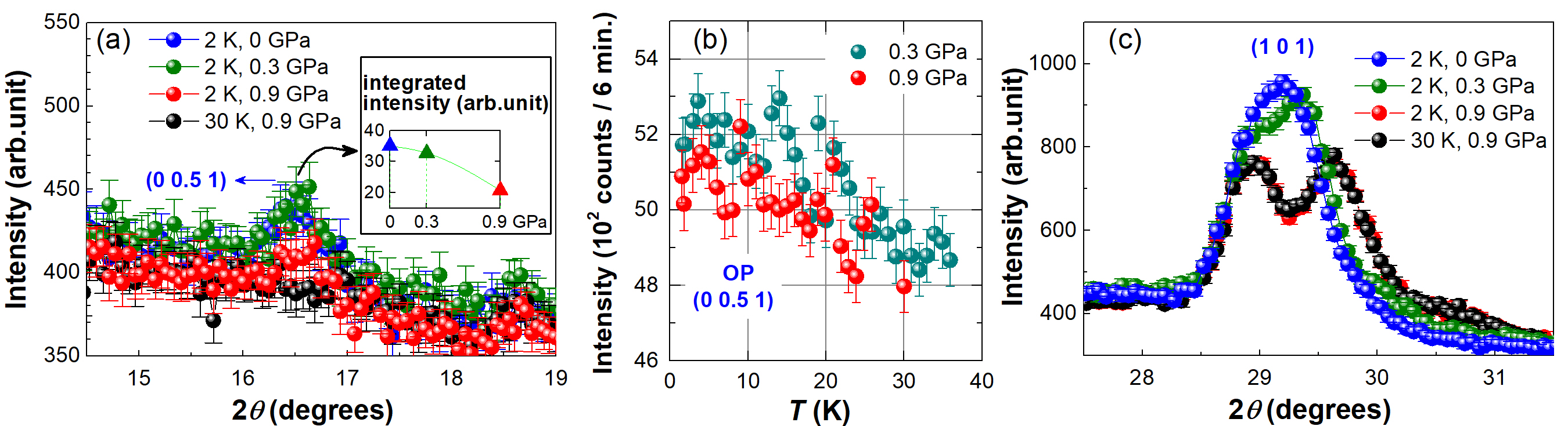
One way to gain additional insight into mechanisms leading to the 3/2 molecular ground state in \chBa3LaRu2O9 is to apply external stimuli Brinzari et al. (2012); O’Neal et al. (2014). Since orbital hybridization should increase with decreasing intradimer Ru-Ru distance, our hypothesis is that a small amount of pressure may induce a spin state transition from = 3/2 to = 1/2 O’Neal et al. (2014). Such a transition is expected to take place with a reduction in the Ru ordered moment from 3 to 1 per cluster. We performed a neutron diffraction experiment at HB-2A in a 1 GPa Cu-Be pressure cell to search for evidence of such a high low spin state transition by tracking the intensity of the strongest magnetic Bragg peak under compression. We find significant suppression in the intensity of this peak when the pressure is increased from 0.3 to 0.9 GPa, as shown in Fig. 9(a) and (b), but no significant change in the magnetic transition temperature. Due to a background shift with increasing pressure, we plot the pressure-dependence of the integrated intensity for this peak in the Fig. 9(a) inset. We also searched for new magnetic Bragg peaks at 0.9 GPa by collecting diffraction patterns over a wide angular range at both 2 and 30 K, but none were found. Suppression of this magnetic peak under compression is therefore consistent with a low-lying spin state transition rather than a magnetic structure change in this material. Notably, the crystal structure also appears to be modified at a lower pressure of 0.3 GPa, as the hexagonal (101) peak near 29° splits into three peaks, as shown in Fig. 9(c). The highest crystal symmetry consistent with this three-fold peak splitting is monoclinic. Unfortunately, our data quality is insufficient for refining the crystal structure of this material under compression due to the high, structured background of the pressure cell and the significant neutron beam attenuation through the cell.
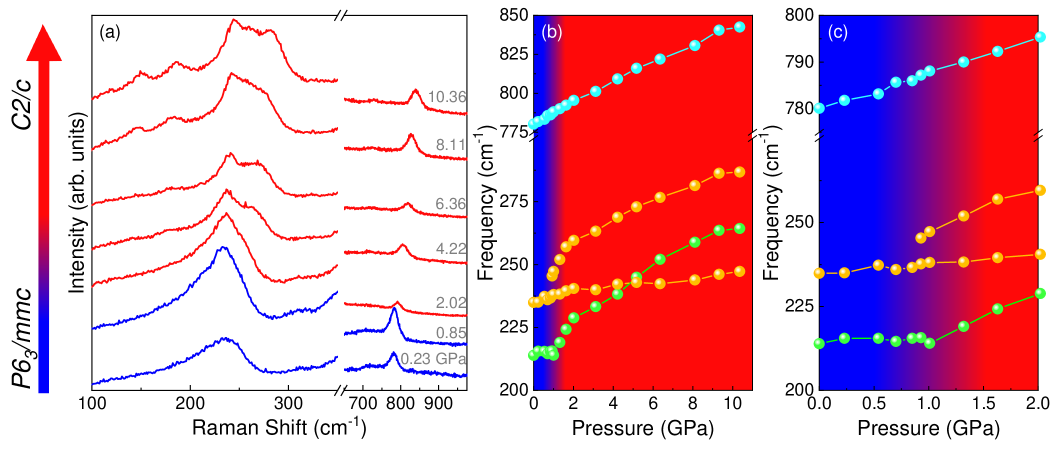
In order to gain additional insight into the pressure-driven transitions, we turn to Raman scattering. This method dovetails well with diamond anvil cell techniques and supports tuning the sample over a much wider pressure range. Figure 10(a) displays the Raman response of \chBa3LaRu2O9 at room temperature under compression. Plots of frequency vs pressure, as shown in Fig. 10(b) and (c), allow us to track the behavior of individual phonons and identify clear changes at P 0.9 GPa. While some modes such as the Ru–O stretch at 776 cm-1 are insensitive to the 0.9 GPa transition, others (for instance at 215 and 235 cm-1) sport inflection points with subsequent hardening as well as strong doublet splitting. This suggests that while the Ru dimer is structurally rigid across the critical pressure, the charge storage layer containing Ba and La is not. This is consistent with findings for structural rigidity of the Ru dimer across the high low spin transition in the bimetallic quantum magnet [Ru2(O2CMe)4]3[Cr(CN)6] Shum et al. (2007); Brinzari et al. (2012); O’Neal et al. (2014). At the same time, the lower frequency modes involving Ba and La motion provide evidence for symmetry breaking across the 0.9 GPa transition.
A correlation group analysis identifies several candidate subgroups of the space group. Recalling that neutron diffraction for 30 K constrains the high pressure phase to a monoclinic structure, these subgroups include , , and . We begin by considering whether the system will have a primitive or centered lattice type in the high-pressure phase. Our system sports a primitive lattice () at ambient conditions and goes through a centered lattice space group () on the way to one of the three candidate monoclinic subgroups. We expect that the final symmetry reduction will retain a centered lattice because the face-centered cell is more dense than the primitive lattice and therefore more stable under pressure. The screw operation is also unlikely to remain intact under these conditions. This eliminates and leaves and as the remaining candidates. Next we consider whether the high pressure phase contains a reflection or glide plane. Here, we proceed by realizing that reflection is a higher symmetry operation and that pressure tends to break mirror planes. This leaves as the most probable space group above the critical pressure, which is consistent with previous work identifying a crystal structure in symmetry-lowering transitions of other 6H-perovskites Kimber et al. (2012); Senn et al. (2013). Further work is required to determine whether the 0.9 GPa structural transition at room temperature is coincident with the spin state transition identified at lower temperatures by neutron powder diffraction.
IV Conclusions
In conclusion, we have used a combination of bulk characterization, muon spin relaxation, neutron diffraction, and inelastic neutron scattering to identify an intermediate 3/2 Ru dimer ground state in \chBa3LaRu2O9 that is generated by orbital-selective Mott insulating behavior at the molecular level. We also find collinear stripe magnetic order below 26(1) K for these spin-3/2 degrees-of-freedom, which is consistent with expectations for an ideal triangular lattice with significant next nearest neighbor in-plane exchange. Finally, we present neutron diffraction and Raman scattering data under applied pressure that reveal low-lying structural and spin state transitions at modest applied pressures P 1 GPa, which highlights the delicate balance between competing energy scales in this material. Interesting future directions for \chBa3LaRu2O9 include identifying the origin of the -dependence of the low-temperature specific heat, determining the magnetic Hamiltonian giving rise to the moment direction of the stripe spin order, solving the high-pressure crystal structure, and carefully mapping out the temperature-pressure phase diagram. Our work highlights the need to develop a comprehensive understanding of the electronic ground state of a heavy transition metal molecular magnet, where large orbital hopping may lead to the breakdown of a simple local moment / double exchange picture, before the collective magnetic properties of the system can be properly identified and characterized.
Acknowledgements.
Research at the University of Tennessee is supported by the National Science Foundation, Division of Materials Research under award NSF-DMR 1350002 (HDZ) and the Department of Energy, Office of Basic Energy Sciences, Materials Science Division under award DE-FG02-01ER45885 (JLM). JGC is supported by the MOST, NSFC and CAS through projects with Grant Nos. 2018YFA0305700, 11874400, 11921004, and QYZDB-SSW-SLH013. GHW and JM are supported by the MOST and NSFC through projects with Grant Nos. 2016YFA0300501, 11774223 and U1732154. JQ acknowledges research funding obtained from NSERC and the FRQNT. A portion of this research used resources at the High Flux Isotope Reactor and the Spallation Neutron Source, which are DOE Office of Science User Facilities operated by Oak Ridge National Laboratory.References
- Anisimov et al. (2002) V. I. Anisimov, I. A. Nekrasov, D. E. Kondakov, T. M. Rice, and M. Sigrist, Eur. Phys. J. B 25, 191 (2002).
- Wang et al. (2004) S. C. Wang, H. B. Yang, A. K. P. Sekharan, S. Souma, H. Matsui, T. Sato, T. Takahashi, C. Lu, J. Zhang, R. Jin, D. Mandrus, E. W. Plummer, Z. Wang, and H. Ding, Phys. Rev. Lett. 93, 177007 (2004).
- Balicas et al. (2005) L. Balicas, S. Nakatsuji, D. Hall, T. Ohnishi, Z. Fisk, Y. Maeno, and D. J. Singh, Phys. Rev. Lett. 95, 196407 (2005).
- Liebsch and Ishida (2007) A. Liebsch and H. Ishida, Phys. Rev. Lett. 98, 216403 (2007).
- Neupane et al. (2009) M. Neupane, P. Richard, Z.-H. Pan, Y.-M. Xu, R. Jin, D. Mandrus, X. Dai, Z. Fang, Z. Wang, and H. Ding, Phys. Rev. Lett. 103, 097001 (2009).
- de’ Medici et al. (2009) L. de’ Medici, S. R. Hassan, M. Capone, and X. Dai, Phys. Rev. Lett. 102, 126401 (2009).
- Koga et al. (2004) A. Koga, N. Kawakami, T. M. Rice, and M. Sigrist, Phys. Rev. Lett. 92, 216402 (2004).
- de’Medici et al. (2005) L. de’Medici, A. Georges, and S. Biermann, Phys. Rev. B 72, 205124 (2005).
- Ferrero et al. (2005) M. Ferrero, F. Becca, M. Fabrizio, and M. Capone, Phys. Rev. B 72, 205126 (2005).
- Liebsch (2005) A. Liebsch, Phys. Rev. Lett. 95, 116402 (2005).
- Biermann et al. (2005) S. Biermann, L. de’ Medici, and A. Georges, Phys. Rev. Lett. 95, 206401 (2005).
- Rincón et al. (2014a) J. Rincón, A. Moreo, G. Alvarez, and E. Dagotto, Phys. Rev. Lett. 112, 106405 (2014a).
- Rincón et al. (2014b) J. Rincón, A. Moreo, G. Alvarez, and E. Dagotto, Phys. Rev. B 90, 241105(R) (2014b).
- Yu and Si (2012) R. Yu and Q. Si, Phys. Rev. B 86, 085104 (2012).
- Yu and Si (2013) R. Yu and Q. Si, Phys. Rev. Lett. 110, 146402 (2013).
- Venturini et al. (2002) F. Venturini, M. Opel, T. P. Devereaux, J. K. Freericks, I. Tüttő, B. Revaz, E. Walker, H. Berger, L. Forró, and R. Hackl, Phys. Rev. Lett. 89, 107003 (2002).
- Laad et al. (2006) M. S. Laad, L. Craco, and E. Müller-Hartmann, Phys. Rev. B 73, 045109 (2006).
- Chen (2018) H. Chen, npj Quantum Materials 3, 57 (2018).
- Mourigal et al. (2015) M. Mourigal, S. Wu, M. B. Stone, J. R. Neilson, J. M. Caron, T. M. McQueen, and C. L. Broholm, Phys. Rev. Lett. 115, 047401 (2015).
- Herbrych et al. (2018) J. Herbrych, N. Kaushal, A. Nocera, G. Alvarez, A. Moreo, and E. Dagotto, Nat. Comm. 9, 3736 (2018).
- Streltsov and Khomskii (2016) S. V. Streltsov and D. I. Khomskii, Proceedings of the National Academy of Sciences 113, 10491 (2016).
- Streltsov and Khomskii (2017) S. V. Streltsov and D. I. Khomskii, Physics-Uspekhi 60, 1121 (2017).
- Streltsov and Khomskii (2014) S. V. Streltsov and D. I. Khomskii, Phys. Rev. B 89, 161112(R) (2014).
- Ziat et al. (2017) D. Ziat, A. A. Aczel, R. Sinclair, Q. Chen, H. D. Zhou, T. J. Williams, M. B. Stone, A. Verrier, and J. A. Quilliam, Phys. Rev. B 95, 184424 (2017).
- Sheckelton et al. (2012) J. P. Sheckelton, J. R. Neilson, D. G. Soltan, and T. M. McQueen, Nature Materials 11, 493 (2012).
- Mourigal et al. (2014) M. Mourigal, W. T. Fuhrman, J. P. Sheckelton, A. Wartelle, J. A. Rodriguez-Rivera, D. L. Abernathy, T. M. McQueen, and C. L. Broholm, Phys. Rev. Lett. 112, 027202 (2014).
- Akbari-Sharbaf et al. (2018) A. Akbari-Sharbaf, R. Sinclair, A. Verrier, D. Ziat, H. D. Zhou, X. F. Sun, and J. A. Quilliam, Phys. Rev. Lett. 120, 227201 (2018).
- Kimber et al. (2012) S. A. J. Kimber, M. S. Senn, S. Fratini, H. Wu, A. H. Hill, P. Manuel, J. P. Attfield, D. N. Argyriou, and P. F. Henry, Phys. Rev. Lett. 108, 217205 (2012).
- Chen et al. (2019) Q. Chen, S. Fan, K. M. Taddei, M. B. Stone, A. I. Kolesnikov, J. Cheng, J. L. Musfeldt, H. D. Zhou, and A. A. Aczel, J. Am. Chem. Soc. 141, 9928 (2019).
- Bechlars et al. (2010) B. Bechlars, D. M. D’Alessandro, D. M. Jenkins, A. T. Iavarone, S. D. Glover, C. P. Kubiak, and J. R. Long, Nature Chemistry 2, 362 (2010).
- Doi et al. (2002) Y. Doi, K. Matsuhira, and Y. Hinatsu, Journal of Solid State Chemistry 165, 317 (2002).
- Senn et al. (2013) M. S. Senn, S. A. J. Kimber, A. M. A. Lopez, A. H. Hill, and J. P. Attfield, Phys. Rev. B 87, 134402 (2013).
- Calder et al. (2018) S. Calder, K. An, R. Boehler, C. R. dela Cruz, M. D. Frontek, M. Guthrie, B. Haberl, A. Huq, S. A. J. Kimber, J. Liu, J. J. Molaison, J. Neuefeind, K. Page, A. M. dos Santos, K. M. Taddei, C. Tulk, and M. G. Tucker, Rev. Sci. Instr. 89, 092701 (2018).
- Rodríguez-Carvajal (1993) J. Rodríguez-Carvajal, Physica B 192, 55 (1993).
- Wills (2000) A. S. Wills, Physica B 276, 680 (2000).
- Granroth et al. (2010) G. E. Granroth, A. I. Kolesnikov, T. E. Sherline, J. P. Clancy, K. A. Ross, J. P. C. Ruff, B. D. Gaulin, and S. E. Nagler, J. Physics: Conference Series 251, 012058 (2010).
- Yaouanc and De Reotier (2011) A. Yaouanc and P. D. De Reotier, Muon spin rotation, relaxation, and resonance: applications to condensed matter, Vol. 147 (Oxford University Press, 2011).
- Mao et al. (1986) H. K. Mao, J. A. Xu, and P. M. Bell, Journal of Geophysical Research: Solid Earth 91, 4673 (1986).
- Furrer et al. (2010) A. Furrer, J. Mesot, and T. Strassle, Neutron scattering in condensed matter physics, Vol. 4 (World Scientific Publishing Co., 2010).
- Kugel et al. (2015) K. I. Kugel, D. I. Khomskii, A. O. Sboychakov, and S. V. Streltsov, Phys. Rev. B 91, 155125 (2015).
- Thirring (1913) H. Thirring, Phys. Z. 14, 867 (1913).
- Gordon et al. (1989) J. E. Gordon, M. L. Tan, R. A. Fisher, and N. E. Phillips, Solid State Commun. 69, 625 (1989).
- Cheng et al. (2005) J. G. Cheng, Y. Sui, X. J. Wang, Z. G. Liu, J. P. Miao, X. Q. Huang, Z. Lü, Z. N. Qian, and W. H. Su, J. Phys.: Cond. Matt. 17, 5869 (2005).
- Holzschuh et al. (1983) E. Holzschuh, A. B. Denison, W. Kundig, P. F. Meier, and B. D. Patterson, Phys. Rev. B 27, 5294 (1983).
- Shimoda et al. (2010) Y. Shimoda, Y. Doi, M. Wakeshima, and Y. Hinatsu, Journal of Solid State Chemistry 183, 33 (2010).
- Parkinson et al. (2003) N. G. Parkinson, P. D. Hatton, J. A. Howard, C. Ritter, F. Z. Chien, and M.-K. Wu, Journal of Materials Chemistry 13, 1468 (2003).
- Seabra and Shannon (2011) L. Seabra and N. Shannon, Phys. Rev. B 83, 134412 (2011).
- Carlo et al. (2013) J. P. Carlo, J. P. Clancy, K. Fritsch, C. A. Marjerrison, G. E. Granroth, J. E. Greedan, H. A. Dabkowska, and B. D. Gaulin, Phys. Rev. B 88, 024418 (2013).
- Aczel et al. (2014) A. A. Aczel, P. J. Baker, D. E. Bugaris, J. Yeon, H. C. zur Loye, T. Guidi, and D. T. Adroja, Phys. Rev. Lett 112, 117603 (2014).
- Taylor et al. (2016) A. E. Taylor, R. Morrow, R. S. Fishman, S. Calder, A. I. Kolesnikov, M. D. Lumsden, P. M. Woodward, and A. D. Christianson, Phys. Rev. B 93, 220408(R) (2016).
- Brinzari et al. (2012) T. V. Brinzari, P. Chen, L. C. Tung, Y. Kim, D. Smirnov, J. Singleton, J. S. Miller, and J. L. Musfeldt, Phys. Rev. B 86, 214411 (2012).
- O’Neal et al. (2014) K. R. O’Neal, Z. Liu, J. S. Miller, R. S. Fishman, and J. L. Musfeldt, Phys. Rev. B 90, 104301 (2014).
- Shum et al. (2007) W. W. Shum, J. H. Her, P. W. Stephens, Y. Lee, and J. S. Miller, Advanced Materials 19, 2910 (2007).