Meta-SVDD: Probabilistic Meta-Learning for One-Class Classification in Cancer Histology Images
Abstract
To train a robust deep learning model, one usually needs a balanced set of categories in the training data. The data acquired in a medical domain, however, frequently contains an abundance of healthy patients, versus a small variety of positive, abnormal cases. Moreover, the annotation of a positive sample requires time consuming input from medical domain experts. This scenario would suggest a promise for one-class classification type approaches. In this work we propose a general one-class classification model for histology, that is meta-trained on multiple histology datasets simultaneously, and can be applied to new tasks without expensive re-training. This model could be easily used by pathology domain experts, and potentially be used for screening purposes.
1 Introduction
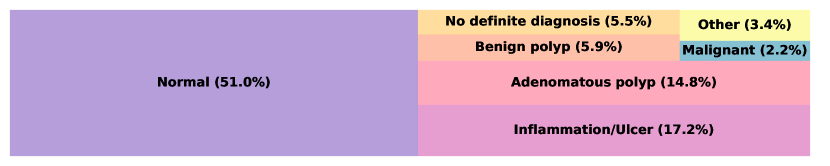
Pathology departments across most countries in the world are experiencing severe under-staffing issues Metter et al. ; Bainbridge et al. . With digital slide scanners becoming increasingly ubiquitous in pathology labs, it is expected that machine learning based decision support systems would substantially reduce the workload for pathology labs Colling et al. . The slide scanners are capable of producing high-resolution multi-gigapixel whole-slide images (generally of the order of pixels), resulting in a wealth of pixel data that could be honed for diagnostic and prognostic purposes. The rapidly growing research community in the area of computational pathology has developed machine learning algorithms for narrow applications such as mitotic counting, cancer grading, cancer detection Balkenhol et al. ; Liu et al. ; Nagpal et al. . These advances are important, but implementing specific but separate approaches for numerous tasks in pathology might be impractical, and in fact may be impossible due to the distribution of target categories in the population (see Figure 1). Furthermore, algorithms are frequently built on datasets that may not be representative of the population, and therefore under-perform in practice Gamper et al. . As such, building general-purpose algorithms, pre-trained on multiple datasets could substantially speed up the application of machine learning tools in practice Hegde et al. . Furthermore, creating general algorithms that learn from few examples, would allow to easily solve very specific and narrow tasks that contain only few learning examples.
In this paper, we propose a general one-class classification model for histology that is meta-trained on multiple datasets and can be applied to new tasks without re-training. We name this approach Meta-SVDD. Past approaches for deep anomaly detection, include deterministic or variational auto-encoding, and adversarial deep generative methods Schlegl et al. ; Schlegl et al. (2019); An and Cho (2015). First, VAE or GAN based methods are computationally intensive, either requiring back propagation at test time, or re-training for new tasks. Second, these generative models are built with the assumption of estimating the underlying data density well, however this assumption has been put under question Nalisnick et al. . Third, both autoencoding and adversarial methods solve optimisation tasks during training that are different to the downstream objectives. To address these vulnerabilities, we propose a one-class classification model that is meta-learned with the explicit loss function for one-class classification. Our novel method allows to adapt to new tasks by only observing few examples without the need for re-training as the task specific parameters inference is amortized using a neural network. The proposed method also uses out-of- as well as in-distribution examples during meta-learning optimisation.
2 Methods & Results
Consider a sample dataset for a given task , which could correspond to a set of patches from histology WSIs, where corresponds to a specific tissue type (colon, lung, breast, etc.). In an ideal supervised learning task with labeled training data and test data pairs, the empirical marginal distribution of class labels is more or less uniform Buda et al. . This allows one to estimate an optimal set of parameters for a function , i.e. a probability vector output from a convolutional neural network, via empirical risk minimization: . Where is a loss function.
2.1 One Class Deep Support Vector Data Description (OC-SVDD)
In the case, when the empirical marginal distribution of categories is not necessarily uniform, as mentioned in Section 1, we can formulate it as a one-class classification problem: (1) reducing the complexity of the task by having only the positive, in-distribution samples, ; (2) adapting the function to map a given sample to a latent encoding; and (3) minimizing the empirical one-class Deep SVDD (OC-SVDD) loss Ruff et al. (a), namely
| (1) |
where one learns a hyper-sphere by minimising the mean distance of all data representations to the center for all positive samples Ruff et al. (a).
2.1.1 OC-SVDD Experiments
In Table A1, we demonstrate the results of applying OC-SVDD loss to histology data. The datasets were obtained from the following sources: Colon from Kather et al. , Lung Alsubaie et al. , Ovary Köbel et al. , Lymphoma Janowczyk and Madabhushi , Oral Shaban et al. , Breast from Chamelyon Challenge111https://camelyon16.grand-challenge.org/, and Meningioma Qureshi et al. .
For these experiments we took an Imagenet pre-trained ResNet18 He et al. , where we replaced the final linear layer to produce the output of dimensionality of the hyper-sphere, 128 for every task. We set batch size to 64, learning rate to , and optimized over 100 epochs using ADAM Kingma and Ba . The hyper-sphere center is initialised using the first pass through the network. For the preliminary results presented in this paper, we did not optimise any of the hyper-parameters, these were taken from Ruff et al. (a).
For every tissue type, we picked one of the classes and treated it as in-distribution data i.e. one task, and optimised the loss function in Equation 1. For most tasks, the in-distribution data is uni-modal, and only consists of that particular category. However, in the case of Breast tissue, the category Other contains healthy tissue, lymphocytes and other tissue phenotypes, which demonstrates the potential of OC-SVDD for tumor screening purposes.
2.2 Probabilistic meta-learning for SVDD (Meta-SVDD)
While the OC-SVDD method offers an explicit loss function for deep learning in one class classification and could be easily applied at test time, it still requires expensive training for any given task. One has to train 32 networks to produce the results of OC-SVDD in Table A1. We propose to address this issue using meta-learning. We induce a distribution over function , by introducing an amortized distribution Garnelo et al. . We adopt the network architecture and inference method for the parameters according to Gordon et al. . Namely, during optimisation we: (i) select a task at random, (ii) sample some training data , (iii) form the posterior predictive given in-distribution data , where , (iv) next we evaluate the posterior predictive on meta test data using semi-supervised SVDD loss:
| (2) |
where . is the number of samples from predictive posterior. We assume to be Gaussian and use reparameterisation trick during optimisation Kingma and Welling . Compared to the Equation 1, the right hand side learns the inverse of the left hand side, pushing positive samples further from the center Ruff et al. (b), and in Equation 2 is a hyper-parameter. This approach allows us to amortize the parameter learning for new tasks directly to inference network, that predicts the parameterizations for the distribution over that maps test data to the latent space. We name this approach Meta-SVDD.
2.2.1 Meta-SVDD Experiments
Following Gordon et al. . We use the same encoder (ResNet18) for all tasks represented by in predictive posterior. We set to 10. We set a meta batch size to 5, and optimise using gradient accumulation, due to restrained computational resources. The inference network consists of three fully connected layers, with ReLU activation functions. The inference network takes mean of in-distribution features and produces parameterisation for posterior from which parameters of function are sampled. The remaining hyper-parameters are the same as in Section 2.1.1.
We adopt leave-one-out cross-validation setup where we pretrain on 32 tasks, and test on the remaining tasks. The results are presented in Table 1. Note that for inferring the posterior we are using only 10 in-distribution samples. Therefore the results of the proposed method are promising, however at the current stage of work we have faced the computational limitations during meta-training Nichol et al. .
3 Discussion & Future Directions
We present preliminary results for meta-learned one-class classification model for histology tasks, such model does not require expensive training and, parameter inference is done at test time. We demonstrated its potential for screening task in the case of Breast tissue, and flexibility with learning uni-modal tasks in other tissues. Future work would include hyper-parameter optimisation for neural network architecture, and for meta-learning. For example OC-SVDD loss resembles the tasks of self-supervised learning, and as it has been demonstrated benefits significantly from larger networks Kolesnikov et al. . However, that would require a careful treatment of sensitive meta-learning optimisation process Antoniou et al. . Once a stable set of architecture and optimisation hyper-parameters are established, we plan to thoroughly test the proposed meta learning scheme for one-class classification on whole slide images for screening and speeding up annotation. Additionally, we are planning on expanding the tasks for training using existing datasets for meta-learning Triantafillou et al. , this, which we hope would also increase the performance on fine-grained tasks such Lung adenocarcinoma subtypes. By increasing the size of the network, stabilising the optimisation process, and increasing the number of datasets, we aim to significantly improve the performance of Meta-SVDD.
Acknowledgments
This research was partially supported by Philips Pathology.
References
- [1] Najah Alsubaie, Korsuk Sirinukunwattana, Shan E. Ahmed Raza, David Snead, and Nasir Rajpoot. A bottom-up approach for tumour differentiation in whole slide images of lung adenocarcinoma. In Medical Imaging 2018: Digital Pathology, volume 10581, page 105810E. International Society for Optics and Photonics. doi: 10.1117/12.2293316. URL https://www.spiedigitallibrary.org/conference-proceedings-of-spie/10581/105810E/A-bottom-up-approach-for-tumour-differentiation-in-whole-slide/10.1117/12.2293316.short.
- An and Cho [2015] Jinwon An and Sungzoon Cho. Variational autoencoder based anomaly detection using reconstruction probability. Special Lecture on IE, 2(1), 2015.
- [3] Antreas Antoniou, Harrison Edwards, and Amos Storkey. How to train your MAML. URL http://arxiv.org/abs/1810.09502.
- [4] S Bainbridge, R Cake, M Meredith, P Furness, and B Gordon. Testing times to come? an evaluation of digital pathology capacity across the UK.
- [5] Maschenka C. A. Balkenhol, David Tellez, Willem Vreuls, Pieter C. Clahsen, Hans Pinckaers, Francesco Ciompi, Peter Bult, and Jeroen A. W. M. van der Laak. Deep learning assisted mitotic counting for breast cancer. pages 1–11. ISSN 1530-0307. doi: 10.1038/s41374-019-0275-0. URL https://www.nature.com/articles/s41374-019-0275-0.
- [6] Mateusz Buda, Atsuto Maki, and Maciej A. Mazurowski. A systematic study of the class imbalance problem in convolutional neural networks. 106:249–259. ISSN 08936080. doi: 10.1016/j.neunet.2018.07.011. URL http://arxiv.org/abs/1710.05381.
- [7] Richard Colling, Helen Pitman, Karin Oien, Nasir Rajpoot, Philip Macklin, David Snead, Tony Sackville, Clare Verrill, CM-Path AI in Histopathology Working Group, Velicia Bachtiar, Richard Booth, Alyson Bryant, Joshua Bull, Jonathan Bury, Fiona Carragher, Richard Colling, Graeme Collins, Clare Craig, Maria Freitas Silva, Daniel Gosling, Jaco Jacobs, Lena Kajland-Wilén, Johanna Karling, Darragh Lawler, Stephen Lee, Philip Macklin, Keith Miller, Guy Mozolowski, Richard Nicholson, Daniel O’Connor, Mikkel Rahbek, Nasir Rajpoot, Alan Sumner, Dirk Vossen, Kieron White, Charlotte Wing, and Corrina Wright. Artificial intelligence in digital pathology: A roadmap to routine use in clinical practice. ISSN 0022-3417, 1096-9896. doi: 10.1002/path.5310. URL https://onlinelibrary.wiley.com/doi/abs/10.1002/path.5310.
- [8] Jevgenij Gamper, Navid Alemi Koohbanani, Ksenija Benet, Ali Khuram, and Nasir Rajpoot. PanNuke: An open pan-cancer histology dataset for nuclei instance segmentation and classification. In Digital Pathology, pages 11–19. Springer, Cham. doi: 10.1007/978-3-030-23937-4_2. URL https://0-link-springer-com.pugwash.lib.warwick.ac.uk/chapter/10.1007/978-3-030-23937-4_2.
- [9] Marta Garnelo, Jonathan Schwarz, Dan Rosenbaum, Fabio Viola, Danilo J Rezende, S M Ali Eslami, and Yee Whye Teh. Neural processes.
- [10] Jonathan Gordon, John Bronskill, Matthias Bauer, Sebastian Nowozin, and Richard Turner. Meta-learning probabilistic inference for prediction. URL https://openreview.net/forum?id=HkxStoC5F7.
- [11] Kaiming He, Xiangyu Zhang, Shaoqing Ren, and Jian Sun. Deep residual learning for image recognition. URL http://arxiv.org/abs/1512.03385.
- [12] Narayan Hegde, Jason D. Hipp, Yun Liu, Michael Emmert-Buck, Emily Reif, Daniel Smilkov, Michael Terry, Carrie J. Cai, Mahul B. Amin, Craig H. Mermel, Phil Q. Nelson, Lily H. Peng, Greg S. Corrado, and Martin C. Stumpe. Similar image search for histopathology: SMILY. 2(1):1–9. ISSN 2398-6352. doi: 10.1038/s41746-019-0131-z. URL https://www.nature.com/articles/s41746-019-0131-z.
- [13] Andrew Janowczyk and Anant Madabhushi. Deep learning for digital pathology image analysis: A comprehensive tutorial with selected use cases. 7. ISSN 2229-5089. doi: 10.4103/2153-3539.186902. URL https://www.ncbi.nlm.nih.gov/pmc/articles/PMC4977982/.
- [14] Jakob Nikolas Kather, Alexander T. Pearson, Niels Halama, Dirk Jäger, Jeremias Krause, Sven H. Loosen, Alexander Marx, Peter Boor, Frank Tacke, Ulf Peter Neumann, Heike I. Grabsch, Takaki Yoshikawa, Hermann Brenner, Jenny Chang-Claude, Michael Hoffmeister, Christian Trautwein, and Tom Luedde. Deep learning can predict microsatellite instability directly from histology in gastrointestinal cancer. page 1. ISSN 1546-170X. doi: 10.1038/s41591-019-0462-y. URL https://www.nature.com/articles/s41591-019-0462-y.
- [15] Diederik P. Kingma and Jimmy Ba. Adam: A method for stochastic optimization. URL http://arxiv.org/abs/1412.6980.
- [16] Diederik P. Kingma and Max Welling. Auto-encoding variational bayes. URL http://arxiv.org/abs/1312.6114.
- [17] Alexander Kolesnikov, Xiaohua Zhai, and Lucas Beyer. Revisiting self-supervised visual representation learning.
- [18] Martin Köbel, Steve E. Kalloger, Patricia M. Baker, Carol A. Ewanowich, Jocelyne Arseneau, Viktor Zherebitskiy, Soran Abdulkarim, Samuel Leung, Máire A. Duggan, Dan Fontaine, Robin Parker, David G. Huntsman, and C. Blake Gilks. Diagnosis of ovarian carcinoma cell type is highly reproducible: a transcanadian study. 34(7):984–993. ISSN 1532-0979. doi: 10.1097/PAS.0b013e3181e1a3bb.
- [19] Yun Liu, Krishna Gadepalli, Mohammad Norouzi, George E. Dahl, Timo Kohlberger, Aleksey Boyko, Subhashini Venugopalan, Aleksei Timofeev, Philip Q. Nelson, Greg S. Corrado, Jason D. Hipp, Lily Peng, and Martin C. Stumpe. Detecting cancer metastases on gigapixel pathology images. URL http://arxiv.org/abs/1703.02442.
- [20] David M. Metter, Terence J. Colgan, Stanley T. Leung, Charles F. Timmons, and Jason Y. Park. Trends in the US and canadian pathologist workforces from 2007 to 2017. 2(5):e194337–e194337. doi: 10.1001/jamanetworkopen.2019.4337. URL https://jamanetwork.com/journals/jamanetworkopen/fullarticle/2734800.
- [21] Kunal Nagpal, Davis Foote, Yun Liu, Po-Hsuan Cameron Chen, Ellery Wulczyn, Fraser Tan, Niels Olson, Jenny L. Smith, Arash Mohtashamian, James H. Wren, Greg S. Corrado, Robert MacDonald, Lily H. Peng, Mahul B. Amin, Andrew J. Evans, Ankur R. Sangoi, Craig H. Mermel, Jason D. Hipp, and Martin C. Stumpe. Development and validation of a deep learning algorithm for improving gleason scoring of prostate cancer. 2(1):1–10. ISSN 2398-6352. doi: 10.1038/s41746-019-0112-2. URL https://www.nature.com/articles/s41746-019-0112-2.
- [22] Eric Nalisnick, Akihiro Matsukawa, Yee Whye Teh, Dilan Gorur, and Balaji Lakshminarayanan. Do deep generative models know what they don’t know? URL http://arxiv.org/abs/1810.09136.
- [23] Alex Nichol, Joshua Achiam, and John Schulman. On first-order meta-learning algorithms. URL http://arxiv.org/abs/1803.02999.
- [24] Hammad Qureshi, Olcay Sertel, Nasir Rajpoot, Roland Wilson, and Metin Gurcan. Adaptive discriminant wavelet packet transform and local binary patterns for meningioma subtype classification. 11:196–204.
- Ruff et al. [a] Lukas Ruff, Robert Vandermeulen, Nico Goernitz, Lucas Deecke, Shoaib Ahmed Siddiqui, Alexander Binder, Emmanuel Müller, and Marius Kloft. Deep one-class classification. In International Conference on Machine Learning, pages 4393–4402, a. URL http://proceedings.mlr.press/v80/ruff18a.html.
- Ruff et al. [b] Lukas Ruff, Robert A. Vandermeulen, Nico Görnitz, Alexander Binder, Emmanuel Müller, Klaus-Robert Müller, and Marius Kloft. Deep semi-supervised anomaly detection. b. URL http://arxiv.org/abs/1906.02694.
- [27] Thomas Schlegl, Philipp Seeböck, Sebastian M. Waldstein, Ursula Schmidt-Erfurth, and Georg Langs. Unsupervised anomaly detection with generative adversarial networks to guide marker discovery. URL http://arxiv.org/abs/1703.05921.
- Schlegl et al. [2019] Thomas Schlegl, Philipp Seeböck, Sebastian M Waldstein, Georg Langs, and Ursula Schmidt-Erfurth. f-anogan: Fast unsupervised anomaly detection with generative adversarial networks. Medical image analysis, 54:30–44, 2019.
- [29] Muhammad Shaban, Syed Ali Khurram, Mariam Hassan, Sajid Mushtaq, Asif Loya, and Nasir Rajpoot. Prognostic significance of automated score of tumor infiltrating lymphocytes in oral cancer. 36(15):e18036–e18036. ISSN 0732-183X. doi: 10.1200/JCO.2018.36.15_suppl.e18036. URL https://ascopubs.org/doi/abs/10.1200/JCO.2018.36.15_suppl.e18036.
- [30] Eleni Triantafillou, Tyler Zhu, Vincent Dumoulin, Pascal Lamblin, Kelvin Xu, Ross Goroshin, Carles Gelada, Kevin Swersky, Pierre-Antoine Manzagol, and Hugo Larochelle. Meta-dataset: A dataset of datasets for learning to learn from few examples. URL http://arxiv.org/abs/1903.03096.
Appendix A Preliminary results table
| Tissue | Category | AUC | |
|---|---|---|---|
| OC-SVDD | META-SVDD | ||
| Colon | Adipose | 0.98 | 0.71 |
| Background | 0.94 | 0.84 | |
| Debris | 0.77 | 0.66 | |
| Lymphocytes | 0.94 | 0.68 | |
| Mucus | 0.94 | 0.64 | |
| Muscle | 0.76 | 0.67 | |
| Normal | 0.76 | 0.69 | |
| Stroma | 0.83 | 0.68 | |
| Tumor | 0.80 | 0.59 | |
| Lung | Solid | 0.57 | 0.52 |
| Acinar | 0.73 | 0.63 | |
| Papillary | 0.75 | 0.58 | |
| Lepidic | 0.62 | 0.57 | |
| Micropapillary | 0.67 | 0.57 | |
| Other | 0.71 | 0.60 | |
| Ovary | High grade serous | 0.71 | 0.66 |
| Low grade serous | 0.76 | 0.65 | |
| Endometrioid | 0.60 | 0.47 | |
| Mucinous | 0.65 | 0.59 | |
| Clear cell | 0.52 | 0.56 | |
| Meningioma | Fibr | 0.85 | 0.73 |
| Meningioma | 0.85 | 0.74 | |
| Psam | 0.77 | 0.72 | |
| Trans | 0.82 | 0.73 | |
| Lymphoma | Chronic lymphocytic leukemia | 0.71 | 0.69 |
| Follicular | 0.57 | 0.48 | |
| Mantle cell | 0.60 | 0.55 | |
| Oral | Tumor | 0.79 | 0.66 |
| Lymphocytes | 0.83 | 0.68 | |
| Breast | Tumor | 0.61 | 0.57 |
| Other | 0.79 | 0.60 | |