High-purity free-electron momentum states prepared by three-dimensional optical phase modulation
Abstract
We demonstrate the quantized transfer of photon energy and transverse momentum to a high-coherence electron beam. In an ultrafast transmission electron microscope, a three-dimensional phase modulation of the electron wavefunction is induced by transmitting the beam through a laser-illuminated thin graphite sheet. This all-optical free-electron phase space control results in high-purity superpositions of linear momentum states, providing an elementary component for optically programmable electron phase plates and beam splitters.
The coherent manipulation of particle wave functions forms the basis of nanoscale and interference-enhanced precision measurements, ranging from matter-wave interferometry 1; 2 and free-electron lasers 3, to advanced electron imaging and holography 4. In electron microscopy, phase plates and diffractive elements produce tailored quantum states, such as electron vortex beams 5; 6; 7; 8; 9 and self-healing Airy 10 or non-diffractive Bessel beams 11; 12; 13, with prospects for nanoscale chiral sensing 14; 15 and manipulations 16; 17.
Currently, optical field control of electron beams is experiencing growing interest from an applied and fundamental perspective 18; 19; 20; 3; 21. Recently, a Zernicke phase plate based on the ponderomotive potential in optical fields was demonstrated 22; 23. Furthermore, high-intensity laser pulses in free space or enhanced by cavities, waveguides and nanostructures facilitate electron pulse characterization 24; 25; 26; 27; 28, compression 29; 30, acceleration 31; 32, and attosecond structuring 33; 34; 28. Electron phase-space manipulation within the cycle of the electromagnetic field is accessible in the Terahertz 35; 36; 37; 38; 39; 40; 41; 42; 43 and radiofrequency domains 44; 45; 46; 47; 48.
Ultrafast transmission electron microscopy (UTEM) 49; 50; 51, an emerging approach to study nanoscale dynamics, is particularly suited for the study of coherent electron-light interactions, including near-field mediated inelastic scattering 52; 53; 54; 55. In UTEM, a number of experimental studies employed longitudinal momentum transfer to conduct near-field imaging 52; 56; 57; 55; 56; 58, characterize ultrashort electron pulses 52; 59; 51, carry out plasmon excitation spectroscopy 60 and probe cavity modes 61; 62. In these experiments, electron spectroscopy reveals quantized energy transfer in multiples of the photon energy. This direct access to the longitudinal phase space enabled the observation of free-electron Rabi oscillations 55, coherent quantum state control 63; 33; 64 and attosecond bunching 33.
Addressing the transverse beam direction, recent experiments reported on the optical deflection 64; 65 and streaking 28; 34, and on vortex phase shaping 66 by chiral plasmonic fields 67; 68. However, the observation of quantized momentum transfer induced by visible light is challenging. To date, experiments on the Kapitza-Dirac effect 69 remain the only demonstrations of a quantized transverse electron beam deflection by optical fields which leads to individual, angularly separated beams.
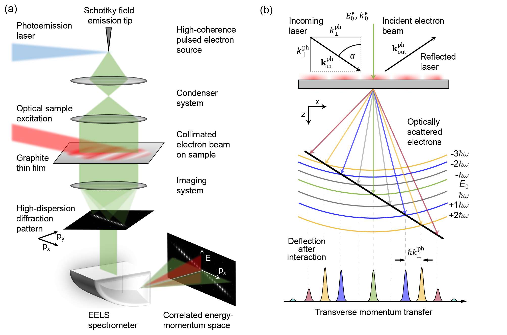
Here, we report the optical preparation and observation of high-purity quantum coherent linear momentum superposition states in the transverse and longitudinal direction of an electron microscope beam. Low-emittance ultrashort electron pulses, generated by nanotip photoemission, are collimated to a micrometer-scale transverse coherence length and transmitted through a laser-illuminated graphite thin film. The imprinted three-dimensional sinusoidal phase modulation yields a coherent superposition of correlated energy-momentum ladder states, which is mapped by its far-field scattering distribution.
We begin by considering the theoretical basis for our study. Inelastic electron-light scattering is well-understood, and several approaches have been presented to derive analytical expressions for PINEM sideband amplitudes, including a Green’s function formalism 54, direct integration of the Schrödinger equation 53 and a ladder operator formalism 55.
In the following, setting a general framework for transverse beam shaping, we offer an alternative, rather concise derivation using a path integral representation of the wavefunction 70. In the semiclassical limit, it is expressed in terms of the classical action as , where is the action - a solution of the Hamilton-Jacobi equation, and is the Lagrangian. The integral over is evaluated along a specific classical path that ends at the point . The initial point of the path at time is specified by the initial velocity . Consequently, the phase difference between the phases of the initial plane-wave state and the final state at position and at time can be written as:
| (1) |
where the second and third terms subtract the phase acquired by the initial state along the path of integration. Equation (1) is the exact semiclassical limit for the phase modulation. It can be further simplified under the assumption that the energy change is small compared to the initial kinetic energy of the electron. Treating the interaction as a perturbation, one obtains in the first order the following expression 71:
| (2) |
where is the interaction Hamiltonian (with elementary charge and electron rest mass ), and is the vector potential in the propagation direction of the electron beam (which we choose along ). With the same accuracy, can be approximated by a straight-line path: , where is the initial velocity.
The spatial and temporal structure of the phase modulation depends on the specific field geometry in a given experiment. A field distribution breaking translational invariance along the electron propagation direction, e.g., at an optical nanostructure, can induce transitions between states separated by the photon energy 52. For translational invariance in the direction perpendicular to the beam path, such transitions will entail quantized transverse momentum transfer 72. The simplest geometry fulfilling both requirements is given by light reflection from a planar surface 59; 33; 64; 34. This allows for a time-harmonic electromagnetic field that abruptly varies in the -direction but is propagating along the -axis.
Consequently, one can choose the form , where denotes the -dependent amplitude of the electric field, with angular frequency and transverse wavevector component .
Hereby, equation (2) can be concisely written in terms of the Fourier transform of , as follows:
| (3) |
where describes the strength of electron-light interaction:
| (4) |
It is convenient to deal with the phase modulation in a shifted frame (given by the substitution into Eq. (High-purity free-electron momentum states prepared by three-dimensional optical phase modulation)) because, in this frame, both the phase and the initial state are independent of time . Finally, we arrive at
| (5) |
Electron scattering at such a time-dependent phase grating results in the formation of distinct photon orders in far-field diffraction, with amplitudes given by Bessel functions of the first kind 53; 55. Physically, the absorption of a photon with energy necessarily involves also absorption of its transverse momentum , with wavelength and angle of incidence for a sample plane normal to the electron beam (cf. Fig. 1b). As a consequence, a one-to-one correspondence of the scattering distribution in the total kinetic energy and the transverse momentum subspace is expected (Fig. 1b) 72. We note that transverse optical phase modulation with a uniform coupling constant acts as a coherent inelastic beam splitter for free-electron beams, if the individual diffraction orders are fully separated in angle. For visible optical frequencies and near-relativistic electrons, the corresponding angular separation amounts to only a few microradians. Therefore, the experimental characterization of such phase-shaped beams, and in particular the resolution of individual scattering orders, requires electron beams of sufficiently low divergence, connected to a micrometer-scale transverse coherence length , with root-mean-square (rms) angular spread 73.
As we now demonstrate, such conditions are achieved in our ultrafast transmission electron microscope, which is based on laser-driven photoelectron emission from a Schottky field emission source 51; 74.
In the experiment (Fig. 1a), we generate ultrashort, high-coherence electron pulses by localized single-photon photoemission from a nanoscopic, Schottky-type field emitter tip 51. After acceleration to a kinetic energy of 200 keV (2.51-pm de-Broglie wavelength), the electron beam is collimated to a FWHM full beam divergence of µ, resulting in a lateral coherence length of µm (cf. Fig. 3a, top), and radially limited to a diameter of about 13 µm. Passing the electrons through the optical field of ultrashort laser pulses (800-nm center wavelength, µm focal spot size, up to 150-nJ pulse energy, dispersively stretched to 3.4-ps duration), that are reflected from the surface of a 120-nm thick single crystalline graphite flake (882.5-nm effective period of the transverse optical phase grating) 222The sample is prepared by cleaving a graphite single crystal perpendicular to the c-axis. In the experiment, the relative angle between laser and electron beam is and the sample was tilted away from the surface normal and laser incidence by , precisely determined by convergent beam electron diffraction, imprints a sinusoidal phase modulation onto the electron wavefunction in the transverse and longitudinal beam directions (Fig. 1b).
The scattered electron distribution is analyzed by operating the electron microscope in diffraction mode (effective camera length of 33 m). We record maps of kinetic energy and momentum using an electron spectrometer (Fig. 2), or of the full transverse momentum distribution in the far-field (Fig. 3).
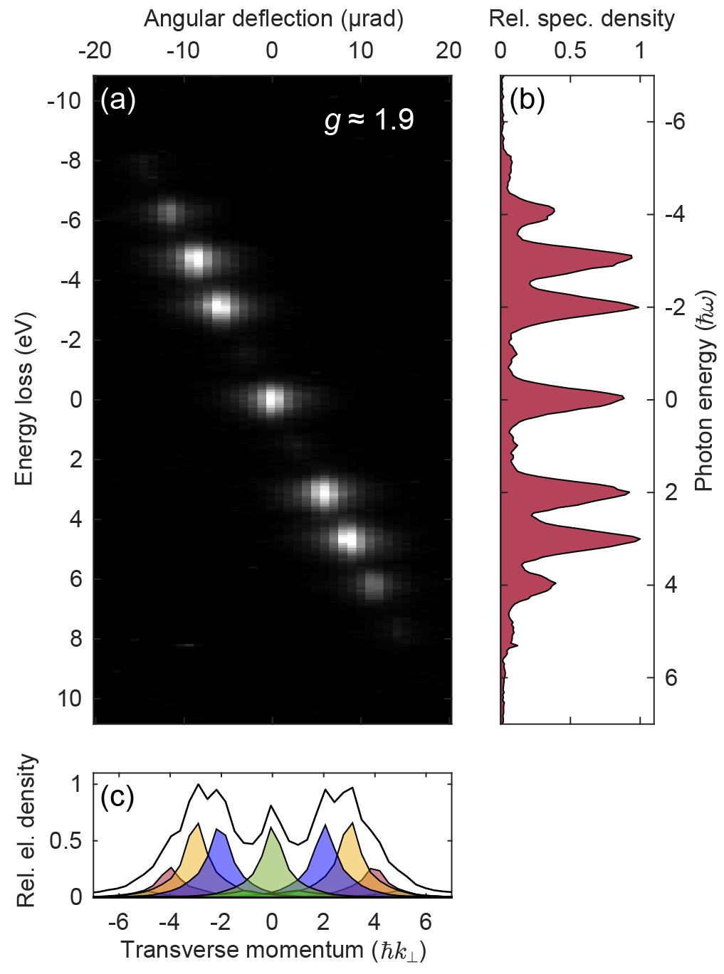
Figure 2a shows the kinetic energy and angle-resolved far-field scattering distribution 333diffraction angles calibrated with a grating replica of 463-nm spacing, clearly demonstrating the creation of an equally-spaced comb of energy-momentum states (Fig. 2b). The transitions in this two-dimensional energy-momentum ladder (cf. Fig. 1b) lead to a correlated quantized gain or loss of kinetic energy and transverse photon momentum (Fig. 2c), with a corresponding single-order deflection angle of µrad.
Notably, while the sidebands are fully separated in energy, with a visibility of ( - FWHM energy spread of the electron beam), the optics of the EEL spectrometer and the employed scintillator-coupled CCD camera limit the detected angular separation to ( - FWHM of detected transverse electron momentum distribution).
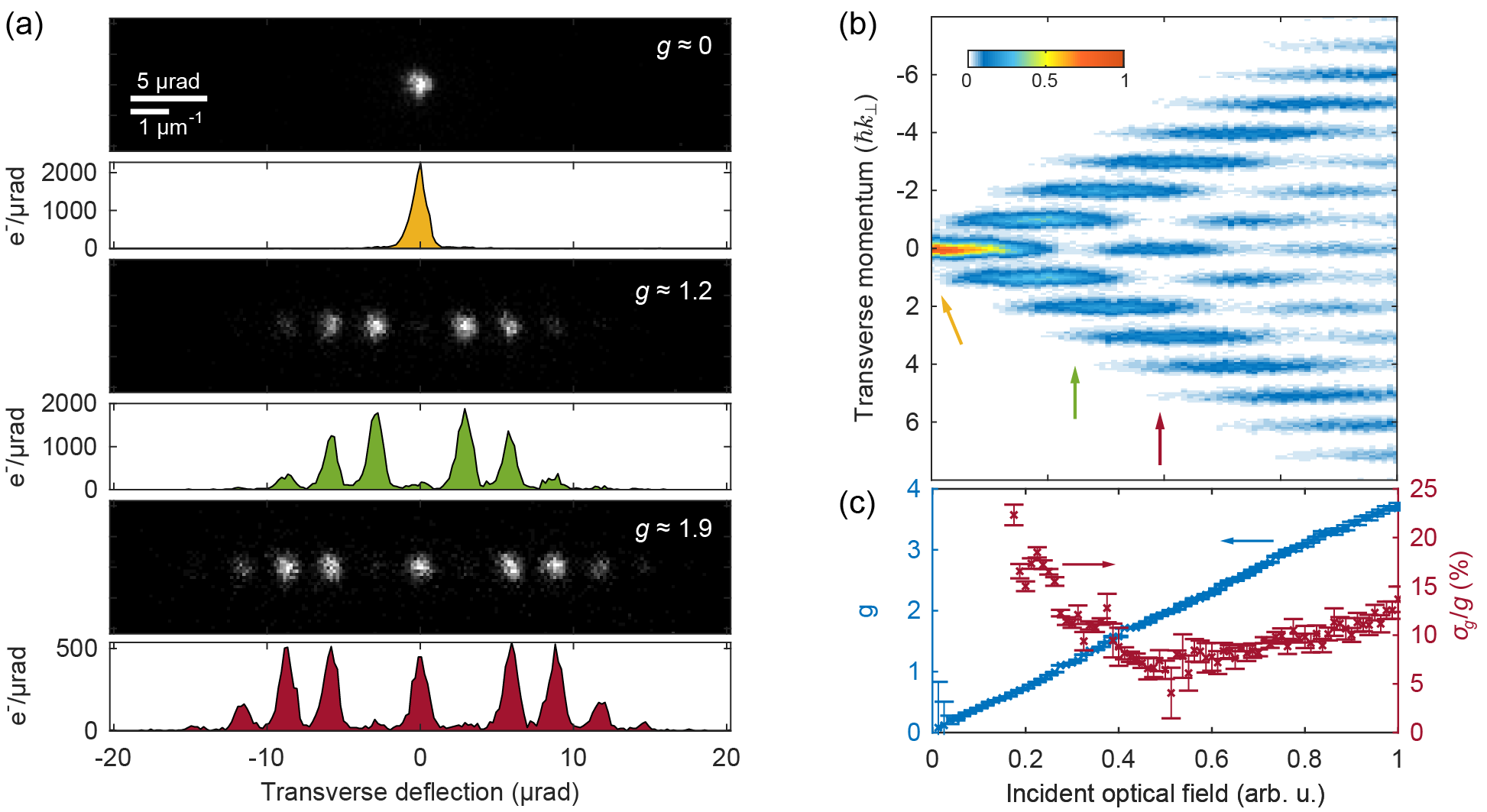
Facilitated by the low-divergence electron beams used, in the following, we directly resolve individual scattering orders by their deflection angles, and without employing energy-dispersive electron optics. We map the full two-dimensional transverse phase space by recording diffraction patterns after the optical interaction. Figure 3 shows the far-field scattering distribution using the same experimental conditions as for Fig. 2, but recording the diffracted beam by counted electron imaging with a direct detection CMOS camera (Direct Electron DE16). For increasing optical fields (cf. Fig. 3a), the initial single-peak transverse momentum distribution splits into an equally spaced comb, separated by the transverse photon momentum and well resolved with a visibility of , with ( - FWHM transverse momentum spread of the electron beam). The purity of the quantum superposition state prepared at a single optical interaction is encoded in the optical-field dependent sideband populations (Fig. 3b, top), which are, for a non-mixed state, analytically given by Bessel functions of the first kind 53; 55. The experimentally observed occupations are well reproduced by considering a Gaussian distributed coupling constant with standard deviation (Fig. 3c). Beyond a value of , we find a relative uncertainty between and , which indicates a high purity of the generated superposition quantum state 444Lower values and a smaller number of populated states lead to a higher relative uncertainty in the fit. A unique fingerprint of such a state – and validation of its action as a coherent electron beam splitter – is the full suppression of specific sideband orders, e.g., in the zero order beam () for the first zero crossing of , which is achieved for , as clearly visible in Fig. 3a (middle).
Generally, multiple sources of incoherence are of relevance, which reduce the purity of the quantum superposition state. In the current experiment, there are two main contributions, which are minimized by the design of the experiment. Firstly, incoherent ensemble averages in the preparation of the initial quantum state (i.e., the initial spread in transverse momentum and energy), are reduced by collimating our high-coherence beam, enabling a full separation of the photon orders. Secondly, temporal and spatial variations in the field strength lead to averaging in , which we minimize by limiting the electron beam diameter and temporally stretching the optical pulse. The simultaneous need of a low electron beam divergence and small diameter require a picometer-scale transverse beam emittance . In our experiment, we achieve a value of (apertured, at the sample stage, with relativistic parameters and ). Such high-brightness/low-emittance ultrashort electron pulses are readily available in field emitter-based UTEM instruments 51; 75; 47; 76.
In conclusion, we have realized a coherent inelastic beam splitter for free-electron beams and demonstrate the optical preparation of high-purity transverse electron beam states.
These capabilities extend the toolset of matter-wave interferometry by providing coherent optical beam manipulation, e.g., as an element for new forms of interference-enhanced multi-path electron microscopes 77. Furthermore, taking advantage of the spectral state separation, inelastic electron holography in a STEM-type geometry 78 may give access to non-local information in fundamental material excitations 79; 54. Exploiting the advanced transverse phase-space control in the sub-optical cycle temporal structuring of electron beams will facilitate electron microscopy with isolated attosecond electron pulses, reminiscent of the attosecond lighthouse in optics 80. Finally, the concept of high-coherence transverse and longitudinal phase modulation can be extended to complex plasmonic light fields, thus, providing for an externally programmable three-dimensional all-optical phase plate for electrons.
Acknowledgement
We thank the Göttingen UTEM team for ongoing support and fruitful collaboration. We acknowledge insightful discussions with Katharina E. Priebe, Thomas Rittmann, Nara Rubiano da Silva, Tyler Harvey and Hugo Lourenço-Martins. This work was funded by the Deutsche Forschungsgemeinschaft (DFG) in the Collaborative Research Center “Atomic Scale Control of Energy Conversion” (DFG-SFB 1073, project A05), the Priority Program “Quantum Dynamics in Tailored Intense Fields” (DFG-SPP 1840) and via funds of the Leibniz Program.
References
- 1 A. D. Cronin, J. Schmiedmayer, and D. E. Pritchard, “Optics and interferometry with atoms and molecules,” Reviews of Modern Physics, vol. 81, no. 3, pp. 1051–1129, 2009.
- 2 F. Hasselbach, “Progress in electron- and ion-interferometry,” Reports on Progress in Physics, vol. 73, no. 1, p. 016101, 2010.
- 3 C. Pellegrini, A. Marinelli, and S. Reiche, “The physics of x-ray free-electron lasers,” Reviews of Modern Physics, vol. 88, no. 1, p. 015006, 2016.
- 4 P. A. Midgley and R. E. Dunin-Borkowski, “Electron tomography and holography in materials science,” Nature Materials, vol. 8, no. 4, pp. 271–280, 2009.
- 5 M. Uchida and A. Tonomura, “Generation of electron beams carrying orbital angular momentum.,” Nature, vol. 464, no. 7289, pp. 737–739, 2010.
- 6 J. Verbeeck, H. Tian, and P. Schattschneider, “Production and application of electron vortex beams,” Nature, vol. 467, no. 7313, pp. 301–304, 2010.
- 7 B. J. McMorran, A. Agrawal, I. M. Anderson, A. A. Herzing, H. J. Lezec, J. J. McClelland, and J. Unguris, “Electron vortex beams with high quanta of orbital angular momentum,” Science, vol. 331, no. 6014, pp. 192–195, 2011.
- 8 V. Grillo, G. Carlo Gazzadi, E. Karimi, E. Mafakheri, R. W. Boyd, and S. Frabboni, “Highly efficient electron vortex beams generated by nanofabricated phase holograms,” Applied Physics Letters, vol. 104, no. 4, p. 043109, 2014.
- 9 K. Bliokh, I. Ivanov, G. Guzzinati, L. Clark, R. Van Boxem, A. Béché, R. Juchtmans, M. Alonso, P. Schattschneider, F. Nori, and J. Verbeeck, “Theory and applications of free-electron vortex states,” Physics Reports, vol. 690, no. 2017, pp. 1–70, 2017.
- 10 N. Voloch-Bloch, Y. Lereah, Y. Lilach, A. Gover, and A. Arie, “Generation of electron Airy beams,” Nature, vol. 494, no. 7437, pp. 331–335, 2013.
- 11 F. Courvoisier, A. Mathis, L. Froehly, R. Giust, L. Furfaro, P. A. Lacourt, M. Jacquot, and J. M. Dudley, “Sending femtosecond pulses in circles: highly nonparaxial accelerating beams,” Optics Letters, vol. 37, no. 10, p. 1736, 2012.
- 12 I. Kaminer, R. Bekenstein, J. Nemirovsky, and M. Segev, “Nondiffracting Accelerating Wave Packets of Maxwell’s Equations,” Physical Review Letters, vol. 108, no. 16, p. 163901, 2012.
- 13 V. Grillo, E. Karimi, G. C. Gazzadi, S. Frabboni, M. R. Dennis, and R. W. Boyd, “Generation of Nondiffracting Electron Bessel Beams,” Physical Review X, vol. 4, no. 1, p. 011013, 2014.
- 14 A. Asenjo-Garcia and F. J. García de Abajo, “Dichroism in the Interaction between Vortex Electron Beams, Plasmons, and Molecules,” Physical Review Letters, vol. 113, no. 6, p. 066102, 2014.
- 15 S. M. Lloyd, M. Babiker, G. Thirunavukkarasu, and J. Yuan, “Electron vortices: Beams with orbital angular momentum,” Reviews of Modern Physics, vol. 89, no. 3, p. 035004, 2017.
- 16 J. Verbeeck, H. Tian, and G. Van Tendeloo, “How to Manipulate Nanoparticles with an Electron Beam?,” Advanced Materials, vol. 25, no. 8, pp. 1114–1117, 2013.
- 17 J. Verbeeck, A. Béché, K. Müller-Caspary, G. Guzzinati, M. A. Luong, and M. Den Hertog, “Demonstration of a 2 × 2 programmable phase plate for electrons,” Ultramicroscopy, vol. 190, no. 2018, pp. 58–65, 2018.
- 18 F. J. García de Abajo, “Optical excitations in electron microscopy,” Reviews of Modern Physics, vol. 82, no. 1, pp. 209–275, 2010.
- 19 E. Hemsing, G. Stupakov, D. Xiang, and A. Zholents, “Beam by design: Laser manipulation of electrons in modern accelerators,” Reviews of Modern Physics, vol. 86, no. 3, pp. 897–941, 2014.
- 20 R. J. England, R. J. Noble, K. Bane, D. H. Dowell, C.-K. Ng, J. E. Spencer, S. Tantawi, Z. Wu, R. L. Byer, E. Peralta, K. Soong, C.-M. Chang, B. Montazeri, S. J. Wolf, B. Cowan, J. Dawson, W. Gai, P. Hommelhoff, Y.-C. Huang, C. Jing, C. McGuinness, R. B. Palmer, B. Naranjo, J. Rosenzweig, G. Travish, A. Mizrahi, L. Schachter, C. Sears, G. R. Werner, and R. B. Yoder, “Dielectric laser accelerators,” Reviews of Modern Physics, vol. 86, no. 4, pp. 1337–1389, 2014.
- 21 M. F. Ciappina, J. A. Pérez-Hernández, A. S. Landsman, W. A. Okell, S. Zherebtsov, B. Förg, J. Schötz, L. Seiffert, T. Fennel, T. Shaaran, T. Zimmermann, A. Chacón, R. Guichard, A. Zaïr, J. W. G. Tisch, J. P. Marangos, T. Witting, A. Braun, S. A. Maier, L. Roso, M. Krüger, P. Hommelhoff, M. F. Kling, F. Krausz, and M. Lewenstein, “Attosecond physics at the nanoscale,” Reports on Progress in Physics, vol. 80, no. 5, p. 054401, 2017.
- 22 H. Müller, J. Jin, R. Danev, J. Spence, H. Padmore, and R. M. Glaeser, “Design of an electron microscope phase plate using a focused continuous-wave laser,” New Journal of Physics, vol. 12, no. 7, p. 073011, 2010.
- 23 O. Schwartz, J. J. Axelrod, S. L. Campbell, C. Turnbaugh, R. M. Glaeser, and H. Müller, “Laser phase plate for transmission electron microscopy,” Nature Methods, vol. 16, no. 10, pp. 1016–1020, 2019.
- 24 P. H. Bucksbaum, M. Bashkansky, and T. J. McIlrath, “Scattering of electrons by intense coherent light,” Physical Review Letters, vol. 58, no. 4, pp. 349–352, 1987.
- 25 B. J. Siwick, A. A. Green, C. T. Hebeisen, and R. J. D. Miller, “Characterization of ultrashort electron pulses by electron-laser pulse cross correlation,” Optics Letters, vol. 30, no. 9, p. 1057, 2005.
- 26 C. T. Hebeisen, G. Sciaini, M. Harb, R. Ernstorfer, T. Dartigalongue, S. G. Kruglik, and R. J. D. Miller, “Grating enhanced ponderomotive scattering for visualization and full characterization of femtosecond electron pulses,” Optics Express, vol. 16, no. 5, p. 3334, 2008.
- 27 M. Gao, H. Jean-Ruel, R. R. Cooney, J. Stampe, M. de Jong, M. Harb, G. Sciaini, G. Moriena, and R. J. Dwayne Miller, “Full characterization of RF compressed femtosecond electron pulses using ponderomotive scattering,” Optics Express, vol. 20, no. 11, p. 12048, 2012.
- 28 M. Kozák, J. McNeur, K. J. Leedle, H. Deng, N. Schönenberger, A. Ruehl, I. Hartl, J. S. Harris, R. L. Byer, and P. Hommelhoff, “Optical gating and streaking of free electrons with sub-optical cycle precision,” Nature Communications, vol. 8, no. 1, p. 14342, 2017.
- 29 G. H. Kassier, N. Erasmus, K. Haupt, I. Boshoff, R. Siegmund, S. M. M. Coelho, and H. Schwoerer, “Photo-triggered pulsed cavity compressor for bright electron bunches in ultrafast electron diffraction,” Applied Physics B, vol. 109, no. 2, pp. 249–257, 2012.
- 30 L. J. Wong, B. Freelon, T. Rohwer, N. Gedik, and S. G. Johnson, “All-optical three-dimensional electron pulse compression,” New Journal of Physics, vol. 17, no. 1, p. 013051, 2015.
- 31 J. Breuer and P. Hommelhoff, “Laser-Based Acceleration of Nonrelativistic Electrons at a Dielectric Structure,” Physical Review Letters, vol. 111, no. 13, p. 134803, 2013.
- 32 E. A. Peralta, K. Soong, R. J. England, E. R. Colby, Z. Wu, B. Montazeri, C. McGuinness, J. McNeur, K. J. Leedle, D. Walz, E. B. Sozer, B. Cowan, B. Schwartz, G. Travish, and R. L. Byer, “Demonstration of electron acceleration in a laser-driven dielectric microstructure,” Nature, vol. 503, no. 7474, pp. 91–94, 2013.
- 33 K. E. Priebe, C. Rathje, S. V. Yalunin, T. Hohage, A. Feist, S. Schäfer, and C. Ropers, “Attosecond electron pulse trains and quantum state reconstruction in ultrafast transmission electron microscopy,” Nature Photonics, vol. 11, no. 12, pp. 793–797, 2017.
- 34 Y. Morimoto and P. Baum, “Diffraction and microscopy with attosecond electron pulse trains,” Nature Physics, vol. 14, no. 3, pp. 252–256, 2018.
- 35 L. Wimmer, G. Herink, D. R. Solli, S. V. Yalunin, K. E. Echternkamp, and C. Ropers, “Terahertz control of nanotip photoemission,” Nature Physics, vol. 10, no. 6, pp. 432–436, 2014.
- 36 E. A. Nanni, W. R. Huang, K.-H. Hong, K. Ravi, A. Fallahi, G. Moriena, R. J. Dwayne Miller, and F. X. Kärtner, “Terahertz-driven linear electron acceleration,” Nature Communications, vol. 6, no. 1, p. 8486, 2015.
- 37 C. Kealhofer, W. Schneider, D. Ehberger, A. Ryabov, F. Krausz, and P. Baum, “All-optical control and metrology of electron pulses,” Science, vol. 352, no. 6284, pp. 429–433, 2016.
- 38 E. Curry, S. Fabbri, J. Maxson, P. Musumeci, and A. Gover, “Meter-Scale Terahertz-Driven Acceleration of a Relativistic Beam,” Physical Review Letters, vol. 120, no. 9, p. 094801, 2018.
- 39 D. Zhang, A. Fallahi, M. Hemmer, X. Wu, M. Fakhari, Y. Hua, H. Cankaya, A.-L. Calendron, L. E. Zapata, N. H. Matlis, and F. X. Kärtner, “Segmented terahertz electron accelerator and manipulator (STEAM),” Nature Photonics, vol. 12, no. 6, pp. 336–342, 2018.
- 40 R. K. Li, M. C. Hoffmann, E. A. Nanni, S. H. Glenzer, M. E. Kozina, A. M. Lindenberg, B. K. Ofori-Okai, A. H. Reid, X. Shen, S. P. Weathersby, J. Yang, M. Zajac, and X. J. Wang, “Terahertz-based subfemtosecond metrology of relativistic electron beams,” Physical Review Accelerators and Beams, vol. 22, no. 1, p. 012803, 2019.
- 41 D. Zhang, A. Fallahi, M. Hemmer, H. Ye, M. Fakhari, Y. Hua, H. Cankaya, A.-L. Calendron, L. E. Zapata, N. H. Matlis, and F. X. Kärtner, “Femtosecond phase control in high-field terahertz-driven ultrafast electron sources,” Optica, vol. 6, no. 7, p. 872, 2019.
- 42 H. W. Kim, N. A. Vinokurov, I. H. Baek, K. Y. Oang, M. H. Kim, Y. C. Kim, K.-H. Jang, K. Lee, S. H. Park, S. Park, J. Shin, J. Kim, F. Rotermund, S. Cho, T. Feurer, and Y. U. Jeong, “Towards jitter-free ultrafast electron diffraction technology,” Nature Photonics, pp. 6–11, 2019.
- 43 L. Zhao, Z. Wang, H. Tang, R. Wang, Y. Cheng, C. Lu, T. Jiang, P. Zhu, L. Hu, W. Song, H. Wang, J. Qiu, R. Kostin, C. Jing, S. Antipov, P. Wang, J. Qi, Y. Cheng, D. Xiang, and J. Zhang, “Terahertz Oscilloscope for Recording Time Information of Ultrashort Electron Beams,” Physical Review Letters, vol. 122, no. 14, p. 144801, 2019.
- 44 R. P. Chatelain, V. R. Morrison, C. Godbout, and B. J. Siwick, “Ultrafast electron diffraction with radio-frequency compressed electron pulses,” Applied Physics Letters, vol. 101, no. 8, p. 081901, 2012.
- 45 A. Gliserin, M. Walbran, F. Krausz, and P. Baum, “Sub-phonon-period compression of electron pulses for atomic diffraction,” Nature Communications, vol. 6, no. 1, p. 8723, 2015.
- 46 J. Maxson, D. Cesar, G. Calmasini, A. Ody, P. Musumeci, and D. Alesini, “Direct Measurement of Sub-10 fs Relativistic Electron Beams with Ultralow Emittance,” Physical Review Letters, vol. 118, no. 15, p. 154802, 2017.
- 47 W. Verhoeven, J. van Rens, E. Kieft, P. Mutsaers, and O. Luiten, “High quality ultrafast transmission electron microscopy using resonant microwave cavities,” Ultramicroscopy, vol. 188, pp. 85–89, 2018.
- 48 C. Jing, Y. Zhu, A. Liu, K. Schliep, X. Fu, Y. Zhao, E. Montgomery, W. Rush, A. Kanareykin, M. Katz, and J. Lau, “Tunable electron beam pulser for picoseconds stroboscopic microscopy in transmission electron microscopes,” Ultramicroscopy, vol. 207, no. August, p. 112829, 2019.
- 49 A. H. Zewail, “4D ultrafast electron diffraction, crystallography, and microscopy,” Annual Review of Physical Chemistry, vol. 57, no. 1, pp. 65–103, 2006.
- 50 D. J. Flannigan and A. H. Zewail, “4D Electron Microscopy: Principles and Applications,” Accounts of Chemical Research, vol. 45, no. 10, pp. 1828–1839, 2012.
- 51 A. Feist, N. Bach, N. Rubiano da Silva, T. Danz, M. Möller, K. E. Priebe, T. Domröse, J. G. Gatzmann, S. Rost, J. Schauss, S. Strauch, R. Bormann, M. Sivis, S. Schäfer, and C. Ropers, “Ultrafast transmission electron microscopy using a laser-driven field emitter: Femtosecond resolution with a high coherence electron beam,” Ultramicroscopy, vol. 176, pp. 63–73, 2017.
- 52 B. Barwick, D. J. Flannigan, and A. H. Zewail, “Photon-induced near-field electron microscopy,” Nature, vol. 462, no. 7275, pp. 902–906, 2009.
- 53 S. T. Park, M. Lin, and A. H. Zewail, “Photon-induced near-field electron microscopy (PINEM): theoretical and experimental,” New Journal of Physics, vol. 12, no. 12, p. 123028, 2010.
- 54 F. J. García de Abajo, A. Asenjo-Garcia, and M. Kociak, “Multiphoton Absorption and Emission by Interaction of Swift Electrons with Evanescent Light Fields,” Nano Letters, vol. 10, no. 5, pp. 1859–1863, 2010.
- 55 A. Feist, K. E. Echternkamp, J. Schauss, S. V. Yalunin, S. Schäfer, and C. Ropers, “Quantum coherent optical phase modulation in an ultrafast transmission electron microscope,” Nature, vol. 521, no. 7551, pp. 200–203, 2015.
- 56 L. Piazza, T. Lummen, E. Quiñonez, Y. Murooka, B. Reed, B. Barwick, and F. Carbone, “Simultaneous observation of the quantization and the interference pattern of a plasmonic near-field,” Nature Communications, vol. 6, no. 1, p. 6407, 2015.
- 57 A. Yurtsever, R. M. van der Veen, and A. H. Zewail, “Subparticle Ultrafast Spectrum Imaging in 4D Electron Microscopy,” Science, vol. 335, no. 6064, pp. 59–64, 2012.
- 58 I. Madan, G. M. Vanacore, E. Pomarico, G. Berruto, R. J. Lamb, D. Mcgrouther, T. T. A. Lummen, T. Latychevskaia, F. J. García De Abajo, and F. Carbone, “Holographic imaging of electromagnetic fields via electron-light quantum interference,” Science Advances, vol. 5, no. (to appear), pp. 1–8, 2019.
- 59 F. O. Kirchner, A. Gliserin, F. Krausz, and P. Baum, “Laser streaking of free electrons at 25 keV,” Nature Photonics, vol. 8, no. 1, pp. 52–57, 2014.
- 60 E. Pomarico, I. Madan, G. Berruto, G. M. Vanacore, K. Wang, I. Kaminer, F. J. García de Abajo, and F. Carbone, “meV Resolution in Laser-Assisted Energy-Filtered Transmission Electron Microscopy,” ACS Photonics, vol. 5, no. 3, pp. 759–764, 2018.
- 61 O. Kfir, H. Lourenço-Martins, G. Storeck, M. Sivis, T. R. Harvey, T. J. Kippenberg, A. Feist, and C. Ropers, “Controlling free electrons with optical whispering-gallery modes,” arXiv:physics.optics, pp. 1–11, 2019.
- 62 K. Wang, R. Dahan, M. Shentcis, Y. Kauffmann, S. Tsesses, and I. Kaminer, “Coherent interaction between free electrons and cavity photons,” arXiv:physics.optics, pp. 1–16, 2019.
- 63 K. E. Echternkamp, A. Feist, S. Schäfer, and C. Ropers, “Ramsey-type phase control of free-electron beams,” Nature Physics, vol. 12, no. 11, pp. 1000–1004, 2016.
- 64 G. M. Vanacore, I. Madan, G. Berruto, K. Wang, E. Pomarico, R. J. Lamb, D. McGrouther, I. Kaminer, B. Barwick, F. J. García de Abajo, and F. Carbone, “Attosecond coherent control of free-electron wave functions using semi-infinite light fields,” Nature Communications, vol. 9, no. 1, p. 2694, 2018.
- 65 J. Krehl, G. Guzzinati, J. Schultz, P. Potapov, D. Pohl, J. Martin, J. Verbeeck, A. Fery, B. Büchner, and A. Lubk, “Spectral field mapping in plasmonic nanostructures with nanometer resolution,” Nature Communications, vol. 9, no. 1, p. 4207, 2018.
- 66 G. M. Vanacore, G. Berruto, I. Madan, E. Pomarico, P. Biagioni, R. J. Lamb, D. McGrouther, O. Reinhardt, I. Kaminer, B. Barwick, H. Larocque, V. Grillo, E. Karimi, F. J. García de Abajo, and F. Carbone, “Ultrafast generation and control of an electron vortex beam via chiral plasmonic near fields,” Nature Materials, vol. 18, no. 6, pp. 573–579, 2019.
- 67 H. Kim, J. Park, S.-W. Cho, S.-Y. Lee, M. Kang, and B. Lee, “Synthesis and Dynamic Switching of Surface Plasmon Vortices with Plasmonic Vortex Lens,” Nano Letters, vol. 10, no. 2, pp. 529–536, 2010.
- 68 G. Spektor, D. Kilbane, A. K. Mahro, B. Frank, S. Ristok, L. Gal, P. Kahl, D. Podbiel, S. Mathias, H. Giessen, F. J. Meyer Zu Heringdorf, M. Orenstein, and M. Aeschlimann, “Revealing the subfemtosecond dynamics of orbital angular momentum in nanoplasmonic vortices,” Science, vol. 355, no. 6330, pp. 1187–1191, 2017.
- 69 D. L. Freimund, K. Aflatooni, and H. Batelaan, “Observation of the Kapitza-Dirac effect,” Nature, vol. 413, no. 6852, pp. 142–143, 2001.
- 70 L. S. Schulman, Techniques and Applications of Path Integration. Dover Publications Inc., 2005.
- 71 A. Lubk, “Paraxial Quantum Mechanics,” in Advances in Imaging and Electron Physics (Volume 206) (P. W. Hawkes, ed.), ch. 2, pp. 15–58, London: Academic Press, 2018.
- 72 F. J. García de Abajo, B. Barwick, and F. Carbone, “Electron diffraction by plasmon waves,” Physical Review B, vol. 94, no. 4, p. 041404, 2016.
- 73 T. Van Oudheusden, E. F. De Jong, S. B. van der Geer, W. P. E. M. Op ’T Root, O. J. Luiten, B. J. Siwick, W. P. E. M. Op’t Root, O. J. Luiten, and B. J. Siwick, “Electron source concept for single-shot sub-100 fs electron diffraction in the 100 keV range,” Journal of Applied Physics, vol. 102, no. 9, p. 093501, 2007.
- 74 N. Bach, T. Domröse, A. Feist, T. Rittmann, S. Strauch, C. Ropers, and S. Schäfer, “Coulomb interactions in high-coherence femtosecond electron pulses from tip emitters,” Structural Dynamics, vol. 6, no. 1, p. 014301, 2019.
- 75 F. Houdellier, G. Caruso, S. Weber, M. Kociak, and A. Arbouet, “Development of a high brightness ultrafast Transmission Electron Microscope based on a laser-driven cold field emission source,” Ultramicroscopy, vol. 186, pp. 128–138, 2018.
- 76 C. Zhu, D. Zheng, H. Wang, M. Zhang, Z. Li, S. Sun, P. Xu, H. Tian, Z. Li, H. Yang, and J. Li, “Development of analytical ultrafast transmission electron microscopy based on laser-driven Schottky field emission,” Ultramicroscopy, vol. 209, no. October 2019, p. 112887, 2020.
- 77 P. Kruit, R. Hobbs, C.-s. Kim, Y. Yang, V. Manfrinato, J. Hammer, S. Thomas, P. Weber, B. Klopfer, C. Kohstall, T. Juffmann, M. Kasevich, P. Hommelhoff, and K. Berggren, “Designs for a quantum electron microscope,” Ultramicroscopy, vol. 164, pp. 31–45, 2016.
- 78 F. S. Yasin, T. R. Harvey, J. J. Chess, J. S. Pierce, and B. J. McMorran, “Path-separated electron interferometry in a scanning transmission electron microscope,” Journal of Physics D: Applied Physics, vol. 51, no. 20, p. 205104, 2018.
- 79 H. Lichte and B. Freitag, “Inelastic electron holography,” Ultramicroscopy, vol. 81, no. 3-4, pp. 177–186, 2000.
- 80 K. T. Kim, C. Zhang, T. Ruchon, J.-F. Hergott, T. Auguste, D. M. Villeneuve, P. B. Corkum, and F. Quéré, “Photonic streaking of attosecond pulse trains,” Nature Photonics, vol. 7, no. 8, pp. 651–656, 2013.