Blue moon ensemble simulation of aquation free energy profiles applied to mono and bifunctional platinum anticancer drugs
Abstract
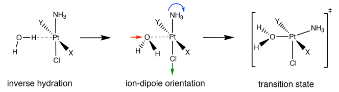
Aquation free energy profiles of neutral cisplatin and cationic monofunctional derivatives, including triaminochloroplatinum(II) and cis-diammine(pyridine)chloroplatinum(II), were computed using state of the art thermodynamic integration, for which temperature and solvent were accounted for explicitly using density functional theory based canonical molecular dynamics (DFT-MD). For all the systems the ”inverse-hydration” where the metal center acts as an acceptor of hydrogen bond has been observed. This has motivated to consider the inversely bonded solvent molecule in the definition of the reaction coordinate required to initiate the constrained DFT-MD trajectories. We found that there exists little difference in free enthalpies of activations, such that these platinum-based anticancer drugs are likely to behave the same way in aqueous media. Detailed analysis of the microsolvation structure of the square-planar complexes, along with the key steps of the aquation mechanism are discussed.
I Introduction
The discovery of the anticancer activity of cisplatin (Scheme 1) during the 60’s has promoted fast development of platinum(II)-based drugs which are currently found in chemotherapy regimens (see Refs. 1; 2; 3; 4 for reviews). Despite few successes for some of them in curing specific cancers, eg. the treatment of testicular cancer with cisplatin, their efficiency against the broad spectrum of carcinoma remains limited. The main challenges to overcome are: (1) the elimination of the severe side effects related to their poor selectivity with respect to the tumor cells, and (2) the resistance —either intrinsic (static) or evolutionary (dynamic)—, observed for some types of cancer.Brabec and Kasparkova (2005); Chabner and Roberts (2005); Kelland (2007) Solving the first problem requires the design of new drug delivery strategies,Apps et al. (2015); Johnstone et al. (2016) which ideally prevents the degradation of platinum complex in the blood stream and allows for a precise targeting of the tumor tissue. Solution to the second problem implies heuristic methods, such as the structure-activity relationship (SAR),Hambley (1997); Johnstone et al. (2014) from which a huge numberHambley (1997) of platinum(II)-based drugs and platinum(IV)-based prodrugs were synthesized and tested as many attempts to mimic the cisplatin’s mechanism of action while trying to improve the cytotoxic properties. As a result of 30 years of trial and error less than 30 platinum drugs have been considered for clinical trials, with only 2 of them (carboplatin and oxaliplatin) approved wordwilde for clinical used.Wheate et al. (2010); Johnstone et al. (2016) Whereas high-throughput synthesis and screeningZiegler et al. (2000) of drug candidates may provide rapid —but partial— solutions to an urgent problem, understanding and controlling every details of cisplatin’s mechanism constitutes a safer —but far more longer— route towards a rational design of a universal platinum-based anticancer molecule. The process by which cisplatin (and bifunctional derivatives) leads to cell death is now rather well understood.Jamieson and Lippard (1999); Dhar and Lippard (2009) It is divided into 4 main steps: (i) the cellular uptake, (ii) the aquation/activation (Scheme 1) in the cellular media, (iii) the DNA platination, that is, bifunctional intra- and interstrand crossed-links via the fomation of 2 covalent bonds between the metal center and the purine bases, and (iv) the DNA-damage recognition initiating apoptosis, or cell-cycle arrest eventually followed by an attempt to repair the lesion. In elucidating each step of the mechanism, atomistic computations and simulations based on either (classical) molecular, quantum, or mixed molecular/quantum mechanics can provide important insights.
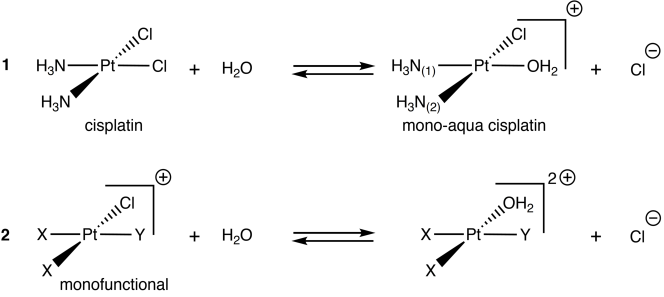
At the pure quantum mechanics level of theory, some of the efforts were devoted to investigate the hydration structure of the square-planar Pt-complexesVidossich et al. (2016); *beret_jctc_2008; *beret_cpc_2009; *vidossich_chemphyschem_2011; Truflandier et al. (2011); Sutter et al. (2011) in response to some experimental evidenceBaidina et al. (1981); Rizzato et al. (2010) showing unusual bonding situation —referred as to inverse-hydration— where the Pt atom acts as a hydrogen-bond acceptor. Energetic contributionsKozelka et al. (2000); Lopes et al. (2008); Aono et al. (2016) and topological analysis,Bergès et al. (2013) along with minute details on the influence of the bulk solvation effects have been extensively discussed in literature.Melchior et al. (2015); Kroutil et al. (2016); Aono et al. (2016) Another interest which is also closely related to our concern is the activation of cisplatin after the cellular uptake of the platinum drug. The aquation reaction (also loosely called hydrolysis) represented on Scheme 1 through the substitution of one or both chloride ligands of cisplatin by water molecules was recognized as a crucial step to initiate DNA platination.Bancroft et al. (1990) The proof of concept is carboplatinKnox et al. (1986); Alberts and Dorr (1998); Johnstone et al. (2014) featuring the cyclobutanedicarboxylate in replacement of the chloride leaving ligands of cisplatin. It demonstrates much more slower aquation and DNA platination rates but displayed similar crossed-link sites.Blommaert et al. (1995)
To access more details on the aquation of cisplatin and derivatives, many theoretical works have focused on computing activation barriers using static molecular clusters and implicit solvation models.Zhang et al. (2001); *robertazzi_hydrogen_2004; *lau_hydrolysis_2006; *alberto_second-generation_2009; *lucas_neutral_2009; *banerjee_cpl_2010; *melchior_comparative_2010 When applied for modelling aqueous reactions, insights arising from this type of computations remain limited since they do not account properly for the dynamic reorganisation of solvation shell surrounding the molecule, especially when the solvent is one of the reactant. More advanced but computationally demanding methods can be envisaged where bulk and microsolvation effect are treated on equal footing using density functional theory molecular dynamics (DFT-MD) along with free energy path evaluation based on metadynamics further refined by umbrella sampling.Carloni et al. (2000); Lau and Ensing (2010) Alternatives to DFT-MD, such as the reference interaction site model self-consistent fieldYokogawa et al. (2007, 2009) (RISM-SCF) can also be employed.Yokogawa et al. (2011)
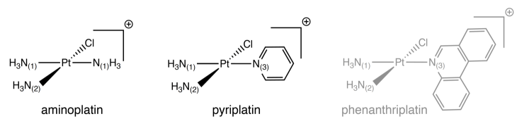
From the last ten-years research focussing on reducing the tumor-cell resistance to bifunctional cisplatin and derivatives, it has been shown that monofunctional analoguesHollis et al. (1989); Brabec (2002); Bursova et al. (2005) can be considered as potent candidates.Lovejoy et al. (2008); Wang et al. (2010); Zhu et al. (2012) Monofunctional platinum(II)-based drugs are cationic square-planar complexes (Scheme. 2) of formula , deriving from the triammine-chloro precursor , later referred as to aminoplatin. In a systematic investigation of the cancer-cell responses with respect to the heterocyclic N-donor ligand bound to the metal, it has been demonstratedPark et al. (2012) that the size and the arrangement of the aromatic rings affects drastically the cytotoxic and selectivity properties of the monofunctional platinum(II) drugs. As a matter of fact, whereas aminoplatin was found to be biologically inactive, phenanthriplatin, and to a lesser extent pyriplatin, presents a remarkable potency,Johnstone et al. (2014) exceeding in the majority of the cases the anticancer activity of cisplatin.Park et al. (2012); Johnstone et al. (2014)
This work aims to investigate the aquation reaction of Pt(II) complexes using state of the art thermodynamic integration coupled to DFT-MD simulations to determine if cationic monofunctional derivatives present differences in the free energy profiles and mechanism of reaction. The paper is organized as following: Section 2 provides methodological and computational aspects related to the DFT-BOMD simulation and blue moon ensemble (BME) integration. Results and discussion are presented in Section 3. The paper concludes with a brief summary of the key findings and an outlook.
II Computational details
II.1 DFT-BOMD simulations
All the DFT-MD simulations reported in this article were performed on the Born-Oppenheimer (BO) surface, where the Kohn-Sham (KS) self-consistent-field (SCF) equations are solved at each step of the dynamics using the Conquest code.Bowler et al. (2006); Bowler and Miyazaki (2010); Hernández et al. (1996) A strictly localized (atomic-like and finite range) numerical double- basis setSankey and Niklewski (1989); Junquera et al. (2001); Torralba et al. (2008) including polarisation functions (DZP) for expanding the valence wavefunctions along with norm-conserving pseudopotentialsTroullier and Martins (1991) (NCPP) were especially designed for this work. Thereafter, this basis set will be referred as pseudo-atomic orbitals (PAO). Using a benchmark of isolated Pt complexes, reliability of the PAOs were checked against molecular calculations based on gaussian-type orbitals (GTO). Additional computational parameters related to the PAOs and NCPPs generation along with their assessments can be found in the Sec. S1 of the Supporting Information (SI). The KS-SCF equations were solved within the framework of the generalized gradient approximation (GGA) of the exchange-correlation functional proposed by Perdew, Burke, and Ernzerhof (PBE).Perdew et al. (1996) This choice of GGA functional was more pragmatic than idealistic. It is well established (see Refs. 59; 60 for recent studies) that the PBE functional yields to a ”glassy” state of liquid water at ambient temperature preventing a fully quantitative agreement with experiments. Nevertheless, also important for this work, it has been demonstrated that the same functional is able to reproduce the inverse-hydration feature.Truflandier and Autschbach (2010)
Solvation of the platinum complexes has been modeled using cubic simulation boxes with 63 water molecules adapted in size to reach an average density of 1.0 gcm-3. For cationic complexes, the excess of positive charge was balanced by substituting water molecule with hydroxide anion keeping the overall charge of the supercell neutral. For the DFT-BOMD simulations, ionic cores were propagated using the velocity Verlet algorithmVerlet (1967) with a time step of 0.5 fs, for which a SCF convergence criteria of 10-7 on the residual of the electronic density, and a grid spacing of about 0.20 au were found sufficient to prevent any energy drift during microcanonical simulations. The same set of convergence parameters were used for NVT-ensemble simulations using a Nosé–Hoover chainMartyna et al. (1992); Hirakawa et al. (2017) of 5 thermostats with a frequency of 500 cm-1.
II.2 Blue moon ensemble integration
The hydrolysis free energy profiles of cisplatin and its derivatives were calculated using thermodynamic integration via the blue moon ensemble (BME) technique.Carter et al. (1989) Given a reaction coordinate, , which in general depends on a subset of the atomic positions , the variation of free energy between some initial state, , up to the current value of can be expressed as:
| (1) |
Integration is performed numerically through the sampling of the reaction coordinate using a discret set of target values . The free energy gradient —also referred to as (minus) the mean force— in Eq. (1) is evaluated for each from a constrained MD trajectory where is enforced. Following BME Lagrangian formulation of Sprik and Ciccotti,Sprik and Ciccotti (1998) the mean force writes
| (2) |
for which and are the Boltzmann constant and temperature, respectively. The Lagrange multiplier, , associated with constrained coordinate , defines the strength the constraint force updated at each MD step.Ryckaert et al. (1977); Andersen (1983); Ciccotti and Ryckaert (1986) The remaining terms are given by,
| (3) | |||||
| (4) |
where designates the mass of the th nucleus. In Eq. (1), the notation stands for the canonical ensemble average performed for each target value . Around 20 values were used for the reaction path discretization. Ensemble averages performed at K were obtained from production runs of 2.5 ps subsequent to 1.0 ps of equilibration. Free energy errors were estimated by the method of Jacucci and Rahman.Jacucci and Rahman (1984)
III Results and discussion
III.1 MD analysis and reliability
Complex Method Basis Distance (Å) Angle (deg) cisplatin Pt–N Pt–Cl N–Pt–N Cl–Pt–Cl BOMD-NVE PAO-DZP CPMD-NVEa PW Static-PCMb GTO-DZP Static GTO-DZP Static PAO-DZP mono-aqua cisplatin Pt–N(1)[Pt–N(2)] Pt–Cl Pt–O N–Pt–N BOMD-NVE PAO-DZP Static-PCM GTO-DZP Static GTO-DZP Static PAO-DZP aminoplatin Pt–N(1)[Pt–N(2)] Pt–Cl N(1)–Pt–N(2) N(1)–Pt–Cl BOMD-NVE PAO-DZP Static-PCM GTO-DZP Static GTO-DZP Static PAO-DZP pyriplatin Pt–N(1)[Pt–N(2)] Pt–Cl Pt–N(3) N(1)–Pt–N(2) BOMD-NVE PAO-DZP Static-PCM GTO-DZP Static GTO-DZP Static PAO-DZP
aResults obtained in Ref. 16 using the Car-Parrinello (CP) approach as implemented in Quantum ESPRESSOGiannozzi and et al. (2009). The implementation is based on planewave (PW) basis set and pseudopotentials. The same model has been used for the BOMD and CPMD simulations, eg. box size and number of atoms. b’Static’ refers to gas-phase optimized structures including (or not) an implicit water-solvent treatment based on the Polarizable Continuum Model (PCM) as implemented in GAUSSIAN09Frisch and et al. .
To assess the quality of our DFT-BOMD simulations, the liquid structure of the solvated Pt-complexes were analysed via the radial distribution function (RDF) —leading to the pair correlation function — and the power spectral density (PSD) of the velocity autocorrelation function (VACF) —leading to the vibrational density of states (VDOS). The methodology used for the calculations of these properties can be found in the SI of Ref. 61. For each system, RDFs (VDOS) were computed from a NVT (NVE) production run of 10 ps performed after a NVT equilibration step of 15 ps. The PSD was refined using the Welch window function with 8 non-overlapping segments of 512 data points.Press et al. (1992) Analysis and a brief discussion of the solvent RDFs and VDOS in light of previous studies are given in Sec. S2 of the SI. Below we shall concentrate on the solute structural and vibrational signatures.
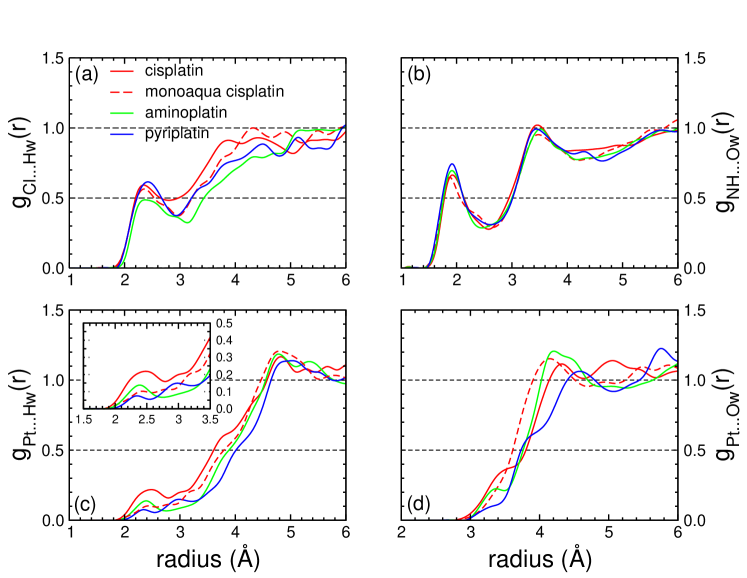
Selected mean bond distances and angles obtained from the microcanonical BOMD simulations are collected in Table 1 for all the Pt-based reactants investigated in this work. For cisplatin, when compared to previous DFT-MD simulations using the same XC functional but a different implementation,Truflandier et al. (2011) a nice agreement is observed. By comparing across the set of platinum complexes the deviations of the Pt-ligand mean bond distances and angles with respect to gas phase optimized strutures, we can isolate two systematic effects of aqueous solvent: () an increase of the PtCl bond length, () a net decrease of the NPtN angles. Analyses of the associated to the solvent-solute interactions given in Figure 3 allows for interpreting the systematic effects in terms of ligand hydration shells.
Complex Peaka first second first second first second first second 2.3 (3.9/4.4)* 2.3/2.7s (4.3/4.9)* 2.4/2.9s (5.1)* 2.4 (4.5/5.1)* 2.8 5.0 3.0 5.0 3.1 5.0 2.9 5.0 1.8 22.7 2.2 22.6 2.1 19.0 1.9 19.6 1.9 3.4 1.9/2.4s 3.5 1.9 3.5/3.9s 1.9 3.4/4.3s 2.6 4.5 2.6 4.5 2.5 4.5 2.6 4.5 0.7 7.7 0.8 7.6 0.6 7.6 0.6 6.7
aAsterisks indicate that the peak(s) is(are) not well resolved. Peak shoulder is indicated by s. bFor the first integration, was fixed to the closest minimum following the first maximum. Beyond these radii fixed values of and 4.5 Å for the Cl and NH coordination numbers respectively, were considered.
As already discussed in Ref. 61 for cisplatin, the NHO shows that the first and second hydration shells of each NH3 group integrates around 1 and 8 water molecules, respectively. This also applies to the other platinum complexes (cf. Table 2). The fact that the ligand solvation shell is only weakly affected by the variety of ligands bonded to the Pt atom and the charge state of the solute is confirmed by the ClH plotted on Figure 3, where each Cl atom integrates in the first neighbor region 2 waters molecules, independently of the system. The second shell is more difficult to discussed due to the limited resolution of the peaks. Nevertheless, in light of this results, () can be directly attributed to the ClHw hydrogen bonds perturbing the PtCl bond strength, whereas () is an indirect consequence of the ammonia group hydration where the preferential arrangement of NH3 from a static gas-phase optimisation —with one of the hydrogen atom pointing towards the closest Cl lone pair— is lost, relaxing the constraint on the ClPtN angles of the square planar complexes. Note that, as shown in Table 1, a polarizable continuum model (PCM) applied to static isolated solute is able to simulate these two features, demonstrating the concomitant contributions of specific and non-specific solvent effect. In Table 3 is compared selected structural parameters extracted from the RDFs for cisplatin and the mono-aqua derivative in regard to the previous studies based on DFT-MD.Lau and Ensing (2010); Truflandier et al. (2011); Kroutil et al. (2016) Even if the results were obtained from different implementations and exchange-correlation functionals —preventing an unbiased comparison— we observe an overall agreement which, besides validating the reliability of our implementation, suggests minor impact of the GGA functional (cf. Ref. 39 DFT-MD simulations performed with BLYP) and inclusion of the empirical dispersion correction (cf. Ref. 25 ; DFT-MD simulations performed with PBE-D3) on the qualitative description of the ligand first hydration shells.
Method PBE/BOMD PBE/CPMD BLYP/CPMD PBE+D3/BOMD PBE/BOMD BLYP/CPMD PBE+D3/BOMD Basis PAO/NCPP PW/USPP PW/NCPP mixed GTO-PW/NCPP PAO/NCPP PW/NCPP mixed GTO-PW/NCPP Reference this work [ 16] [ 74] [ 25] this work [ 74] [ 25] 3.0 3.2* 2.9 3.0 2.9 2.9 3.0 3.3 3.4* – 3.6 3.7 – 3.5 2.4 2.5* 2.9 3.6 3.2 3.5 3.5 1.9 1.9 – – 1.9/2.4s – – 2.6 2.5 – – 2.6 – – 0.7 0.7 – – 0.8 – – 3.4* 3.3 3.2* – 3.5 3.1 – 3.7* 3.7 – – 3.9 – – 2.8* 2.6 2.9* – 3.8 3.4 – 2.3 2.3 2.3 2.3 2.3/2.7s 2.3 2.3 2.8 2.9 – 2.8 3.0 – 2.9 1.8 2.2 1.7 2.2 2.2 2.4 2.2
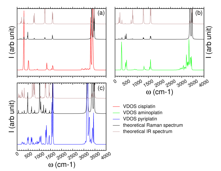
Another important step in the assessment process of our DFT-MD simulations is to ensure that the characterisitic vibrational modes of the solute and solvent were properly activated. Partial VDOS obtained for cisplatin, aminoplatin and pyriplatin are plotted on Figure 4. As external references, Raman and infra-red theoretical spectra of the isolated molecules are also reported. They were calculated on top of the stationary points obtained at the PBE-GTO-DZP level of theory including a PCM.Frisch and et al. Here, it is worth to recall that VDOS heights computed from the Fourier transform of the VACFs are not to be compared with spectroscopic intensities, and to emphasize that even if the underlying semiclassical theory involved in evaluating the VDOS —based on the classical propagation of the nuclei on the BO potential energy surface— is quite different from the response function computed from analytic (or numerical) derivatives —based on the harmonic approximation— their comparisons remain valuable for routine checks. As expected, cisplatin and aminoplatin vibrational spectrum are similar in many respects since the Pt center is surrounded by the same type of ligands. In both cases the bond-stretching modes: NH ( 3200 cm-1), PtN ( 500 cm-1) and PtCl ( 300 cm-1) are observed as well as the NH3 rocking and symmetric/asymetric deformation signatures, found around 800 and 1300/1300 cm-1, respectively.Nakamoto et al. (1965); Wysokiński and Michalska (2001); Truflandier et al. (2011) For pyriplatin, the complexity of the vibrational spectra is increased by the contributions of the pyridine ligand, with the stretching modes: CH ( 3250 cm-1), CC/CN (in the range 1100 to 1500 cm-1), the skeletal bending modes along with the out-of-plane CH wagging ( 1000 cm-1 and 700 cm-1). As shown on Figure 4, VDOSs extracted for the DFT-BOMD is in fair accordance with the theoretical spectra, such that, given the level of theory PAO-DZP-PBE, we can be confident in the reliability of our simulations when applied to free energy profiles calculation.
III.2 Inverse hydration
Complex Peak first second first second first second first second 2.5 3.0 2.4 (2.8/3.2) 2.4 na 2.3 (3.0/4.0) 2.6 3.3 2.6 3.3 2.6 3.3 2.6 3.3 0.5 1.4 0.2 0.9 0.2 0.7 0.1 0.7 3.5 4.3 na 4.2 3.4 (4.2/4.5) 3.3 (3.9/4.6) 3.6 4.6 3.6 4.6 3.6 4.6 3.6 4.6 0.7 6.4 0.7 7.0 0.5 6.7 0.4 5.3
aSince RDFs are poorly-resolved, fixed values were considered for the first and second integration.
We shall now focussed on the inverse-hydration taking place in the axial region of the square-planar Pt(II) complexes (Scheme 5). It can be observed at the beginning of each MD movies provided in the SI. The hydrogen-like bonding between a water molecule and the metal center following a H-ahead orientation is easily revealed by analysing the and available on Figure 3. First we note that for all the systems investigated in this work a non-negligible probability of having a Pt(H2O) contact is observed at an intermolecular distance of around 2.4 Å. The poorly-resolved peaks obtained for the PtH (PtO) in the range 2 to 3 (3 to 4) Å illustrate the motion or/and the exchange of H2O with other solvent molecules in close proximity, eg. those available from the Cl hydration shell(s). Another important insight is brought by the height of the peaks, cf. the PtH (PtO) around 2.4 (3.4) Å, indicating a net decrease of the contact occurrence when going from neutral to positively charged complexes. Quantification of the PtH2O contact is provided in Table 4 for the bi- and monofunctional platinum anticancer drugs, through the evaluation of the Pt(H) and Pt(O) coordination numbers, noted and , respectively. Due to the broad shape of the peaks, a set of two integration radius ( in Table 3) were used independently of the complex. For neutral cisplatin, is evaluated to be within the interval , along with for . Note that for larger , it is likely that the value of the coordination numbers also incorporate H2O molecules from the NH3 and Cl ligand solvent shells. For the set of positively charged complexes, the probability of Pt(H2O) contact is significantly reduced from for mono-aqua cisplatin to for pyriplatin. For this set, variations of seems to be more affected by the type of ligand, for which, mono-aqua cisplatin presents values similar to its neutral parent, whereas for aminoplatin and pyriplatin the coordination number is lowered to and , respectively. These results corroborate the work of Kroutil et al.Kroutil et al. (2016) which showed, among other findings, that: (i) H coordination number decreases with increasing positive charge of the complex, (ii) given a state charge (+1 in our case) the H-ahead orientation toward the Pt atom is strongly dependent on the hydrogen bond network formed by the nearby ligands. We refer the reader to this reference for extended discussion on this topic which also includes energetics consideration.
Concerning now the second part of this work dealing with evaluation of the hydrolysis free energy profiles of reaction (1) and (2), among all the water molecules accessible within the solute first solvation shell, it seems quite reasonable to consider the inversely bounded water molecule as the reactant.
III.3 Hydrolysis free energy profiles
The choice of the reaction coordinate is an important point in BME integration which can be a delicate issue when little is known about the chemical reaction. In this work, we have relied on the broad (experimental and theoretical) litterature agreeing that cisplatin aquation(s) is a one step process, eg. the SN2-like mechanism, for which the reactants (R) and products (P) are connected by a single transition state (TS). From this assumption, a rapid analysis of the corresponding saddle point (optimized with a quantum chemistry code) shows that the characteristic imaginary frequency is related to the ClPtO antisymmetric stretching mode together with the in-plane rotation of the NH3 group in trans position (Scheme 5). This led us to consider the difference of distanceRaugei et al. (1999); Yang et al. (2004); Bühl and Kabrede (2006); Komeiji et al. (2009) for the reaction coordinate:
| (5) |
where O is the oxygen atom of the HPt bonded water molecule. Configurations used to initiate the constrained DFT-BOMD trajectories were extracted from the canonical ensemble simulations discussed in Sec. III.2. Note that variations of (Helmholtz) free energies as computed from Eq. (1) were directly converted to (Gibbs) free enthalpies since we did not observe any strong variation of pressure during the thermodynamic integration which would have significantly modified the final enthalpy values.
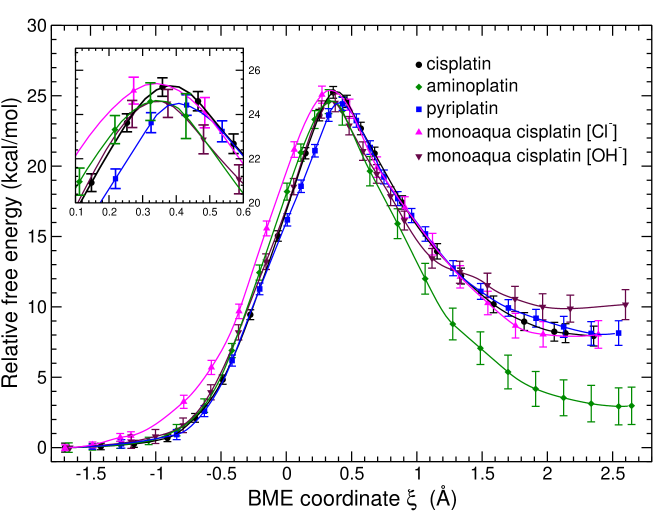
Free energy reaction paths are displayed in Figure 6 along with the free enthalpies of activation and reaction collected in Table 5. Concerning the second hydrolysis of cisplatin performed via the monoaqua complex, two set of BME calculation were produced: the first with Cl- as counterion following the first hydrolysis (later abbreviated by monoaqua[Cl]), the second from afresh NVT equilibration in which Cl- has been replaced by OH- (abbreviated by monoaqua[OH]). From Figure 6 we observe very similar energy profiles from the attack of the water molecule up to the transitions states. Evaluated for cationic monofuntional species are found to be lower of about 1 than the reference value of 25 obtained for the neutral parent. The presence of the Cl- counterion during the second hydrolysis of cisplatin tends to slightly decrease the barrier, but without dramatic effect. Indeed, major deviations between aquation profiles are found after the TS. This lead to noticeable differences in the values of , for which we found that the aminoplatin aquation is endothermic by 3 compared to 8 for cisplatin, pyriplatin and moaqua[Cl], whereas for moaqua[OH] the free enthalpy of reaction reach the value of 10 . It is tempting to compare these values to available experimental data. This should be done with lots of care owing to: the large amount of papers dealing with this subject, which comes generally with the same amount of differences in experimental condition, the limitations of our model in reproducing these conditions.
| Reactant | Counterion | ) | |
|---|---|---|---|
| cisplatin | none | ||
| mono-aqua cisplatin | OH- | ||
| Cl- | |||
| aminoplatin | OH- | ||
| pyriplatin | OH- |
As a result, even if the pH of the aqueous media can impact significantly the kinetic of the reactions —as shown in the comprehensive study of House and coworkersMiller and House (1989, 1989, 1990)— we must emphasize that the precision reach by the BME simulations remains far from the chemical accuracy required to discuss variation in activation energies below 1 .111It is worth to recall that a variation of 1 on the free energy of activation convert roughly to a variation of one order of magnitude on the rate constant. From Refs. 81; 82; 84; 85; 86; 87; 88 we can establish a consensus on the experimental free enthalpy (at ambient temperature) for both, the first and second aquation, to be between 23 and 24 . The experimental values for the cationic aminoplatin and pyriplatin are expected to be within this range.Park et al. (2012) A consensus on the endothermic property of the aquation reactions can also be drawn, with for cisplatin experimentally evaluated to be in the range 3-10 .Miller and House (1989, 1989); Lee and Martin (1976) Our results which are in line with experimental data should not be overinterpreted. Whereas we are quite confident in the reliability of the computed BME activation enthalpies, the thermodynamic quantities must not be considered as definitive. The limited size of cell can not prevent intramolecular ion pair contact to occur (the outermost ClPt distance being of 6.5 Å). For aminoplatin, the larger error bars on the integrated free energy observed at the late stages of the reaction suggest that the final is not fully converged.
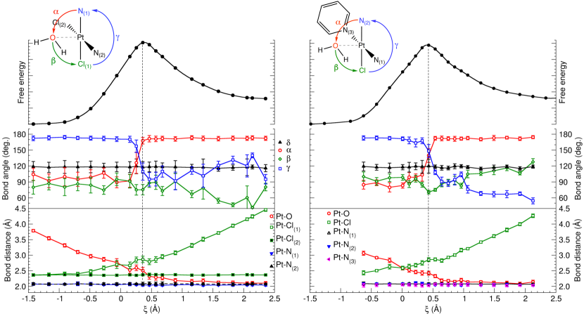
In order to gain some insights into the reactants motion and solvent reorganisation along the reaction path, visual inspection of the constrained MD movies is quite instructive. Representative examples are provided in the SI for cisplatin and pyriplatin. For all the complexes investigated similar trends in the evolution of skeletal bond parameters and solvent shell relative to the reactant constrained dynamics were found. The study cases of cisplatin and pyriplatin aquations are depicted in Figures 7 and 8, where the variation of selected structural parameters are plotted as a function of . At the beginning of the reaction (), the square planar structure with the inverse-hydration is globally conserved in both cases. In this regime, for cisplatin, the net decrease of the PtO bond length ( Å) is accompanied by a weaker increase of the PtCl distance ( Å) whereas the NPtO (), OPtCl () and ClPtN ( ) angles oscillate around mean values of 100, 85 and 175∘, respectively. These observations also hold for pyriplatin with slight differences in values. For cisplatin, magnitude of the variations along observed for and , which are much higher than for , indicates that the constrained water molecule remains free to visit the axial region of the square-planar complex. It seems to be less pronounced for pyriplatin. The difference in lability of the inversely bonded H2O between the two complexes is apparent when comparing the standard deviation (std) of the PtH distances plotted on Figure 8. For cisplatin we occasionally observe that these distances collapse towards a single value, eg. at , indicating H-atom swap through the rotation along the axis perpendicular to the H2O molecular plan. The variations of the HOPt angles with mean values of about for cisplatin, and for pyriplatin confirm the preferential inverse-hydration orientation, and for the later case, the reduced mobility of the H2O-reactant.
When approaching the transition state located at and Å for cisplatin and pyriplatin respectively, the inversely bonded H2O is activated via the ion-dipole orientation depicted in Scheme 5 with and Å. For cisplatin, when ( for pyriplatin), the rotation of the NH3 group is engaged leading to the TS. Note that, within the TS region, reported PtH distances and angles are weakly affected compared to the rotation angles . By identifying on Figure 7 the skeletal parameters corresponding to the maximum of free energy, it seems that the cisplatin TS is closer to a square pyramidal structure than a trigonal bipyramid as observed in gas phase calculations.Zhang et al. (2001); *robertazzi_hydrogen_2004; *lau_hydrolysis_2006; *alberto_second-generation_2009; *lucas_neutral_2009; *banerjee_cpl_2010; *melchior_comparative_2010 With a trigonal pyramid can be clearly assigned to pyriplatin. For , the water molecule is fully coordinated to the metal center with a typical bond length Å, and the Cl anion is released to bulk water. Inspection of the cisplatin BME integration movie at reveals that the singularity on Figure 8 is due to the departure of one of the H2O-ligand proton towards the solvent shell. From Figure 7, looking to the PtN and unconstrained PtCl mean bond lengths, it is interesting to outline that the corresponding ligands are spectator of the reaction in agreement with the in-plane symmetry of the transition state. This is well exemplified by the mean angle which remains fixed to 120∘ with standard deviation not exceeding 10∘ for both cases, showing that there are only weak perturbations from outside the NOCl plane.
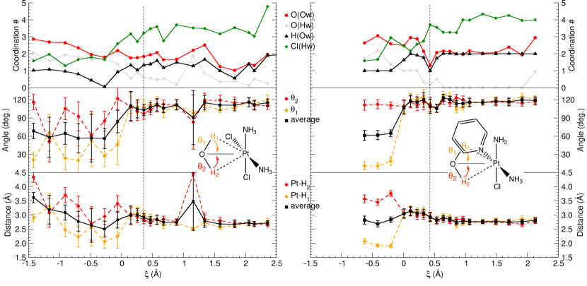
By visualizing the constrained MD trajectories reported in the SI, it is rather clear that the activated water molecule is embedded in a hydrogen-bond network throughout the whole course of the aquation reaction. To quantify the number of first nearest neighbor solvent molecules surrounding the H2O-reactant and Cl-product, the coordination numbers , (,) and are plotted on Figure 8 as a function of . They were evaluated from the ensemble averages (cf. Sec. II.2) by integrating the corresponding RDFs up to standard radiiZhang et al. (2013); DiStasio et al. (2014) of 3.2, 2.5 and 2.9 Å, respectively. Even if at first sight the evolution of is somewhat erratic due to the short time sampling, we can extract some relevant trends. At the beginning of the reaction, the inversely bonded water molecule integrates around 3 solvent molecules respecting its H-bond donor[1]/acceptor[2] capacity. Around the ion-dipole orientation region, as expected, H-bond acceptor (donor) capacity of the H2O-reactant reduces (raises). At the transition state, around 2 and 1 solvent molecule(s) are in close proximity of the activated H2O in cisplatin and pyriplatin, respectively. After this step, the average number acceptor-type H-bond continue to diminish whereas donor-type stabilizes to 2. The evolution of the H2O-reactant H-bond network contrasts with the global increase of the solvent shell size surrounding the Cl-product. Due the limited size of the model never really stabilized to the reference value comprised between 5 and 6 as obtained for free solvated chlorine anion.Zhang et al. (2013)
Finally, the BME aquation profiles and their analysis suggest that the aquation reaction of cisplatin and its monofunctional derivatives presenting a Cl-leaving group is likely to be independent of the charge state the type and number of N-donor ligands despite variations in the mobility and microsolvation of the activated water molecule. Even if conditioned by the choice of coordinate, the inverse hydration followed by the ion-dipole orientation seems to be a prerequisite to reach the transition state.
IV Conclusion and Outlook
In this work, reliable DFT-MD simulations of the aquation process of cisplatin and a set of monofunctional platinum anticancer drugs have been performed using constrained dynamics in conjuction with BME thermodynamic integration. Given the level of theory, accuracy of about 1 has been reached on the activation barrier. For the set of square-planar Pt(II) complexes, it is found that the kinetic of the reaction is independent of the ligands and the charge state. It seems that the free enthalpies of reaction differ from a few from one complex to another, and counterions can impact the thermodynamic quantity with variations in the same range of magnitude. This result need to be confirmed by performing larger scale DFT-MD simulations. We conclude that it is likely that the mono and bifunctional platinum anticancer drugs behave the same way during the time lapse between the cellular uptake and the DNA platination.
From a computational point of view, such kind of approach alleviates many intricacies related to modelling chemical reactions in aqueous solvent which are commonly based on isolated molecule approximation where thermodynamic quantities are computed from the ideal gas approximation. Even if the protocol used here is computationally expensive, we believe that it will become a routine task in near future along with the possibility of increasing the system size and using more sophisticated exchange correlation functionals, especially with the Conquest code which have been developped to perform large scale DFT simulations. This work opens the way for futur high level theoretical investigations of the anticancer activity of monofuntional platinum complexes which are important steps for approaching in silico design of new potent drugs.
References
- Wheate et al. (2010) Wheate, N. J.; Walker, S.; Craig, G. E.; Oun, R. The status of platinum anticancer drugs in the clinic and in clinical trials. Dalton Trans. 2010, 39, 8113–8127
- Johnstone et al. (2014) Johnstone, T. C.; Park, G. Y.; Lippard, S. J. Understanding and Improving Platinum Anticancer Drugs – Phenanthriplatin. Anticancer Res. 2014, 34, 471–476
- Apps et al. (2015) Apps, M. G.; Choi, E. H. Y.; Wheate, N. J. The state-of-play and future of platinum drugs. Endocr. Relat. Cancer 2015, 22, R219–R233
- Johnstone et al. (2016) Johnstone, T. C.; Suntharalingam, K.; Lippard, S. J. The Next Generation of Platinum Drugs: Targeted Pt(II) Agents, Nanoparticle Delivery, and Pt(IV) Prodrugs. Chem. Rev. 2016, 116, 3436–3486
- Brabec and Kasparkova (2005) Brabec, V.; Kasparkova, J. Modifications of DNA by platinum complexes. Drug Resistance Updates 2005, 8, 131–146
- Chabner and Roberts (2005) Chabner, B. A.; Roberts, T. G. Chemotherapy and the war on cancer. Nat. Rev. Cancer 2005, 5, 65–72
- Kelland (2007) Kelland, L. The resurgence of platinum-based cancer chemotherapy. Nat. Rev. Cancer 2007, 7, 573–584
- Hambley (1997) Hambley, T. W. The influence of structure on the activity and toxicity of Pt anti-cancer drugs. Coord. Chem. Rev. 1997, 166, 181–223
- Ziegler et al. (2000) Ziegler, C. J.; Silverman, A. P.; Lippard, S. J. High-throughput synthesis and screening of platinum drug candidates. J. Biol. Inorg. Chem. 2000, 5, 774–783
- Jamieson and Lippard (1999) Jamieson, E. R.; Lippard, S. J. Structure, Recognition, and Processing of Cisplatin-DNA Adducts. Chem. Rev. 1999, 99, 2467–2498
- Dhar and Lippard (2009) Dhar, S.; Lippard, S. J. In Platinum and Other Heavy Metal Compounds in Cancer Chemotherapy; Bonetti, A., Leone, R., Muggia, F. M., Howell, S. B., Eds.; Cancer Drug Discovery and Development; Humana Press, 2009; pp 135–147
- Vidossich et al. (2016) Vidossich, P.; Lledós, A.; Ujaque, G. First-Principles Molecular Dynamics Studies of Organometallic Complexes and Homogeneous Catalytic Processes. Acc. Chem. Res. 2016, 49, 1271–1278
- Beret et al. (2008) Beret, E. C.; Martínez, J. M.; Pappalardo, R. R.; Sánchez Marcos, E.; Doltsinis, N. L.; Marx, D. Explaining Asymmetric Solvation of Pt(II) versus Pd(II) in Aqueous Solution Revealed by Ab Initio Molecular Dynamics Simulations. J. Chem. Theory Comput. 2008, 4, 2108–2121
- Beret et al. (2009) Beret, E. C.; Pappalardo, R. R.; Marx, D.; Sánchez Marcos, E. Characterizing Pt-Derived Anticancer Drugs from First Principles: The Case of Oxaliplatin in Aqueous Solution. ChemPhysChem 2009, 10, 1044–1052
- Vidossich et al. (2011) Vidossich, P.; Ortuño, M. Á.; Ujaque, G.; Lledós, A. Do Metal…Water Hydrogen Bonds Hold in Solution? Insight from Ab Initio Molecular Dynamics Simulations. ChemPhysChem 2011, 12, 1666–1668
- Truflandier et al. (2011) Truflandier, L. A.; Sutter, K.; Autschbach, J. Solvent Effects and Dynamic Averaging of 195Pt NMR Shielding in Cisplatin Derivatives. Inorg. Chem. 2011, 50, 1723–1732
- Sutter et al. (2011) Sutter, K.; Truflandier, L. A.; Autschbach, J. NMR J-Coupling Constants in Cisplatin Derivatives Studied by Molecular Dynamics and Relativistic DFT. ChemPhysChem 2011, 12, 1448–1455
- Baidina et al. (1981) Baidina, I. A.; Podberezskaya, N. V.; Krylova, L. F.; Borisov, S. V. Crystal structure of trans-dichloroammineglycineplatinum (II) monohydrate trans-[PtNH3(H2NCH2COOH)Cl2]·H2O. J. Struct. Chem. 1981, 22, 463–465
- Rizzato et al. (2010) Rizzato, S.; Bergès, J.; Mason, S. A.; Albinati, A.; Kozelka, J. Dispersion-Driven Hydrogen Bonding: Predicted Hydrogen Bond between Water and Platinum(II) Identified by Neutron Diffraction. Angew. Chem. Int. Ed. 2010, 49, 7440–7443
- Kozelka et al. (2000) Kozelka, J.; Bergès, J.; Attias, R.; Fraitag, J. Hydrogen Bond with a Strong Dispersion Component. Angew. Chem. Int. Ed. 2000, 39, 198–201
- Lopes et al. (2008) Lopes, J. F.; Rocha, W. R.; Dos Santos, H. F.; De Almeida, W. B. Theoretical study of the potential energy surface for the interaction of cisplatin and their aquated species with water. J. Chem. Phys. 2008, 128, 165103–14
- Aono et al. (2016) Aono, S.; Mori, T.; Sakaki, S. 3D-RISM-MP2 Approach to Hydration Structure of Pt(II) and Pd(II) Complexes: Unusual H-Ahead Mode vs Usual O-Ahead One. J. Chem. Theory Comput. 2016, 12, 1189–1206
- Bergès et al. (2013) Bergès, J.; Fourré, I.; Pilmé, J.; Kozelka, J. Quantum Chemical Topology Study of the Water-Platinum(II) Interaction. Inorg. Chem. 2013, 52, 1217–1227
- Melchior et al. (2015) Melchior, A.; Tolazzi, M.; Martínez, J. M.; Pappalardo, R. R.; Sánchez Marcos, E. Hydration of Two Cisplatin Aqua-Derivatives Studied by Quantum Mechanics and Molecular Dynamics Simulations. J. Chem. Theory Comput. 2015, 11, 1735–1744
- Kroutil et al. (2016) Kroutil, O.; Předota, M.; Chval, Z. Pt···H Nonclassical Interaction in Water-Dissolved Pt(II) Complexes: Coaction of Electronic Effects with Solvent-Assisted Stabilization. Inorg. Chem. 2016, 55, 3252–3264
- Bancroft et al. (1990) Bancroft, D. P.; Lepre, C. A.; Lippard, S. J. Platinum-195 NMR kinetic and mechanistic studies of cis- and trans-diamminedichloroplatinum(II) binding to DNA. J. Am. Chem. Soc. 1990, 112, 6860–6871
- Knox et al. (1986) Knox, R. J.; Friedlos, F.; Lydall, D. A.; Roberts, J. J. Mechanism of Cytotoxicity of Anticancer Platinum Drugs: Evidence That cis-Diamminedichloroplatinum(II) and cis-Diammine-(1,1-cyclobutanedicarboxylato)platinum(II) Differ Only in the Kinetics of Their Interaction with DNA. Cancer Res. 1986, 46, 1972–1979
- Alberts and Dorr (1998) Alberts, D. S.; Dorr, R. T. New Perspectives on an Old Friend: Optimizing Carboplatin for the Treatment of Solid Tumors. The Oncologist 1998, 3, 15–34
- Johnstone et al. (2014) Johnstone, T. C.; Alexander, S. M.; Wilson, J. J.; Lippard, S. J. Oxidative halogenation of cisplatin and carboplatin: synthesis, spectroscopy, and crystal and molecular structures of Pt(IV) prodrugs. Dalton Trans. 2014, 44, 119–129
- Blommaert et al. (1995) Blommaert, F. A.; van Dijk-Knijnenburg, H. C. M.; Dijt, F. J.; den Engelse, L.; Baan, R. A.; Berends, F.; Fichtinger-Schepman, A. M. J. Formation of DNA Adducts by the Anticancer Drug Carboplatin: Different Nucleotide Sequence Preferences in Vitro and in Cells. Biochemistry 1995, 34, 8474–8480
- Zhang et al. (2001) Zhang, Y.; Guo, Z.; You, X. Hydrolysis Theory for Cisplatin and Its Analogues Based on Density Functional Studies. J. Am. Chem. Soc. 2001, 123, 9378–9387
- Robertazzi and Platts (2004) Robertazzi, A.; Platts, J. A. Hydrogen bonding, solvation, and hydrolysis of cisplatin: A theoretical study. J. Comput. Chem. 2004, 25, 1060–1067
- Lau and Deubel (2006) Lau, J. K.; Deubel, D. V. Hydrolysis of the Anticancer Drug Cisplatin: Pitfalls in the Interpretation of Quantum Chemical Calculations. J. Chem. Theory Comput. 2006, 2, 103–106
- Alberto et al. (2009) Alberto, M. E.; Lucas, M. F. A.; Pavelka, M.; Russo, N. The Second-Generation Anticancer Drug Nedaplatin: A Theoretical Investigation on the Hydrolysis Mechanism. J. Phys. Chem. B 2009, 113, 14473–14479
- Lucas et al. (2009) Lucas, M. F. A.; Pavelka, M.; Alberto, M. E.; Russo, N. Neutral and Acidic Hydrolysis Reactions of the Third Generation Anticancer Drug Oxaliplatin. J. Phys. Chem. B 2009, 113, 831–838
- Banerjee et al. (2010) Banerjee, S.; Sengupta, P. S.; Mukherjee, A. K. Trans Platinum Anticancer Drug AMD443: A Detailed Theoretical Study by DFT-TST Method on the Hydrolysis Mechanism. Chem. Phys. Lett. 2010, 487, 108–115
- Melchior et al. (2010) Melchior, A.; Marcos, E. S.; Pappalardo, R. R.; Martínez, J. M. Comparative study of the hydrolysis of a third- and a first-generation platinum anticancer complexes. Theor. Chem. Acc. 2010, 128, 627–638
- Carloni et al. (2000) Carloni, P.; Sprik, M.; Andreoni, W. Key Steps of the cis-Platin-DNA Interaction: Density Functional Theory-Based Molecular Dynamics Simulations. J. Phys. Chem. B 2000, 104, 823–835
- Lau and Ensing (2010) Lau, J. K.-C.; Ensing, B. Hydrolysis of cisplatin—a first-principles metadynamics study. Phys. Chem. Chem. Phys. 2010, 12, 10348–10355
- Yokogawa et al. (2007) Yokogawa, D.; Sato, H.; Sakaki, S. New generation of the reference interaction site model self-consistent field method: Introduction of spatial electron density distribution to the solvation theory. J. Chem. Phys. 2007, 126, 244504
- Yokogawa et al. (2009) Yokogawa, D.; Sato, H.; Sakaki, S. Analytical energy gradient for reference interaction site model self-consistent field explicitly including spatial electron density distribution. J. Chem. Phys. 2009, 131, 214504
- Yokogawa et al. (2011) Yokogawa, D.; Ono, K.; Sato, H.; Sakaki, S. Theoretical study on aquation reaction of cis-platin complex: RISM–SCF–SEDD, a hybrid approach of accurate quantum chemical method and statistical mechanics. Dalton Trans. 2011, 40, 11125–11130
- Hollis et al. (1989) Hollis, L. S.; Amundsen, A. R.; Stern, E. W. Chemical and biological properties of a new series of cis-diammineplatinum(II) antitumor agents containing three nitrogen donors: cis-[Pt(NH3)2(N-donor) Cl]+. J. Med. Chem. 1989, 32, 128–136
- Brabec (2002) Brabec, V. DNA Modifications by antitumor platinum and ruthenium compounds: Their recognition and repair. Prog. Nucleic Acid Res. Mol. Biol. 2002, 71, 1–68
- Bursova et al. (2005) Bursova, V.; Kasparkova, J.; Hofr, C.; Brabec, V. Effects of Monofunctional Adducts of Platinum(II) Complexes on Thermodynamic Stability and Energetics of DNA Duplexes. Biophys. J. 2005, 88, 1207–1214
- Lovejoy et al. (2008) Lovejoy, K. S.; Todd, R. C.; Zhang, S.; McCormick, M. S.; D’Aquino, J. A.; Reardon, J. T.; Sancar, A.; Giacomini, K. M.; Lippard, S. J. cis-Diammine(pyridine)chloroplatinum(II), a monofunctional platinum(II) antitumor agent: Uptake, structure, function, and prospects. PNAS 2008, 105, 8902–8907
- Wang et al. (2010) Wang, D.; Zhu, G.; Huang, X.; Lippard, S. J. X-ray structure and mechanism of RNA polymerase II stalled at an antineoplastic monofunctional platinum-DNA adduct. PNAS 2010, 107, 9584–9589
- Zhu et al. (2012) Zhu, G.; Myint, M.; Ang, W. H.; Song, L.; Lippard, S. J. Monofunctional Platinum–DNA Adducts Are Strong Inhibitors of Transcription and Substrates for Nucleotide Excision Repair in Live Mammalian Cells. Cancer Res. 2012, 72, 790–800
- Park et al. (2012) Park, G. Y.; Wilson, J. J.; Song, Y.; Lippard, S. J. Phenanthriplatin, a monofunctional DNA-binding platinum anticancer drug candidate with unusual potency and cellular activity profile. PNAS 2012, 109, 11987–11992
- Johnstone et al. (2014) Johnstone, T. C.; Alexander, S. M.; Lin, W.; Lippard, S. J. Effects of Monofunctional Platinum Agents on Bacterial Growth: A Retrospective Study. J. Am. Chem. Soc. 2014, 136, 116–118
- Bowler et al. (2006) Bowler, D. R.; Choudhury, R.; Gillan, M. J.; Miyazaki, T. Recent progress with large-scale ab initio calculations: the CONQUEST code. phys. stat. sol. (b) 2006, 243, 989–1000
- Bowler and Miyazaki (2010) Bowler, D. R.; Miyazaki, T. Calculations for millions of atoms with density functional theory: linear scaling shows its potential. J. Phys.: Condens. Matter 2010, 22, 074207
- Hernández et al. (1996) Hernández, E.; Gillan, M. J.; Goringe, C. M. Linear-scaling density-functional-theory technique: The density-matrix approach. Phys. Rev. B 1996, 53, 7147
- Sankey and Niklewski (1989) Sankey, O. F.; Niklewski, D. J. Ab initio multicenter tight-binding model for molecular-dynamics simulations and other applications in covalent systems. Phys. Rev. B 1989, 40, 3979–3995
- Junquera et al. (2001) Junquera, J.; Paz, Ó.; Sánchez-Portal, D.; Artacho, E. Numerical atomic orbitals for linear-scaling calculations. Phys. Rev. B 2001, 64, 235111
- Torralba et al. (2008) Torralba, A. S.; Todorović, M.; Brázdová, V.; Choudhury, R.; Miyazaki, T.; Gillan, M. J.; Bowler, D. R. Pseudo-atomic orbitals as basis sets for the O(N) DFT code CONQUEST. J. Phys.: Condens. Matter 2008, 20, 294206
- Troullier and Martins (1991) Troullier, N.; Martins, J. L. Efficient pseudopotentials for plane-wave calculations. Phys. Rev. B 1991, 43, 1993–2006
- Perdew et al. (1996) Perdew, J. P.; Burke, K.; Ernzerhof, M. Generalized Gradient Approximation Made Simple. Phys. Rev. Lett. 1996, 77, 3865–3868
- Lin et al. (2012) Lin, I.-C.; Seitsonen, A. P.; Tavernelli, I.; Rothlisberger, U. Structure and Dynamics of Liquid Water from ab Initio Molecular Dynamics—Comparison of BLYP, PBE, and revPBE Density Functionals with and without van der Waals Corrections. J. Chem. Theory Comput. 2012, 8, 3902–3910
- DiStasio et al. (2014) DiStasio, R. A.; Santra, B.; Li, Z.; Wu, X.; Car, R. The individual and collective effects of exact exchange and dispersion interactions on the ab initio structure of liquid water. J. Chem. Phys. 2014, 141, 084502
- Truflandier and Autschbach (2010) Truflandier, L. A.; Autschbach, J. Probing the Solvent Shell with 195Pt Chemical Shifts: Density Functional Theory Molecular Dynamics Study of Pt(II) and Pt(IV) Anionic Complexes in Aqueous Solution. J. Am. Chem. Soc. 2010, 132, 3472–3483
- Verlet (1967) Verlet, L. Computer ”Experiments” on Classical Fluids. I. Thermodynamical Properties of Lennard-Jones Molecules. Phys. Rev. 1967, 159, 98–103
- Martyna et al. (1992) Martyna, G. J.; Klein, M. L.; Tuckerman, M. Nosé–Hoover chains: The canonical ensemble via continuous dynamics. J. Chem. Phys. 1992, 97, 2635–2643
- Hirakawa et al. (2017) Hirakawa, T.; Suzuki, T.; Bowler, D. R.; Miyazaki, T. Canonical-ensemble extended Lagrangian Born–Oppenheimer molecular dynamics for the linear scaling density functional theory. J. Phys.: Condens. Matter 2017, 29, 405901
- Carter et al. (1989) Carter, E.; Ciccotti, G.; Hynes, J. T.; Kapral, R. Constrained reaction coordinate dynamics for the simulation of rare events. Chem. Phys. Lett. 1989, 156, 472–477
- Sprik and Ciccotti (1998) Sprik, M.; Ciccotti, G. Free energy from constrained molecular dynamics. J. Chem. Phys. 1998, 109, 7737–7744
- Ryckaert et al. (1977) Ryckaert, J.-P.; Ciccotti, G.; Berendsen, H. J. Numerical integration of the cartesian equations of motion of a system with constraints: molecular dynamics of n-alkanes. J. Comput. Phys. 1977, 23, 327–341
- Andersen (1983) Andersen, H. C. Rattle: A “velocity” version of the shake algorithm for molecular dynamics calculations. J. Comput. Phys. 1983, 52, 24–34
- Ciccotti and Ryckaert (1986) Ciccotti, G.; Ryckaert, J.-P. Molecular dynamics simulation of rigid molecules. Comp. Phys. Rep. 1986, 4, 346–392
- Jacucci and Rahman (1984) Jacucci, G.; Rahman, A. Comparing the efficiency of Metropolis Monte Carlo and molecular-dynamics methods for configuration space sampling. Il Nuovo Cimento D 1984, 4, 341–356
- Giannozzi and et al. (2009) Giannozzi, P.; et al., J. Phys.: Condens. Matter 2009, 21, 395502
- (72) Frisch, M. J.; et al., Gaussian09. Gaussian 09, Revision A.02; Gaussian, Inc., Wallingford, CT, 2009
- Press et al. (1992) Press, W. H.; Flannery, B. P.; Teukolsky, S. A.; Vetterling, W. T. Numerical Recipes in Fortran 77: The Art of Scientific Computing, 2nd ed.; Cambridge University Press: Cambridge England ; New York, NY, USA, 1992
- Lau and Ensing (2010) Lau, J. K.; Ensing, B. Hydrolysis of cisplatin—a first-principles metadynamics study. Phys. Chem. Chem. Phys. 2010,
- Nakamoto et al. (1965) Nakamoto, K.; McCarthy, P. J.; Fujita, J.; Condrate, R. A.; Behnke, G. T. Infrared Studies of Ligand-Ligand Interaction in Dihalogenodiammineplatinum(II) Complexes. Inorg. Chem. 1965, 4, 36–43
- Wysokiński and Michalska (2001) Wysokiński, R.; Michalska, D. The performance of different density functional methods in the calculation of molecular structures and vibrational spectra of platinum(II) antitumor drugs: cisplatin and carboplatin. J. Comput. Chem. 2001, 22, 901–912
- Raugei et al. (1999) Raugei, S.; Cardini, G.; Schettino, V. An ab initio molecular dynamics study of the SN2 reaction Cl-+CH3BrCH3Cl+Br-. The Journal of Chemical Physics 1999, 111, 10887–10894
- Yang et al. (2004) Yang, S.-Y.; Fleurat-Lessard, P.; Hristov, I.; Ziegler, T. Free Energy Profiles for the Identity SN2 Reactions Cl- + CH3Cl and NH3 + H3BNH3: A Constraint Ab Initio Molecular Dynamics Study. J. Phys. Chem. A 2004, 108, 9461–9468
- Bühl and Kabrede (2006) Bühl, M.; Kabrede, H. Mechanism of Water Exchange in Aqueous Uranyl(VI) Ion. A Density Functional Molecular Dynamics Study. Inorg. Chem. 2006, 45, 3834–3836
- Komeiji et al. (2009) Komeiji, Y.; Ishikawa, T.; Mochizuki, Y.; Yamataka, H.; Nakano, T. Fragment Molecular Orbital method-based Molecular Dynamics (FMO-MD) as a simulator for chemical reactions in explicit solvation. J. Comput. Chem. 2009, 30, 40–50
- Miller and House (1989) Miller, S. E.; House, D. A. The hydrolysis products of cis-diamminedichloroplatinum(II). 1. The kinetics of formation and anation of the cis-diammine(aqua)chloroplatinum(II) cation in acidic aqueous solution. Inorg. Chim. Acta 1989, 161, 131–137
- Miller and House (1989) Miller, S. E.; House, D. A. The hydrolysis products of cis-dichlorodiammineplatinum(II) 2. The kinetics of formation and anation of the cis-diamminedi(aqua)platinum(II) cation. Inorg. Chim. Acta 1989, 166, 189–197
- Miller and House (1990) Miller, S. E.; House, D. A. The hydrolysis products of cis-dichlorodiammineplatinum(II) 3. Hydrolysis kinetics at physiological pH. Inorg. Chim. Acta 1990, 173, 53–60
- Reishus and Martin (1961) Reishus, J. W.; Martin, D. S. cis-Dichlorodiammineplatinum(II). Acid Hydrolysis and Isotopic Exchange of the Chloride Ligands. J. Am. Chem. Soc. 1961, 83, 2457–2462
- Marti et al. (1998) Marti, N.; Bon Hoa, G. H.; Kozelka, J. Reversible hydrolysis of [PtCl(dien)]+ and [PtCl(NH3)3]+. Determination of the rate constants using UV spectrophotometry. Inorg. Chem. Comm. 1998, 1, 439–442
- Davies et al. (2000) Davies, M. S.; Berners-Price, S. J.; Hambley, T. W. Slowing of Cisplatin Aquation in the Presence of DNA but Not in the Presence of Phosphate: Improved Understanding of Sequence Selectivity and the Roles of Monoaquated and Diaquated Species in the Binding of Cisplatin to DNA. Inorg. Chem. 2000, 39, 5603–5613
- Lee and Martin (1976) Lee, K. W.; Martin, D. S. Cis-dichlorodiammineplatinum(II). Aquation equilibria and isotopic exchange of chloride ligands with free chloride and tetrachloroplatinate(II). Inorg. Chim. Acta 1976, 17, 105–110
- Vinje et al. (2005) Vinje, J.; Sletten, E.; Kozelka, J. Influence of dT20 and [d(AT)10]2 on Cisplatin Hydrolysis Studied by Two-Dimensional [1H,15N] HMQC NMR Spectroscopy. Chem. Eur. J. 2005, 11, 3863–3871
- Zhang et al. (2013) Zhang, C.; Pham, T. A.; Gygi, F.; Galli, G. Communication: Electronic structure of the solvated chloride anion from first principles molecular dynamics. J. Chem. Phys. 2013, 138, 181102