Inhibition of the Main Protease 3CLPro of the Coronavirus Disease 19 via Structure-Based Ligand Design and Molecular Modeling.
Supporting Information for “Inhibition of the Main Protease 3CLpro of the Coronavirus Disease 19 via Structure-Based Ligand Design and Molecular Modeling”
Abstract
We have applied a computational strategy, based on the synergy of virtual screening, docking and molecular dynamics techniques, aimed at identifying possible lead compounds for the non-covalent inhibition of the main protease 3CLpro of the SARS-Cov2 Coronavirus. Based on the recently resolved 6LU7 PDB structure, ligands were generated using a multimodal structure-based design and then optimally docked to the 6LU7 monomer. Docking calculations show that ligand-binding is strikingly similar in SARS-CoV and SARS-CoV2 main proteases, irrespectively of the protonation state of the catalytic CYS-HIS dyad. The most potent docked ligands are found to share a common binding pattern with aromatic moieties connected by rotatable bonds in a pseudo-linear arrangement. Molecular dynamics calculations fully confirm the stability in the 3CLpro binding pocket of the most potent binder identified by docking, namely a chlorophenyl-pyridyl-carboxamide derivative.
keywords:
American Chemical Society, LaTeXCOVID-19, 3CLPro, Coronavirus inhibitors, Docking, ligand design, playmolecule, main protease, autodock At the beginning of this year, the world was dismayed by the outbreak of a new severe viral acute respiratory syndrome (SARS), currently known as COVID-19, that rapidly spreads from its origin in the Hubei Chinese district to virtually whole China and, as of today, to more than thirty nations in five continents.1 The new coronavirus, named SARS-CoV2 and believed to have a zoonotic origin, has infected thus far about 80000 people worldwide with nearly 10000 in critical conditions, causing the death of more than 3000 people. The SARS-CoV2’s genome2, 3 has a large identity4 with that of the SARS-CoV whose epidemic started in early in 2003 and ended in the summer of the same year.
Most of the Coronaviridae genome encodes two large polyproteins, pp1a and, through ribosomal frameshifting during translation5, pp1ab. These polyproteins are cleaved and transformed in mature non-structural proteins (NSPs) by the two proteases 3CLpro (3C-like protease) and PLpro (Papain Like Protease) encoded by the open reading frame 1.6 NSPs, in turn, play a fundamental role in the transcription/replication during the infection.5 Targeting these proteases may hence constitute a valid approach for antiviral drug design. The catalytically active 3CLpro is a dimer. Cleavage by 3CLpro occurs at the glutamine residue in the P1 position of the substrate via the protease CYS-HIS dyad in which the cysteine thiol functions as the nucleophile in the proteolytic process.7 While dimerization is believed to provide a substrate-binding cleft between the two monomers,8 in the dimer the solvent-exposed CYS-HYS dyads are symmetrically located at the opposite edges the cleft, probably acting independently.9 As no host-cell proteases are currently known with this specificity, early drug discovery was directed towards the so-called covalent Michael inhibitors,10 via electrophilic attack to the cysteinate of the 3CLpro dyad. On the other hand, the consensus in drug discovery leads to excluding electrophiles from drug candidates for reasons primarily relating to safety and adverse effects such as allergies, tissue destruction, or carcinogenesis.11
In spite of the initial effort in developing small-molecule compounds (SMC) with anti-coronavirus activity immediately after the SARS outbreak,12 no anti-viral drug was ever approved or even reached the clinical stage due to a sharp decline in funding of coronavirus research after 2005-2006, based on the erroneous conviction by policy-makers and scientists that chance of a repetition of a new zoonotic transmission was extremely unlikely. The most potent non-covalent inhibitor for 3CLpro, ML188, was reported nearly ten years ago13 with moderate activity in the low micromolar range.14
According to the latest report of the structure of 3CLpro from SARS-CoV215 (PDB code 6LU7) and the available structure of 3CLpro from SARS-CoV,12 (PDB code 1UK4), the two main proteases differ by only 12 amino acids, with carbon atoms all lying at least 1 nm away from the 3CLpro active site (see Figure 1a).
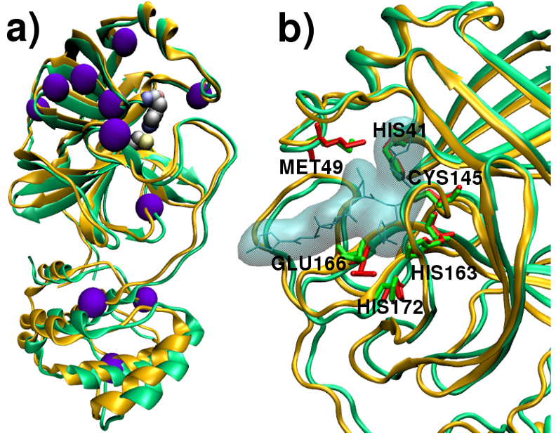
The substrate-binding pockets of two coronavirus main proteases are compared in Figure 1b, exhibiting a strikingly high level of alignment of the key residues involved in substrate binding, including the CYS145HIS41 dyad, and HIS163/HIS172/GLU166. The latter residues are believed to provide the opening gate for the substrate in the active state of the protomer.12
Figure 1(a,b) strongly suggest that effective non-covalent inhibitors for SARS-CoV and SARS-CoV2 main proteases should share the same structural and chemical features. In order to investigate this matter, we have performed a molecular modeling study on both the 6LU7 and 1UK4 PDB structures. 6LU7 is the monomer of the main protease in the active state with the N3 peptidomimetic inhibitor15 while 1UK4 is the dimer with the protomer chain A in the active state.12 The main protease monomer contains three domains. Domains I and II (residues 8-101 and residues 102-184) are made of antiparallel -barrel structures in a chymotrypsin-like fold responsible for catalysis.16
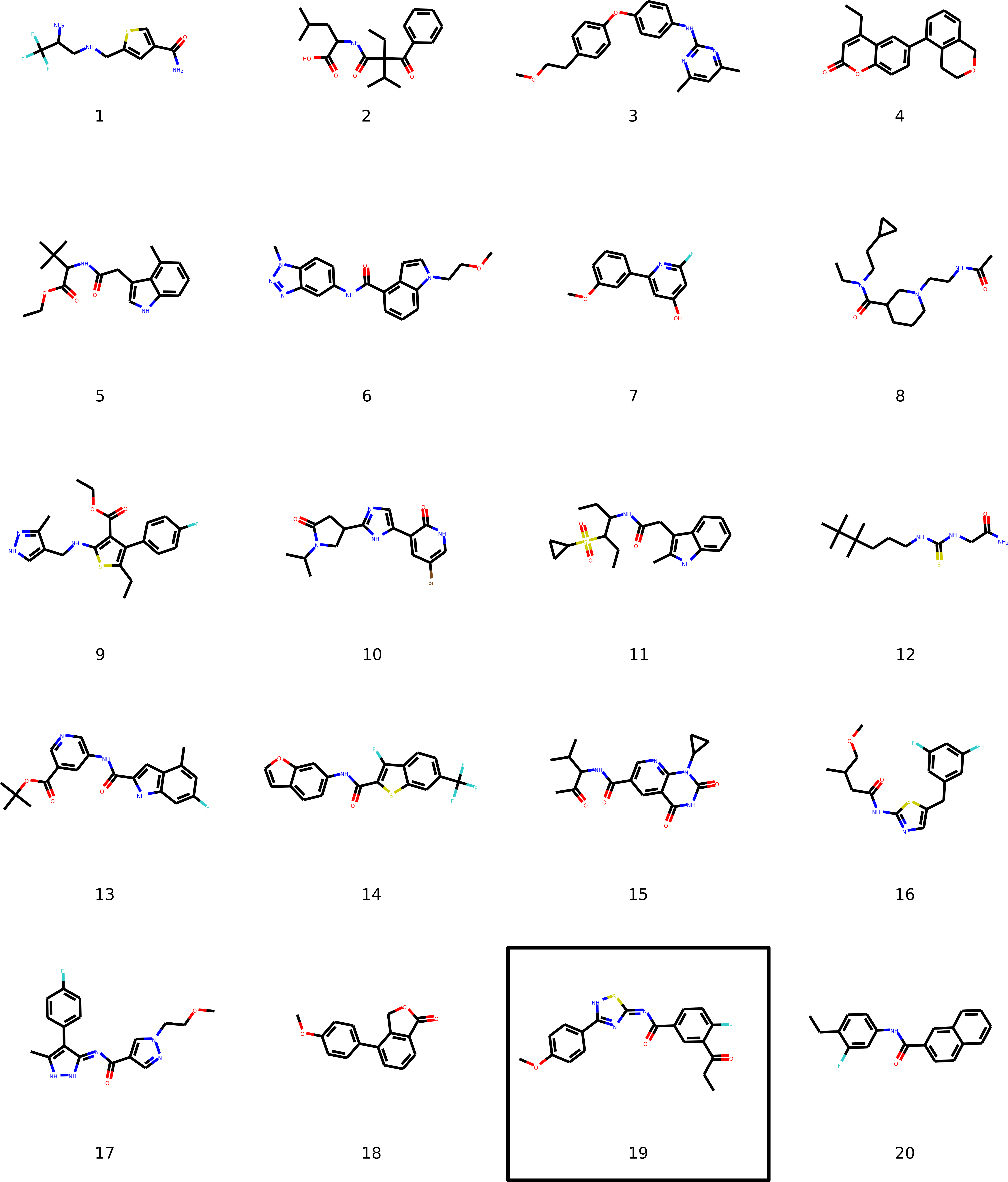
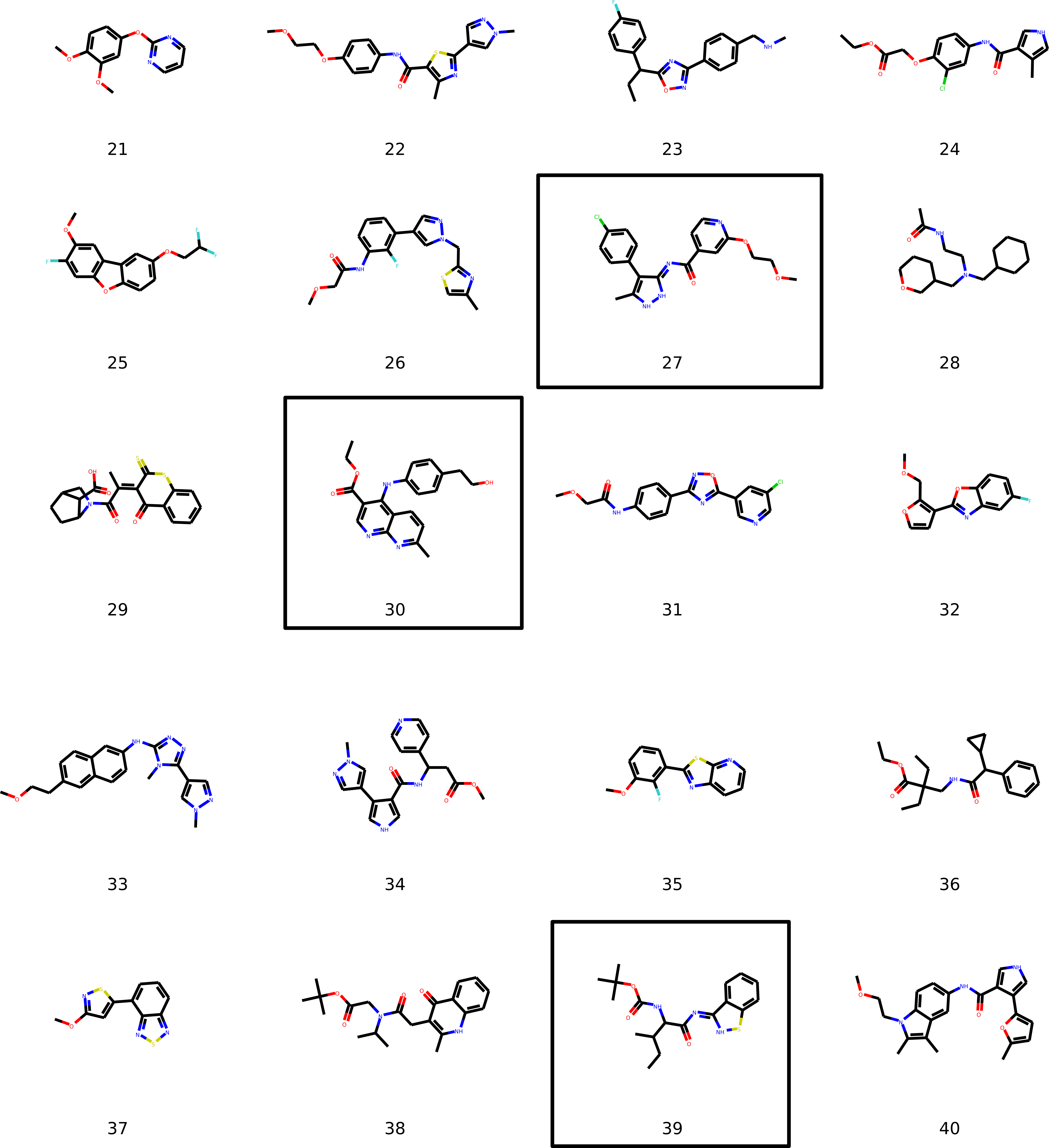
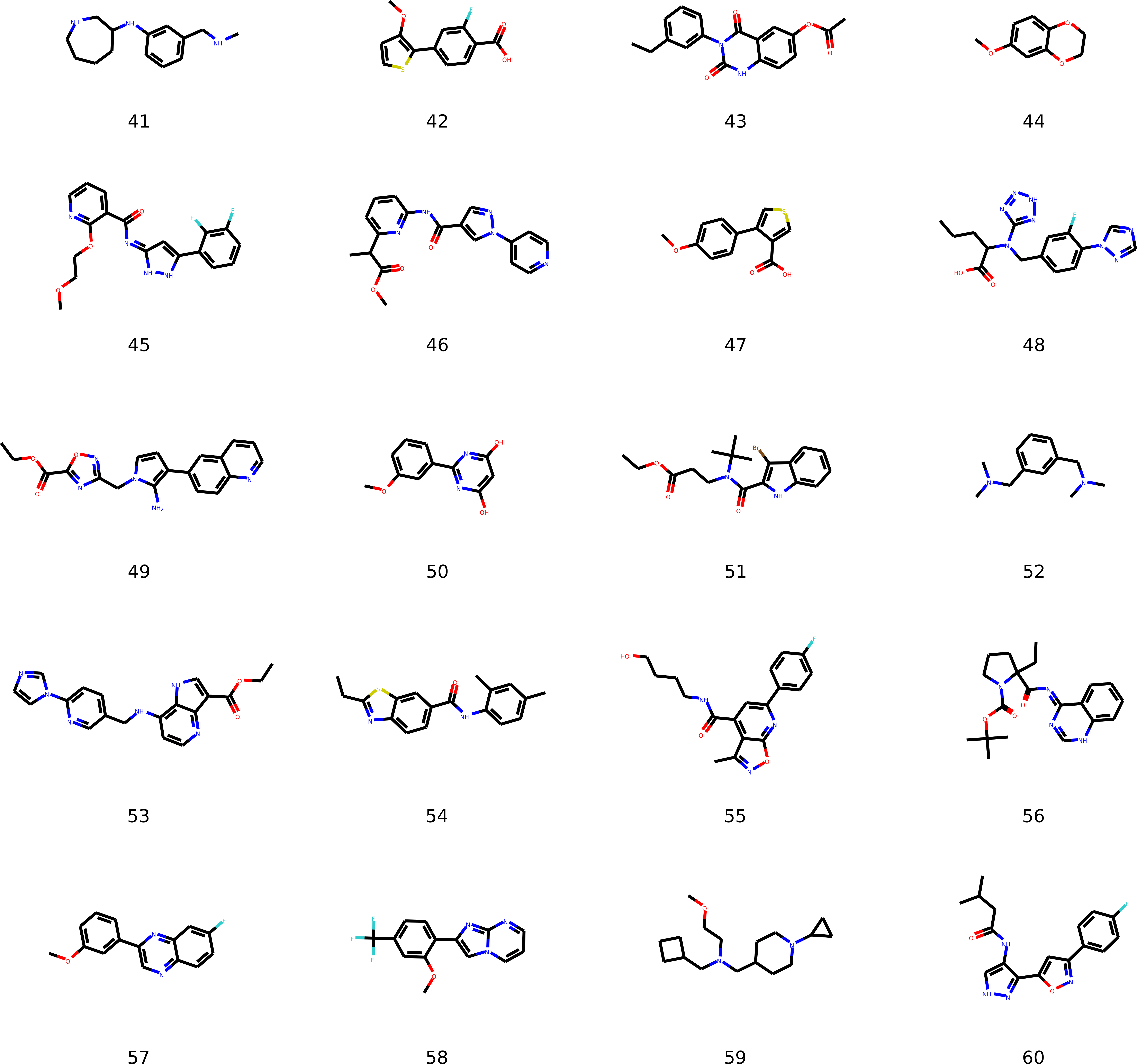
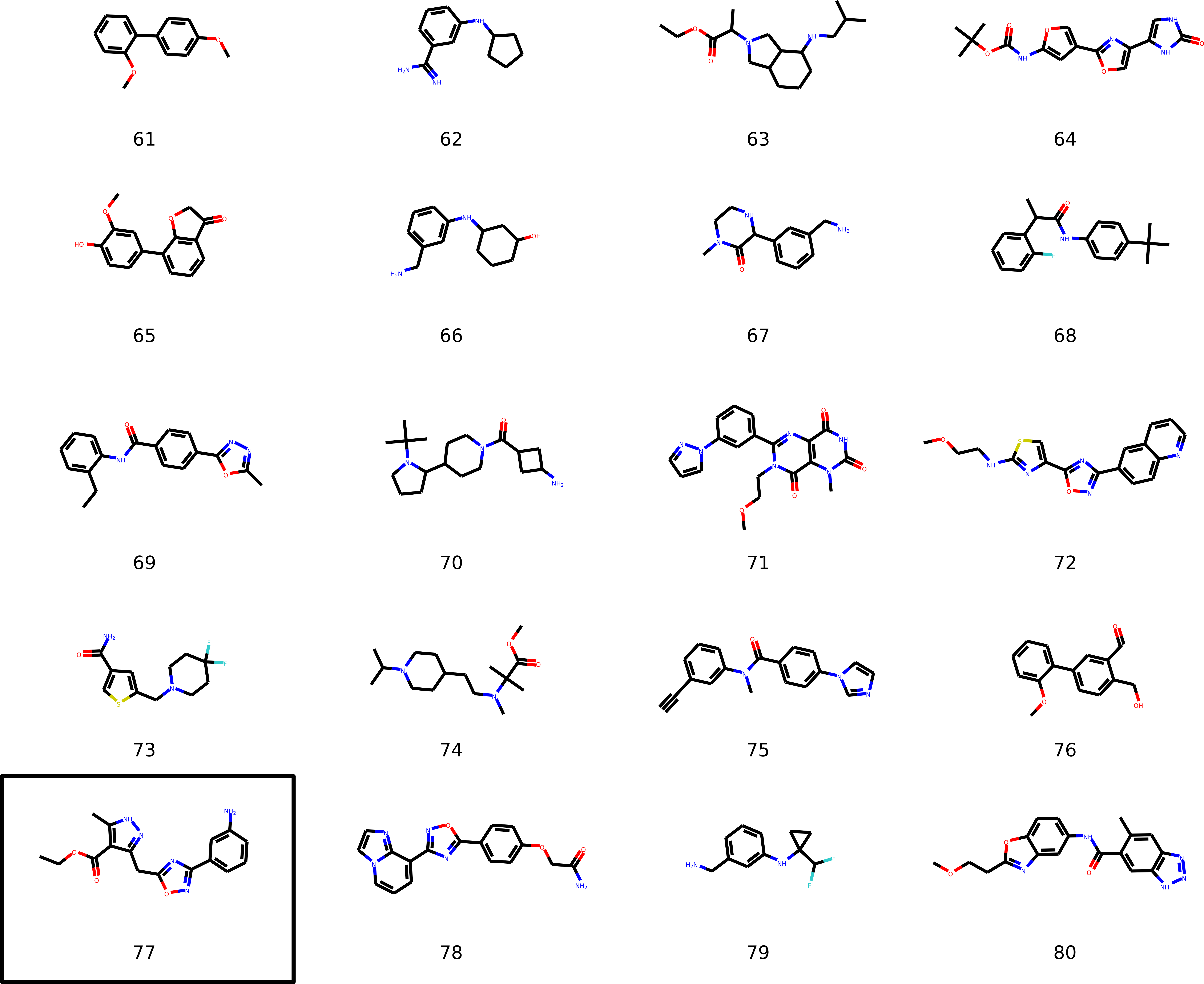
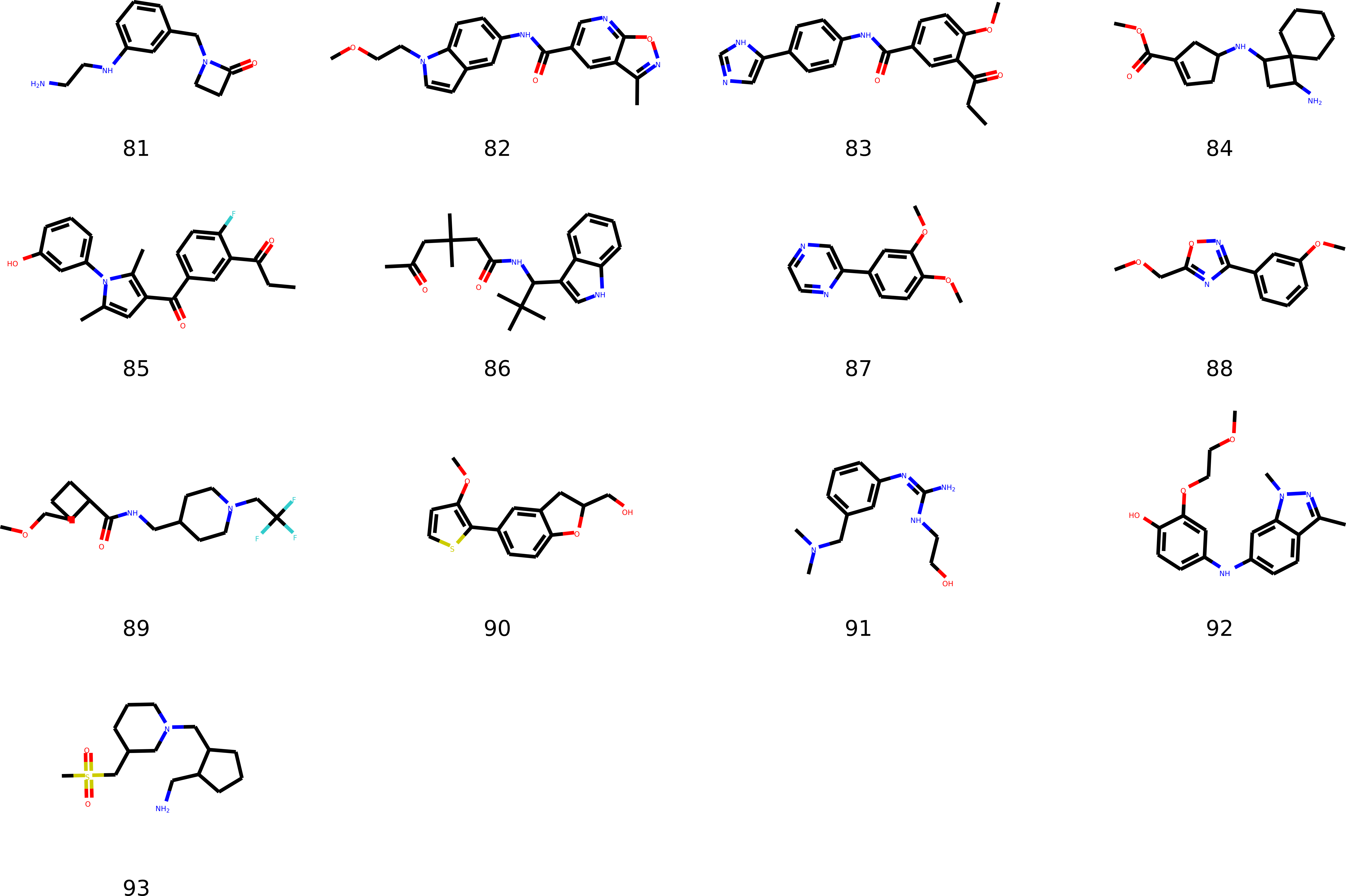
The 6LU7 structure was first fed to the PlayMolecule web application17 using a novel virtual screening technique for the multimodal structure-based ligand design18, called Ligand Generative Adversarial Network (LIGANN). Ligands in LIGANN are generated so as to match the shape and chemical attributes of the binding pocket and decoded into a sequence of SMILES enabling directly the structure-based de novo drug design. SMILES codes for ligands were obtained using the default LIGANN values for shapes and channels with the cubic box center set at the midpoint vector connecting the SH and NE atoms of the CYS-HIS dyad in the 6LU7 PDB structure. The PlayMolecule interface delivered 93 optimally fit non-congeneric compounds, spanning a significant portion of the chemical space, whose SMILES and structures are reported in the Supporting Information (SI).
Each of these compounds was docked to the 6LU7 and to the 1UK4 structures, using Autodock419 with full ligand flexibility. For both structures, the docking was repeated by setting the dyad with the residue in their neutral (CYS-HIS) and charged state (CYS-/HIS+). Further details on Docking parameters are given in the SI.
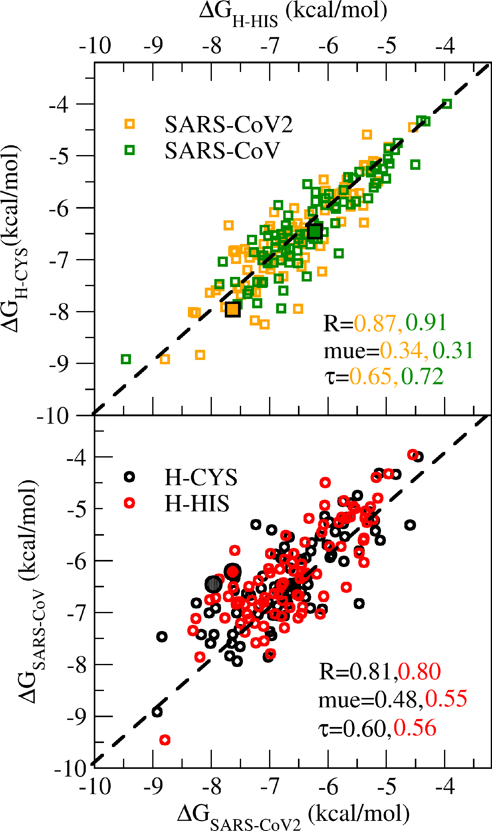
Results for the binding free energies of the 93 LIGGAN-determined 3CLpro ligands are reported in Figure 2. Binding free energies are comprised in the range 4-9 kcal/mol and are found to be strongly correlated for the two protonation states of the CYS-HIS dyad. Correlation is still high when ligand binding free energies for the main proteases are compared, confirming that good binders for SARS-CoV are, in general, also good binders for SARS-CoV2 3CLpro.
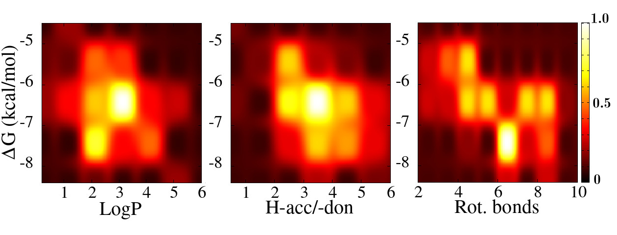
For each of these compounds, using the knowledge-based XLOGP3 methodology20, we computed the octanol/water partition coefficient (LogP) to assess the distribution in hydrophobic and cytosolic environments. LogP values range from -0.5 to a maximum of 5 with a number of rotatable bonds from 2 to a maximum of 12. Most of the LIGGAN compounds bear from 2 to 5 H-bond acceptor or donors (see Table 1 of the SI). In Figure 3 we show the 2D probability distributions for correlated in turn to the LogP, number of H-bond donor/acceptors and number of rotatable bonds. We note, on the left and central panel, sharp maxima for , and for , , respectively, suggestive of a ligand-protein association driven mostly by hydrophobic interactions. We must stress here that the computed pertains to the associations of the ligand with one protein whatever the state of association of the protein. At free ligand concentration equal to , i.e. when half of the protein molecules are inhibited, the probability to have both monomers inhibited is equal to 1/4, whatever the dissociation constant of the dimer,21 hence the need for identifying nanomolar or subnanomolar inhibitors of 3CLpro.
Figure 4 shows the chemical structures of the five compounds exhibiting the highest binding affinity to the 6LU7 main protease of SARS-CoV2 when the CYS-HIS dyad is in the neutral state. None of these compounds is commercially available, although some of them (27, 31, 40) show a high degree of similarity with known structures according to the Tanimoto metrics.22 The LIGGAN-determined structures of Figure 4, as well as many of those reported in Figures 1-5 of the SI, seem to share a common pattern with aromatic moieties connected by rotatable bonds in a pseudo-linear arrangement. In Table 1, the binding free energy data of these five best ligands are shown for both CoV proteases and both protonation states of the catalytic dyad.

Inspection of Table 1 confirms that SARS-CoV2 best binders 27, 29, 39, 77, 19 are also good binders for SARS-CoV 3CLpro. Remarkably, compound 27 is consistently the most potent ligand for the two proteases, irrespective of the dyad protonation state. In the Table 1 we also report the Autodock4-computed binding free energy for ML188. The Autodock4-predicted binding free energy for the association of ML188-SARS-Cov protease is -6.2 and -6.5 kcal/mol for the H-HIS and H-CYS tautomers, not too distant from the experimentally determined value of -8 kcal/mol, hence lending support for the LIGGAN-Autodock4 protocol used in identifying the lead compounds of Table 1.
| CoV19 | SARS | ||||
|---|---|---|---|---|---|
| Comp. | H-CYS | H-HIS | H-CYS | H-HIS | LogP |
| 27 | -8.92 | -8.79 | -8.92 | -9.46 | 4.90 |
| 30 | -8.84 | -8.19 | -7.47 | -7.86 | 3.74 |
| 39 | -8.25 | -7.08 | -6.82 | -6.72 | 6.06 |
| 77 | -8.17 | -7.25 | -7.43 | -7.21 | 2.03 |
| 19 | -8.03 | -8.26 | -7.01 | -7.12 | 5.58 |
| ML188 | -7.96 | 7.63 | 6.46 | 6.22a | 4.97 |
aExperimental value for ML188 is14 kcal/mol.
In order to assess the stability of the 3CLpro-27 association, we have performed extensive molecular dynamics simulations2, 3 of the bound state with explicit solvent. The overall structural information was obtained by combining data from three independent simulations (for a total of about 120 ns), all started from the best docking pose of 27 on the 6LU7 monomeric structure. Further methodological aspects25 are provided in the Supporting Information.
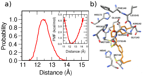
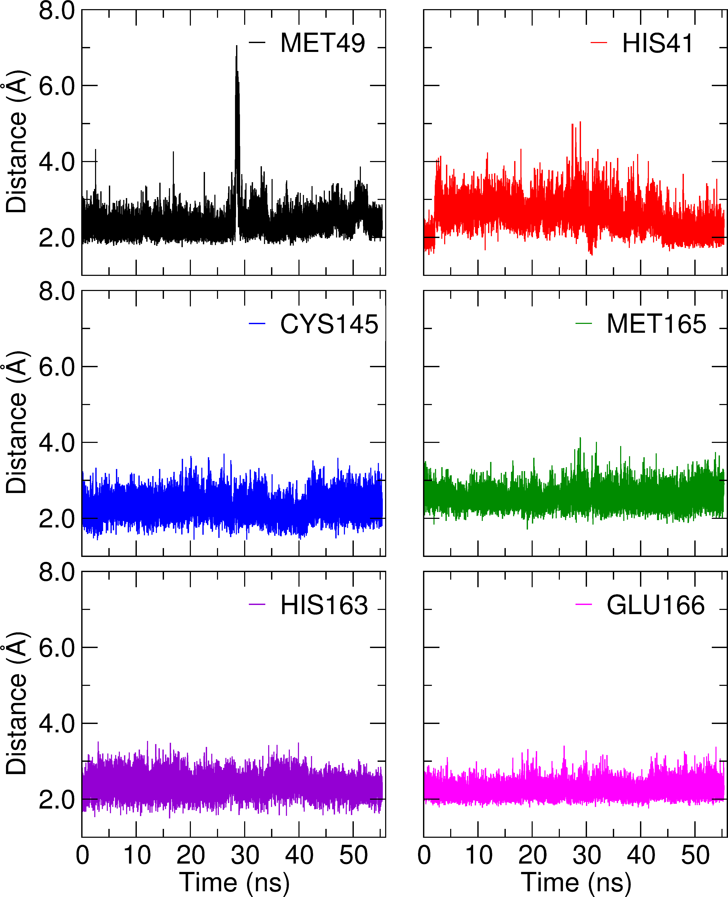
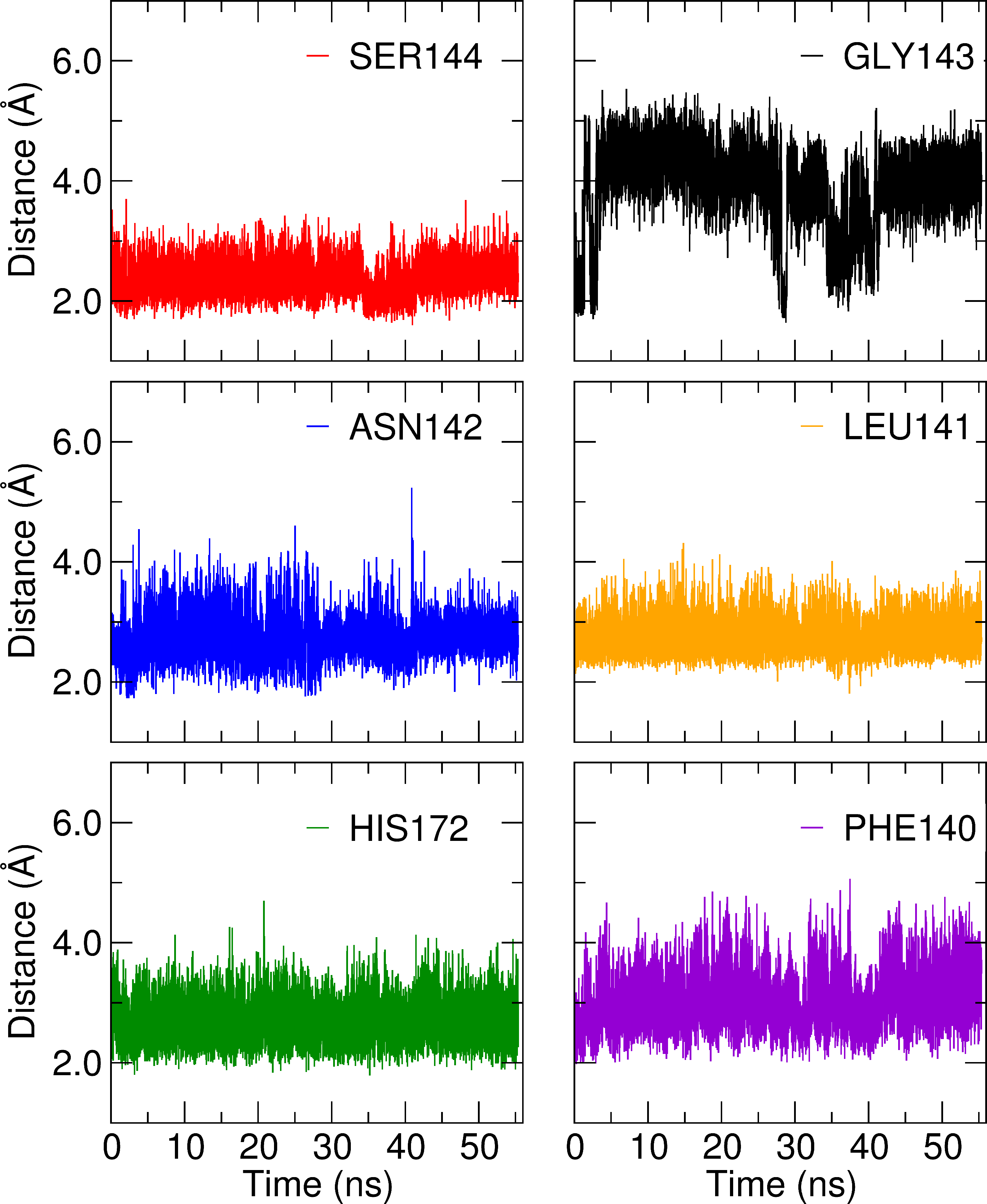
In Figure 5, we show the probability distribution, , of the distance between the center of mass (CoM) of the ligand and that of the domains I+II. The distribution has nearly a Gaussian shape with a half-width of about 1 Å, exhibiting only a minor positive skewness and defining a tight binding site volume of few Å3 at most.26 The MD-determined shows that the ligand never leaves the binding pocket at any stage during the whole simulation. In the inset of Figure 5a, we show the potential of mean force (PMF) along the ligand-protein CoM distance , computed as . As = ,27, the steepness of the curve is suggestive of a profound minimum and hence of a large association constant, confirming the indication obtained from the Docking calculations. Figure 5b shows polar and hydrophobic residues found in at least 90% of the simulation time within 4.5 and 5.5 Å distance, respectively, from any atom of the ligand 27. All essential residues for binding are included, with the important addition of Met165, Phe140 and Leu141 hydrophobic residues that consistently linger near the pyrazolic or the chlorinated phenyl rings of 27, in agreement with the hydrophobic character of the interaction.
Figures 3 and 5 shows possible avenues for improvement. For example, forcing the L-shaped binding structure (see Figure 5b) in bulk also, possibly by redesigning the rotatable connectors in the ligand, may reduce the penalty due conformational entropy loss upon binding,26 hence boosting the ligand affinity for 3CLpro. Building upon this knowledge, we hence plan to optimize the lead using MD simulations coupled to efficient relative binding free energy calculation via free energy perturbation on congeneric variants 28, eventually providing in silico determined anti-viral compounds to be synthesized an experimentally tested in vitro and in vivo.
While the infection rate for COVID 19 in China is currently declining for days, new shocking outbreaks are developing in the South Corea, the Middle East and Europe, with high risk for a pandemic. The scientific community is hence called to an extraordinary and collaborative effort for a rapid delivering of an effective anti-COVID 19 drug, hoping that our contribution can be of help in such a worldwide endeavor.
References
- Dong et al. 2020 Dong, E.; Du, H.; Gardner, L. An Interactive Web-Based Dashboard to Track COVID-19 in Real Time. Lancet Infect. Dis. 2020,
- vir 2020 2020; Viralzone News, https://viralzone.expasy.org
- ncb 2020 2020; The National Center for Biotechnology Information, https://www.ncbi.nlm.nih.gov
- Shanker et al. 2020 Shanker, A.; Bhanu, D.; Alluri, A. Analysis of Whole Genome Sequences and Homology Modelling of a 3C Like Peptidase and a Non-Structural Protein of the Novel Coronavirus COVID-19 Shows Protein Ligand Interaction with an Aza-Peptide and a Noncovalent Lead Inhibitor with Possible Antiviral Properties. ChemRxiv 2020,
- Thiel et al. 2003 Thiel, V.; Ivanov, K. A.; Putics, A.; Hertzig, T.; Schelle, B.; Bayer, S.; Weißbrich, B.; Snijder, E. J.; Rabenau, H.; Doerr, H. W.; Gorbalenya, A. E.; Ziebuhr, J. Mechanisms and Enzymes Involved in SARS Coronavirus Genome Expression. J. Gen. Virol. 2003, 84, 2305–2315
- Hilgenfeld 2014 Hilgenfeld, R. From SARS to MERS: Crystallographic Studies on Coronaviral Proteases Enable Antiviral Drug Design. FEBS J. 2014, 281, 4085–4096
- Anand et al. 2003 Anand, K.; Ziebuhr, J.; Wadhwani, P.; Mesters, J. R.; Hilgenfeld, R. Coronavirus Main Proteinase (3CLpro) Structure: Basis for Design of Anti-SARS Drugs. Science 2003, 300, 1763–1767
- Chuck et al. 2010 Chuck, C.-P.; Chong, L.-T.; Chen, C.; Chow, H.-F.; Wan, D. C.-C.; Wong, K.-B. Profiling of Substrate Specificity of SARS-CoV 3CL. PloS One 2010, 5, e13197–e13197
- Shi et al. 2008 Shi, J.; Sivaraman, J.; Song, J. Mechanism for Controlling the Dimer-Monomer Switch and Coupling Dimerization to Catalysis of the Severe Acute Respiratory Syndrome Coronavirus 3C-Like Protease. J. Virol. 2008, 82, 4620–4629
- Johansson 2012 Johansson, M. H. Reversible Michael Additions: Covalent Inhibitors and Prodrugs. Mini-Rev. Med. Chem. 2012, 12, 1330–1344
- Vasudevan et al. 2019 Vasudevan, A.; Argiriadi, M. A.; Baranczak, A.; Friedman, M. M.; Gavrilyuk, J.; Hobson, A. D.; Hulce, J. J.; Osman, S.; Wilson, N. S. In Chapter One - Covalent Binders in Drug Discovery; Witty, D. R., Cox, B., Eds.; Prog. Med. Chem.; Elsevier, 2019; Vol. 58; pp 1 – 62
- Yang et al. 2003 Yang, H.; Yang, M.; Ding, Y.; Liu, Y.; Lou, Z.; Zhou, Z.; Sun, L.; Mo, L.; Ye, S.; Pang, H.; Gao, G. F.; Anand, K.; Bartlam, M.; Hilgenfeld, R.; Rao, Z. The Crystal Structures of Severe Acute Respiratory Syndrome Virus Main Protease and Its Complex with an Inhibitor. Proc. Natl. Acad. Sci. USA 2003, 100, 13190–13195
- Jacobs et al. 2010 Jacobs, J.; Zhou, S.; Dawson, E.; Daniels, J. S.; Hodder, P.; Tokars, V.; Mesecar, A.; Lindsley, C. W.; ; Stauffer., S. R. Discovery of Non-Covalent Inhibitors of the SARS Main Proteinase 3CLpro. Probe Reports from the NIH Molecular Libraries Program 2010, https://www.ncbi.nlm.nih.gov/books/NBK133447/
- Jacobs et al. 2013 Jacobs, J.; Grum-Tokars, V.; Zhou, Y.; Turlington, M.; Saldanha, S. A.; Chase, P.; Eggler, A.; Dawson, E. S.; Baez-Santos, Y. M.; Tomar, S.; Mielech, A. M.; Baker, S. C.; Lindsley, C. W.; Hodder, P.; Mesecar, A.; Stauffer, S. R. Discovery, Synthesis, and Structure-Based Optimization of a Series of N-(tert-Butyl)-2-(N-arylamido)-2-(pyridin-3-yl) Acetamides (ML188) as Potent Noncovalent Small Molecule Inhibitors of the Severe Acute Respiratory Syndrome Coronavirus (SARS-CoV) 3CL Protease. J. Med. Chem. 2013, 56, 534–546
- 15 Liu, X.; Zhang, B.; Jin, Z.; Yang, H.; Rao, Z. The Crystal Structure of 2019-nCoV Main Protease in Complex with an Inhibitor N3. RSCB PDB, pdbode: 6LU7
- Hu et al. 2009 Hu, T.; Zhang, Y.; Li, L.; Wang, K.; Chen, S.; Chen, J.; Ding, J.; Jiang, H.; Shen, X. Two Adjacent Mutations on the Dimer Interface of SARS Coronavirus 3C-like Protease Cause Different Conformational Changes in Crystal Structure. Virology 2009, 388, 324 – 334
- 17 PlayMolecule™, https://www.acellera.com, accessed 20 February 2020
- Skalic et al. 2019 Skalic, M.; Sabbadin, D.; Sattarov, B.; Sciabola, S.; De Fabritiis, G. From Target to Drug: Generative Modeling for the Multimodal Structure-Based Ligand Design. Mol. Pharm. 2019, 16, 4282–4291
- Morris et al. 2009 Morris, G. M.; Huey, R.; Lindstrom, W.; Sanner, M. F.; Belew, R. K.; Goodsell, D. S.; Olson, A. J. AutoDock4 and AutoDockTools4: Automated Docking with Selective Receptor Flexibility. J. Comput. Chem. 2009, 30, 2785–2791
- Cheng et al. 2007 Cheng, T.; Zhao, Y.; Li, X.; Lin, F.; Xu, Y.; Zhang, X.; Li, Y.; Wang, R.; Lai, L. Computation of Octanol-Water Partition Coefficients by Guiding an Additive Model with Knowledge. J. Chem. Inf. Model. 2007, 47, 2140–2148
- Graziano et al. 2006 Graziano, V.; McGrath, W. J.; Yang, L.; Mangel, W. F. SARS CoV Main Proteinase: The Monomer-Dimer Equilibrium Dissociation Constant. Biochemistry 2006, 45, 14632–14641
- Kim et al. 2016 Kim, S.; Thiessen, P. A.; Bolton, E. E.; Chen, J.; Fu, G.; Gindulyte, A.; Han, L.; He, J.; He, S.; Shoemaker, B. A.; Wang, J.; Yu, B.; Zhang, J.; Bryant, S. H. PubChem Substance and Compound Databases. Nucleic Acids Res. 2016, 44, D1202–D1213
- Pronk et al. 2013 Pronk, S.; Páll, S.; Schulz, R.; Larsson, P.; Bjelkmar, P.; Apostolov, R.; Shirts, M. R.; Smith, J. C.; Kasson, P. M.; van der Spoel, D.; Hess, B.; Lindahl, E. GROMACS 4.5: a High-Throughput and Highly Parallel Open Source Molecular Simulation Toolkit. Bioinformatics 2013, 29, 845
- Van Der Spoel et al. 2005 Van Der Spoel, D.; Lindahl, E.; Hess, B.; Groenhof, G.; Mark, A. E.; Berendsen, H. J. C. GROMACS: Fast, Flexible, and Free. J. Comput. Chem. 2005, 26, 1701–1718
- Macchiagodena et al. 2019 Macchiagodena, M.; Pagliai, M.; Andreini, C.; Rosato, A.; Procacci, P. Upgrading and Validation of the AMBER Force Field for Histidine and Cysteine Zinc(II)-Binding Residues in Sites with Four Protein Ligands. J. Chem. Inf. Model. 2019, 59, 3803–3816
- Procacci and Chelli 2017 Procacci, P.; Chelli, R. Statistical Mechanics of Ligand-Receptor Noncovalent Association, Revisited: Binding Site and Standard State Volumes in Modern Alchemical Theories. J. Chem. Theory Comput. 2017, 13, 1924–1933
- Gilson et al. 1997 Gilson, M. K.; Given, J. A.; Bush, B. L.; McCammon, J. A. The Statistical-Thermodynamic Basis for Computation of Binding Affinities: A Critical Review. Biophys. J. 1997, 72, 1047–1069
- Shirts and Mobley 2013 Shirts, M. R.; Mobley, D. L. An Introduction to Best Practices in Free Energy Calculations. Methods Mol. Biol. 2013, 924, 271–311
| G (kcal/mol) | |||||||||
|---|---|---|---|---|---|---|---|---|---|
| SMILES | Comp. | logP | Hacc | Hdon | SARS-CoV-2 | SARS-CoV | |||
| H-CYS | H-HID | H-CYS | H-HID | ||||||
| NC(=O)c1csc(CNCC(N)C(F)(F)F)c1 | 1 | 0.120 | 3 | 1 | 5 | -5.46 | -5.68 | -6.83 | -6.52 |
| CCC(C(=O)NC(CC(C)C)C(=O)O)(C(=O)c1ccccc1)C(C)C | 2 | 4.450 | 2 | 4 | 11 | -6.64 | -6.49 | -6.15 | -6.68 |
| COCCc1ccc(Oc2ccc(Nc3nc(C)cc(C)n3)cc2)cc1 | 3 | 4.520 | 1 | 2 | 7 | -7.56 | -7.55 | -7.01 | -6.80 |
| CCc1cc(=O)oc2ccc(-c3cccc4c3CCOC4)cc12 | 4 | 2.870 | 0 | 1 | 2 | -6.34 | -7.70 | -6.46 | -6.69 |
| CCOC(=O)C(NC(=O)Cc1c[nH]c2cccc(C)c12)C(C)(C)C | 5 | 3.670 | 1 | 2 | 8 | -6.35 | -6.99 | -6.30 | -6.24 |
| COCCn1ccc2c(C(=O)Nc3ccc4c(c3)nnn4C)cccc21 | 6 | 1.810 | 1 | 3 | 6 | -7.32 | -6.76 | -6.67 | -6.43 |
| COc1cccc(-c2cc(O)cc(F)n2)c1 | 7 | 2.560 | 1 | 2 | 3 | -5.10 | -5.22 | -5.61 | -5.21 |
| CCN(CCC1CC1)C(=O)C1CCCN(CCNC(C)=O)C1 | 8 | 1.370 | 1 | 2 | 10 | -6.46 | -6.19 | -6.07 | -6.26 |
| CCOC(=O)c1c(NCc2c[nH]nc2C)sc(CC)c1-c1ccc(F)cc1 | 9 | 5.440 | 1 | 2 | 8 | -7.95 | -6.51 | -7.43 | -7.11 |
| CC(C)N1CC(c2ncc(-c3cc(Br)c[nH]c3=O)[nH]2)CC1=O | 10 | 2.150 | 0 | 3 | 3 | -6.74 | -6.74 | -5.74 | -5.98 |
| CCC(NC(=O)Cc1c(C)[nH]c2ccccc12)C(CC)S(=O)(=O)C1CC1 | 11 | 3.510 | 1 | 3 | 9 | -7.20 | -7.38 | -7.03 | -7.03 |
| CC(C)(C)C(C)(C)CCCNC(=S)NCC(N)=O | 12 | 2.540 | 3 | 1 | 9 | -6.27 | -5.77 | -5.66 | -5.73 |
| Cc1cc(F)cc2[nH]c(C(=O)Nc3cncc(C(=O)OC(C)(C)C)c3)cc12 | 13 | 3.450 | 1 | 3 | 6 | -6.84 | -7.62 | -7.07 | -6.51 |
| O=C(Nc1ccc2ccoc2c1)c1sc2cc(C(F)(F)F)ccc2c1F | 14 | 5.500 | 1 | 1 | 3 | -6.13 | -6.32 | -5.53 | -5.19 |
| CC(=O)C(NC(=O)c1cnc2c(c1)c(=O)[nH]c(=O)n2C1CC1)C(C)C | 15 | 3.020 | 1 | 5 | 6 | -6.37 | -6.61 | -5.95 | -6.04 |
| COCC(C)CC(=O)Nc1ncc(Cc2cc(F)cc(F)c2)s1 | 16 | 3.200 | 1 | 2 | 8 | -6.74 | -6.13 | -7.07 | -7.03 |
| COCCn1cc(C(=O)N=c2[nH][nH]c(C)c2-c2ccc(F)cc2)cn1 | 17 | 3.380 | 0 | 2 | 6 | -6.68 | -6.90 | -6.84 | -7.03 |
| COc1ccc(-c2cccc3c2COC3=O)cc1 | 18 | 2.940 | 0 | 1 | 2 | -5.73 | -5.68 | -5.20 | -5.19 |
| CCC(=O)c1cc(C(=O)N=c2nc(-c3ccc(OC)cc3)[nH]s2)ccc1F | 19 | 5.580 | 0 | 3 | 6 | -8.03 | -8.26 | -7.01 | -7.12 |
| CCc1ccc(NC(=O)c2ccc3ccccc3c2)cc1F | 20 | 4.900 | 1 | 1 | 4 | -7.23 | -6.10 | -5.31 | -5.46 |
| COc1ccc(Oc2ncccn2)cc1OC | 21 | 2.010 | 0 | 2 | 4 | -5.74 | -5.56 | -4.86 | -4.96 |
| COCCOc1ccc(NC(=O)c2sc(-c3cnn(C)c3)nc2C)cc1 | 22 | 2.320 | 1 | 3 | 8 | -6.91 | -7.19 | -6.51 | -6.39 |
| CCC(c1ccc(F)cc1)c1nc(-c2ccc(CNC)cc2)no1 | 23 | 3.910 | 1 | 2 | 6 | -7.02 | -7.09 | -7.86 | -7.55 |
| CCOC(=O)COc1ccc(NC(=O)c2c[nH]cc2C)cc1Cl | 24 | 2.800 | 1 | 2 | 8 | -7.63 | -7.23 | -7.43 | -7.59 |
| COc1cc2c(cc1F)oc1ccc(OCC(F)F)cc12 | 25 | 4.560 | 0 | 0 | 4 | -5.98 | -5.61 | -5.26 | -5.16 |
| COCC(=O)Nc1cccc(-c2cnn(Cc3nc(C)cs3)c2)c1F | 26 | 2.080 | 1 | 3 | 7 | -6.57 | -6.45 | -6.47 | -6.07 |
| COCCOc1cc(C(=O)N=c2[nH][nH]c(C)c2-c2ccc(Cl)cc2)ccn1 | 27 | 4.900 | 0 | 2 | 7 | -8.92 | -8.79 | -8.92 | -9.46 |
| CC(=O)NCCN(CC1CCCCC1)CC1CCCOC1 | 28 | 2.550 | 1 | 1 | 8 | -6.89 | -6.72 | -5.88 | -5.52 |
| CC(C(=O)N1CC2CCC1C2C(=O)O)=C1C(=O)c2ccccc2SC1=S | 29 | 2.730 | 1 | 4 | 4 | -6.61 | -6.07 | -6.44 | -6.69 |
| CCOC(=O)c1cnc2nc(C)ccc2c1Nc1ccc(CCO)cc1 | 30 | 3.740 | 2 | 4 | 8 | -8.84 | -8.19 | -7.47 | -7.86 |
| COCC(=O)Nc1ccc(-c2noc(-c3cncc(Cl)c3)n2)cc1 | 31 | 2.080 | 1 | 4 | 6 | -7.55 | -7.51 | -7.94 | -6.80 |
| COCc1occc1-c1nc2cc(F)ccc2o1 | 32 | 2.280 | 0 | 1 | 3 | -5.29 | -5.27 | -5.06 | -4.97 |
| COCCc1ccc2cc(Nc3nnc(-c4cnn(C)c4)n3C)ccc2c1 | 33 | 2.640 | 1 | 3 | 6 | -6.94 | -7.08 | -7.06 | -6.93 |
| COC(=O)CC(NC(=O)c1c[nH]cc1-c1cnn(C)c1)c1ccncc1 | 34 | 0.210 | 1 | 4 | 8 | -6.80 | -7.42 | -5.95 | -6.85 |
| COc1cccc(-c2nc3cccnc3s2)c1F | 35 | 3.380 | 0 | 2 | 2 | -5.60 | -5.72 | -4.89 | -4.85 |
| CCOC(=O)C(CC)(CC)CNC(=O)C(c1ccccc1)C1CC1 | 36 | 4.120 | 1 | 2 | 11 | -7.14 | -6.45 | -6.54 | -7.02 |
| COc1cc(-c2cccc3nsnc23)sn1 | 37 | 2.820 | 0 | 3 | 2 | -5.93 | -5.97 | -5.89 | -5.73 |
| Cc1[nH]c2ccccc2c(=O)c1CC(=O)N(CC(=O)OC(C)(C)C)C(C)C | 38 | 4.360 | 0 | 3 | 8 | -7.64 | -8.02 | -7.60 | -6.76 |
| CCC(C)C(NC(=O)OC(C)(C)C)C(=O)N=c1[nH]sc2ccccc12 | 39 | 6.060 | 1 | 2 | 8 | -8.25 | -7.08 | -6.82 | -6.72 |
| COCCn1c(C)c(C)c2cc(NC(=O)c3c[nH]cc3-c3ccc(C)o3)ccc21 | 40 | 3.300 | 1 | 1 | 7 | -7.19 | -6.98 | -7.04 | -7.80 |
| CNCc1cccc(NC2CCCCNC2)c1 | 41 | 1.760 | 3 | 0 | 4 | -6.09 | -6.92 | -6.94 | -6.64 |
| COc1ccsc1-c1ccc(C(=O)O)c(F)c1 | 42 | 2.860 | 1 | 2 | 4 | -5.12 | -5.17 | -4.32 | -4.40 |
| CCc1cccc(-n2c(=O)[nH]c3ccc(OC(C)=O)cc3c2=O)c1 | 43 | 5.120 | 0 | 3 | 4 | -6.57 | -6.79 | -6.52 | -6.48 |
| COc1ccc2c(c1)OCCO2 | 44 | 1.610 | 0 | 0 | 1 | -4.45 | -4.54 | -4.00 | -3.96 |
| COCCOc1ncccc1C(=O)N=c1cc(-c2cccc(F)c2F)[nH][nH]1 | 45 | 4.100 | 0 | 2 | 7 | -6.70 | -7.10 | -6.51 | -6.80 |
| COC(=O)C(C)c1cccc(NC(=O)c2cnn(-c3ccncc3)c2)n1 | 46 | 1.320 | 1 | 5 | 7 | -6.70 | -6.43 | -6.76 | -6.90 |
| COc1ccc(-c2cscc2C(=O)O)cc1 | 47 | 2.730 | 1 | 2 | 4 | -5.29 | -5.35 | -5.25 | -5.05 |
| CCCC(C(=O)O)N(Cc1ccc(-n2cncn2)c(F)c1)c1nn[nH]n1 | 48 | 2.140 | 1 | 7 | 9 | -5.96 | -5.39 | -5.94 | -5.61 |
| CCOC(=O)c1nc(Cn2ccc(-c3ccc4ncccc4c3)c2N)no1 | 49 | 2.610 | 1 | 4 | 6 | -7.68 | -7.49 | -7.84 | -7.21 |
| COc1cccc(-c2nc(O)cc(O)n2)c1 | 50 | 1.790 | 2 | 4 | 4 | -5.72 | -5.46 | -6.02 | -5.86 |
| CCOC(=O)CCN(C(=O)c1[nH]c2ccccc2c1Br)C(C)(C)C | 51 | 3.710 | 0 | 2 | 8 | -7.02 | -6.84 | -6.79 | -6.65 |
| CN(C)Cc1cccc(CN(C)C)c1 | 52 | 1.590 | 0 | 0 | 4 | -4.59 | -5.33 | -5.32 | -5.26 |
| CCOC(=O)c1c[nH]c2c(NCc3ccc(-n4ccnc4)nc3)ccnc12 | 53 | 1.780 | 1 | 4 | 7 | -6.41 | -7.01 | -6.35 | -6.16 |
| CCc1nc2ccc(C(=O)Nc3ccc(C)cc3C)cc2s1 | 54 | 4.780 | 1 | 2 | 4 | -6.46 | -6.38 | -6.60 | -6.24 |
| Cc1noc2nc(-c3ccc(F)cc3)cc(C(=O)NCCCCO)c12 | 55 | 2.440 | 2 | 4 | 8 | -6.99 | -7.57 | -7.37 | -6.84 |
| CCC1(C(=O)N=c2nc[nH]c3ccccc23)CCCN1C(=O)OC(C)(C)C | 56 | 4.460 | 0 | 3 | 6 | -6.96 | -7.34 | -7.45 | -7.13 |
| COc1cccc(-c2cnc3ccc(F)cc3n2)c1 | 57 | 2.940 | 0 | 2 | 2 | -6.05 | -6.05 | -5.15 | -4.94 |
| COc1cc(C(F)(F)F)ccc1-c1cn2cccnc2n1 | 58 | 3.630 | 0 | 2 | 2 | -5.49 | -5.14 | -4.75 | -4.80 |
| COCCN(CC1CCC1)CC1CCN(C2CC2)CC1 | 59 | 2.740 | 0 | 0 | 8 | -6.66 | -6.47 | -5.55 | -6.35 |
| CC(C)CC(=O)Nc1c[nH]nc1-c1cc(-c2ccc(F)cc2)no1 | 60 | 2.560 | 1 | 3 | 6 | -7.59 | -7.92 | -6.34 | -6.72 |
| COc1ccc(-c2ccccc2OC)cc1 | 61 | 3.490 | 0 | 0 | 3 | -5.20 | -5.36 | -5.23 | -5.18 |
| N=C(N)c1cccc(NC2CCCC2)c1 | 62 | 2.140 | 2 | 0 | 3 | -5.71 | -5.76 | -5.54 | -5.17 |
| CCOC(=O)C(C)N1CC2CCCC(NCC(C)C)C2C1 | 63 | 3.050 | 1 | 1 | 7 | -6.86 | -7.35 | -6.60 | -6.50 |
| CC(C)(C)OC(=O)Nc1cc(-c2nc(-c3c[nH]c(=O)[nH]3)co2)co1 | 64 | 2.510 | 1 | 3 | 6 | -7.17 | -7.10 | -6.86 | -7.27 |
| COc1cc(-c2cccc3c2OCC3=O)ccc1O | 65 | 2.720 | 1 | 2 | 3 | -6.01 | -6.58 | -5.70 | -6.21 |
| NCc1cccc(NC2CCCC(O)C2)c1 | 66 | 1.410 | 3 | 1 | 4 | -6.54 | -6.74 | -6.62 | -7.30 |
| CN1CCNC(c2cccc(CN)c2)C1=O | 67 | -0.350 | 2 | 1 | 2 | -6.93 | -6.82 | -7.06 | -6.66 |
| CC(C(=O)Nc1ccc(C(C)(C)C)cc1)c1ccccc1F | 68 | 5.040 | 1 | 1 | 5 | -6.68 | -6.44 | -5.88 | -6.21 |
| CCc1ccccc1NC(=O)c1ccc(-c2nnc(C)o2)cc1 | 69 | 3.440 | 1 | 3 | 5 | -6.77 | -6.65 | -6.53 | -6.91 |
| CC(C)(C)N1CCCC1C1CCN(C(=O)C2CC(N)C2)CC1 | 70 | 1.630 | 1 | 1 | 4 | -6.30 | -6.52 | -6.92 | -6.66 |
| COCCn1c(-c2cccc(-n3cccn3)c2)nc2c(=O)[nH]c(=O)n(C)c2c1=O | 71 | 3.360 | 0 | 5 | 5 | -6.76 | -6.78 | -6.72 | -6.77 |
| COCCNc1nc(-c2nc(-c3ccc4ncccc4c3)no2)cs1 | 72 | 2.970 | 1 | 4 | 6 | -7.60 | -7.30 | -6.58 | -6.77 |
| NC(=O)c1csc(CN2CCC(F)(F)CC2)c1 | 73 | 1.510 | 1 | 1 | 3 | -5.67 | -6.04 | -5.17 | -4.50 |
| COC(=O)C(C)(C)N(C)CCC1CCN(C(C)C)CC1 | 74 | 2.710 | 0 | 1 | 7 | -7.11 | -6.98 | -6.30 | -5.87 |
| C#Cc1cccc(N(C)C(=O)c2ccc(-n3ccnc3)cc2)c1 | 75 | 2.940 | 0 | 2 | 5 | -6.45 | -6.58 | -5.98 | -5.87 |
| COc1ccccc1-c1ccc(CO)c(C=O)c1 | 76 | 2.100 | 1 | 2 | 5 | -6.38 | -6.41 | -6.71 | -6.30 |
| CCOC(=O)c1c(Cc2nc(-c3cccc(N)c3)no2)n[nH]c1C | 77 | 2.030 | 1 | 4 | 6 | -8.17 | -7.25 | -7.43 | -7.21 |
| NC(=O)COc1ccc(-c2nc(-c3cccn4ccnc34)no2)cc1 | 78 | 2.220 | 1 | 4 | 5 | -7.52 | -7.86 | -7.33 | -6.36 |
| NCc1cccc(NC2(C(F)F)CC2)c1 | 79 | 1.990 | 2 | 0 | 4 | -5.18 | -5.38 | -5.43 | -5.57 |
| COCCc1nc2cc(NC(=O)c3cc4[nH]nnc4cc3C)ccc2o1 | 80 | 2.290 | 1 | 4 | 6 | -8.01 | -8.31 | -7.60 | -7.35 |
| NCCNc1cccc(CN2CCC2=O)c1 | 81 | -0.030 | 2 | 1 | 5 | -6.29 | -5.38 | -5.82 | -6.04 |
| COCCn1ccc2cc(NC(=O)c3cnc4onc(C)c4c3)ccc21 | 82 | 2.070 | 1 | 3 | 6 | -7.92 | -7.76 | -6.92 | -7.11 |
| CCC(=O)c1cc(C(=O)Nc2ccc(-c3cnc[nH]3)cc2)ccc1OC | 83 | 2.680 | 1 | 3 | 7 | -6.82 | -7.60 | -6.46 | -6.19 |
| COC(=O)C1=CCC(NC2CC(N)C23CCCCC3)C1 | 84 | 2.140 | 2 | 1 | 4 | -7.39 | -7.18 | -6.64 | -6.89 |
| CCC(=O)c1cc(C(=O)c2cc(C)n(-c3cccc(O)c3)c2C)ccc1F | 85 | 4.360 | 1 | 3 | 6 | -7.50 | -7.42 | -7.66 | -7.38 |
| CC(=O)CC(C)(C)CC(=O)NC(c1c[nH]c2ccccc12)C(C)(C)C | 86 | 3.700 | 1 | 2 | 8 | -6.55 | -6.79 | -6.43 | -7.08 |
| COc1ccc(-c2cnccn2)cc1OC | 87 | 1.390 | 0 | 2 | 3 | -4.83 | -4.96 | -4.34 | -4.33 |
| COCc1nc(-c2cccc(OC)c2)no1 | 88 | 1.460 | 0 | 2 | 4 | -5.73 | -5.81 | -5.29 | -5.26 |
| COCCC1(C(=O)NCC2CCN(CC(F)(F)F)CC2)CCC1 | 89 | 2.470 | 1 | 1 | 8 | -6.96 | -6.39 | -5.41 | -5.65 |
| COc1ccsc1-c1ccc2c(c1)CC(CO)O2 | 90 | 2.620 | 1 | 1 | 4 | -5.87 | -5.53 | -5.44 | -5.00 |
| CN(C)Cc1cccc(N=C(N)NCCO)c1 | 91 | -0.040 | 3 | 1 | 7 | -5.46 | -6.07 | -5.82 | -5.31 |
| COCCOc1cc(Nc2ccc3c(C)nn(C)c3c2)ccc1O | 92 | 2.980 | 2 | 2 | 7 | -6.64 | -6.82 | -6.60 | -6.17 |
| CS(=O)(=O)CC1CCCN(CC2CCCC2CN)C1 | 93 | 0.990 | 1 | 2 | 5 | -6.86 | -7.59 | -6.88 | -5.81 |
1 Computational Details
1.1 Molecular Docking
Following the two input file used for molecular docking.
-
•
file.gpf
npts 60 60 60
gridfld 6lu7.maps.fld
spacing 0.375
receptor_types A C HD N OA SA
ligand_types ...
receptor 6lu7.pdbqt
gridcenter -10.18 20.65 66.75
smooth 0.5
map 6lu7.A.map
map ...
...
elecmap 6lu7.e.map
dsolvmap 6lu7.d.map
dielectric -0.1465
-
•
file.dpf
autodock_parameter_version 4.2
outlev 1
intelec
seed pid time
ligand_types   ....
fld 6lu7.maps.fld
map 6lu7.A.map
map ...
...
elecmap 6lu7.e.map
desolvmap 6lu7.d.map
move ligand.pdbqt
about 6.66645 -3.33598 -3.92332
tran0 random
quaternion0 random
dihe0 random
torsdof 4
rmstol 2.0
extnrg 1000.0
e0max 0.0 10000
ga_pop_size 150
ga_num_evals 2500000
ga_num_generations 27000
ga_elitism 1
ga_mutation_rate 0.02
ga_crossover_rate 0.8
ga_window_size 10
ga_cauchy_alpha 0.0
ga_cauchy_beta 1.0
set_ga
unbound_model bound
do_global_only 50
analysis
1.2 Molecular Dynamics Simulations
Molecular dynamics (MD) simulations were carried out in a cubic box with periodic boundary conditions, whose side-length was chosen so that the minimum distance between protein atoms belonging to neighboring replicas was larger than 14 Å in any direction. The system (protein+compound) was explicitly solvated with the SPC/E 1 water model at the standard density. The starting configuration was generated using GROMACS2, 3 and PrimadORAC.4 The system was initially minimized at 0 K with a steepest descent procedure and subsequently heated to 298.15 K in an NPT ensemble (P=1 atm) using Berendsen barostat5 and velocity rescaling algorithm6 with an integration time step of 0.1 fs and a coupling constant of 0.1 ps for 250 ps.
Production run in the NPT ensemble were carried out starting three independent simulations with different initial velocities randomization. Each MD run has been performed for 40 ns (for a total of 120 ns) imposing rigid constraints only on the X-H bonds (with X being any heavy atom) by means of the LINCS algorithm (t=2.0 fs).7 Electrostatic interactions were treated by using particle-mesh Ewald (PME)8 method with a grid spacing of 1.2 Å and a spline interpolation of order 4. The cross interactions for Lennard-Jones terms were calculated using the Lorentz-Berthelot9, 10 mixing rules and we excluded intramolecular non-bonded interactions between atom pairs separated up to two bonds. The non-bonded interactions between 1-4 atoms involved in a proper torsion were scaled by the standard AMBER fudge factors (0.8333 and 0.5 for the Coulomb and Lennard-Jones, respectively).
The simulations and the trajectories analysis were carried out using the GROMACS 2018.3 program.2, 3
References
- Berendsen et al. 1987 Berendsen, H. J. C.; Grigera, J. R.; Straatsma, T. P. The Missing Term in Effective Pair Potentials. J. Phys. Chem. 1987, 91, 6269–6271
- Pronk et al. 2013 Pronk, S.; Páll, S.; Schulz, R.; Larsson, P.; Bjelkmar, P.; Apostolov, R.; Shirts, M. R.; Smith, J. C.; Kasson, P. M.; van der Spoel, D.; Hess, B.; Lindahl, E. GROMACS 4.5: a High-Throughput and Highly Parallel Open Source Molecular Simulation Toolkit. Bioinformatics 2013, 29, 845
- Van Der Spoel et al. 2005 Van Der Spoel, D.; Lindahl, E.; Hess, B.; Groenhof, G.; Mark, A. E.; Berendsen, H. J. C. GROMACS: Fast, Flexible, and Free. J. Comput. Chem. 2005, 26, 1701–1718
- Procacci 2017 Procacci, P. PrimaDORAC: A Free Web Interface for the Assignment of Partial Charges, Chemical Topology, and Bonded Parameters in Organic or Drug Molecules. J. Chem. Inf. Model. 2017, 57, 1240–1245
- Berendsen et al. 1984 Berendsen, H. J. C.; Postma, J. P. M.; van Gunsteren, W. F.; Di Nola, A.; Haak, J. R. Molecular Dynamics with Coupling to an External Bath. J. Chem. Phys. 1984, 81, 3684–3690
- Bussi et al. 2007 Bussi, G.; Donadio, D.; Parrinello, M. Canonical Sampling Through Velocity Rescaling. J. Chem. Phys. 2007, 126, 014101
- Hess et al. 1997 Hess, B.; Bekker, H.; Berendsen, H.; Fraaije, J. LINCS: A Linear Constraint Solver for Molecular Simulations. J. Comput. Chem. 1997, 18, 1463–1472
- Darden et al. 1993 Darden, T.; York, D.; Pedersen, L. Particle Mesh Ewald: An N log(N) Method for Ewald Sums in Large Systems. J. Chem. Phys. 1993, 98, 10089–10092
- Antoon 1881 Antoon, L. H. Ueber die Anwendung des Satzes vom Virial in der Kinetischen Theorie der Gase. Ann. Phys 1881, 248, 127–136
- Marcellin 1898 Marcellin, B. Sur Le Mélange Des Gaz. Comptes Rendus Acad. Sci. 1898, 126, 1703–1855