Deep Learning of Movement Intent and Reaction Time for EEG-informed Adaptation of Rehabilitation Robots
Abstract
Mounting evidence suggests that adaptation is a crucial mechanism for rehabilitation robots in promoting motor learning. Yet, it is commonly based on robot-derived movement kinematics, which is a rather subjective measurement of performance, especially in the presence of a sensorimotor impairment. Here, we propose a deep convolutional neural network (CNN) that uses electroencephalography (EEG) as an objective measurement of two kinematics components that are typically used to assess motor learning and thereby adaptation: i) the intent to initiate a goal-directed movement, and ii) the reaction time (RT) of that movement. We evaluated our CNN on data acquired from an in-house experiment where 13 subjects moved a rehabilitation robotic arm in four directions on a plane, in response to visual stimuli. Our CNN achieved average test accuracies of 80.08% and 79.82% in a binary classification of the intent (intent vs. no intent) and RT (slow vs. fast), respectively. Our results demonstrate how individual movement components implicated in distinct types of motor learning can be predicted from synchronized EEG data acquired before the start of the movement. Our approach can, therefore, inform robotic adaptation in real-time and has the potential to further improve one’s ability to perform the rehabilitation task.
I INTRODUCTION
By delivering intensive, challenging and task-specific training, rehabilitation robots [1] have now established themselves as an alternative treatment for sensorimotor impairments [2]. Given the importance of active participation during therapy for motor recovery [3], there is an increasing interest in adapting the robotic therapy and tailoring it to the needs and skills of the patients [4, 5, 6]. The current ’assist-as-needed’ adaptive controllers use movement-related kinematics feedback to alter their control strategies, and subsequently adapt the rehabilitation task components [4, 7]. Despite their demonstrated success in functional motor recovery [8, 9], the robot-derived movement kinematics is a subjective measurement of movement performance as it often becomes masked by the targeted sensorimotor impairments. This evidence-based adaptation [7] limits our ability to study and improve the robotic intervention in rehabilitation.
To overcome this limitation, a fascinating possibility is to have an adaptive strategy targeting specific movement components that are implicated in motor learning, either explicitly or implicitly- depending upon whether or not the patients deliberately aim to improve these attributes . One such component implicated in explicit motor learning is the intent of patients to initiate a movement. It has long been known that when rehabilitation training consists of voluntary movements, a significant improvement in the motor performance is achieved. This has been associated with the cortical reorganization of the motor cortex upon motor training [3]. In our previous work, we used the initiation of voluntary movements as a faithful indicator of explicit motor learning [9], followed by clinical outcome [10]. Another movement component, implicated in implicit motor learning, is the reaction time (RT) of a movement, defined as the time passed between the onset of a stimulus and the start of the movement [11, 12]. We have previously shown that there is a decrease in average RT of the patients as the training progresses, further confirming its role as a metric for motor learning that is used to assess motor deficits [13, 14] and improvement [9]. Given their immediate applicability in assessing both implicit and explicit motor learning, these two movement components can be used as adaptation criteria.
For the adaptation to become clinically important, one needs to first define the criteria that are indicative of the motor performance and then to objectively measure them. Arguably, the most faithful representation of movement is in its underlying neuronal activity. Indeed, for more than half a century, a significant amount of studies have identified specialized neurons in the motor cortex and other cortical and sub-cortical areas that encode movement components, including direction [15], amplitude [16], force [17] and speed [18]. Interestingly enough, a recent study showed that the motor thalamic neurons encode the movement intent and RT [19]. Overall, neuronal activity offers an objective assessment of movement components in healthy and impaired subjects.
By virtue of their non-invasiveness and high temporal resolution, electroencephalography (EEG) has established itself as an objective measure for assessing motor behavior [20, 21], and has also become a reliable measurement of the cognitive functions involved in the execution of motor tasks in stroke [22, 23]. The first approaches to decode EEG relied on hand-crafted features such as common spatial pattern [24] and classifiers such as support vector machines (SVM) to segregate those features. However, these methods do not generalize well to new subjects [25], and their need for hand-crafted features further impedes their use in real-time. Deep neural networks [26] overcome these limitations through unsupervised feature extraction and powerful generalization [27]. These networks have recently started to be used in classifying EEG signals for various mental decoding tasks with remarkable performance [28].
Here, we propose a deep learning approach to predict two fundamental movement components associated with motor learning, namely the intent to initiate a goal-directed movement and RT, using EEG data as a measurement that circumvents the limitations imposed by the impaired kinematics. To do so, we developed a convolutional neural network (CNN) that was trained to decode EEG activity and predict: i) the intent of the subject to initiate a movement (intent vs. no intent), and ii) RT in initiating the movement (2 classes- fast vs. slow). We evaluated our CNN on data acquired from an in-house experiment where 13 subjects moved a rehabilitation robotic arm in four directions on a plane in response to visual stimuli. The CNN achieved average test accuracies of 80.02% and 79.82% for movement intent and RT classification respectively. These results support our ongoing effort to devise a rehabilitation strategy that adapts based on an objective measurement of specific movement components, further personalizing therapy.
II Methods
II-A Subjects and Experimental Apparatus
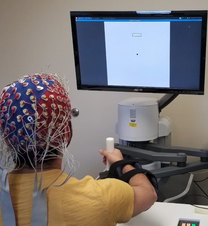
Thirteen healthy subjects (age=232, 5 females, right handed) participated in this experiment. Subjects provided written consent and the experimental protocol was approved by the local Institutional Review Board (IRB). The EEG data were recorded using a 128-channel Biosemi ActiveOne EEG system with sampling frequency of 1024 Hz. The motor task was performed on the InMotion Arm Robot (Bionik Laboratories Corp.)(Figure 1), with the robot data collected at a sampling frequency of 180 Hz. Subjects were seated at an appropriate distance from the screen so that they could perform the task comfortably without moving their torso.
II-B Experiment
Building on a previous experimental protocol [29, 12], we developed a goal-directed task and asked the subjects to perform it on the arm rehabilitation robot. The task environment comprised of a pointer and a target box (Figure 1). The pointer indicated the current position of the end-effector of the robotic arm in the 2D plane of motion. Subjects were asked to move the pointer to the target box “as fast as possible”. After the pointer entered the target box, the next target box appeared with a delay of 20ms with small added jitter. The target appeared at random in any of the four orthogonal directions- left, right, up or down. Subjects performed the task in two modes: i) active mode, where subjects performed the motion, and ii) passive mode, where motion was performed by the robot with the subject’s arm affixed to the robotic end effector. Each subject performed 210 active and 210 passive motion trials.
II-C EEG Minimal Preprocessing and Labeling
Minimal data preprocessing was done to get rid of the contamination in the EEG data, as described below. The minimal preprocessing on the one hand ensured the CNN would learn the representations by itself and, on the other hand, made it possible for the CNN to be compatible with a real-time system. A bandpass filter of 1 Hz - 40 Hz was applied to remove the low and high frequency artifacts and drifts. Independent component analysis (ICA) was used to get rid of occular artifacts. We then segmented the data into trials containing the events of interest. The time window for segmentation was chosen by grid search: -0.5s to 0.0s for both movement intent classification and RT (with t=0s indicating the start of the motion). We normalized the segmented trials using z-score normalization and downsampled all the trials to 250 Hz to reduce the computational load. For classifying the movement intent, all active motion trials were labelled as ’Intent’ and all passive motion trials were labelled as ’No Intent’. The movement intent dataset had 210 data points for each class per subject. To compute the RT, we measured the time difference between the onset of a stimulus and the start of the movement, where the movement was said to be started when the velocity exceeded a certain threshold. We discretized the RT into two classes- fast and slow, by choosing suitable thresholds that were determined from the histogram of RTs for each subject separately, based on the distribution of their RT across the experiement. In addition, all trials corresponding to RTs that were outliers, i.e. less that 0.15 seconds and greater than 0.8 seconds were removed from consideration, similarly to our previous studies [12, 30]. This resulted in RT datasets of varying size for each subject, with an average size of 75 data points per class (SD = 14).
II-D Notations
For each subject , we created a dataset , where denotes the number of trials recorded for that subject. For every trial , is a 2 dimensional matrix which was created by stacking recordings from all the channels. is the duration of recording of each trial. The labels of the trial contained a value from corresponding to the two classes.
II-E The Convolutional Neural Network
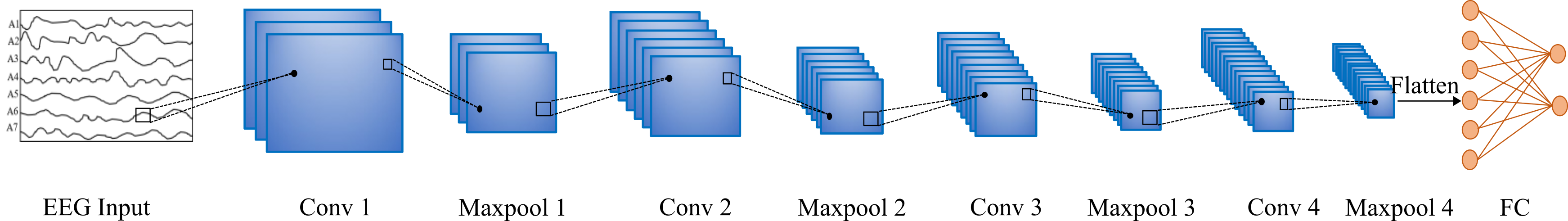
We developed a multi-layered CNN (Figure 2) for classifying movement intent and RT, that performed convolution in spatial and temporal space. The inputs to both networks were preprocessed EEG trials acquired from 128 channels and sampled at 250 Hz. We passed the outputs of each convolutional layer through ReLU non-linearities and then applied batch normalization, to normalize the ReLU outputs to zero mean and unit variance. Batch normalization has regularization properties and is helpful in preventing overfitting [31]. We also applied max pooling at the end of each layer to reduce computational load. Max pooling has also the desirable property of translational invariance which often gives better generalization across subjects. The last layer in both networks was a fully connected layer with softmax that took in the flattened feature vector produced by the last convolutional layer and converted it to class probabilities. The choice of the CNN hyper-parameters, i.e. the number of layers, kernel size, etc. were limited by the training data size and the input dimension, and were found using a grid search over the allowable hyper-parameters space. The CNN architectures for classifying movement intent and RT are shown in Tables I and II.
| Layers | Output Shape |
|---|---|
| Conv1 (5x5) | 32 x 124 x 121 |
| ReLU + BatchNorm | 32 x 124 x 121 |
| MaxPool (3x3) | 32 x 41 x 40 |
| Conv2(5x5) | 64 x 37 x 36 |
| ReLU + BatchNorm | 64 x 37 x 36 |
| MaxPool (3x3) | 64 x 12 x 12 |
| Conv3 (5x5) | 128 x 8 x 8 |
| ReLU + BatchNorm | 128 x 8 x 8 |
| MaxPool (3x3) | 128 x 2 x 2 |
| Fully Connected | 1 x 2 |
| Softmax | 1 x 2 |
| Layers | Output Shape |
|---|---|
| Conv1 (3x5) | 32 x 126 x 121 |
| ReLU + BatchNorm | 32 x 126 x 121 |
| MaxPool (2x2) | 32 x 63 x 60 |
| Conv2(3x5) | 64 x 61 x 56 |
| ReLU + BatchNorm | 64 x 61 x 56 |
| MaxPool (2x2) | 64 x 30 x 28 |
| Conv3 (3x5) | 128 x 28 x 24 |
| ReLU + BatchNorm | 128 x 28 x 24 |
| MaxPool (2x2) | 128 x 14 x 12 |
| Conv4 (3x5) | 256 x 12 x 8 |
| ReLU + BatchNorm | 256 x 12 x 8 |
| MaxPool (2x2) | 256 x 6 x 4 |
| Fully Connected | 1 x 2 |
| Softmax | 1 x 2 |
II-F Network Training
The CNN computed a mapping from the EEG trial to the labels, where were the trainable parameters of the network. The network was trained to minimize the average loss over all training examples:
| (1) |
where denotes the number of training examples and is the loss function, which in our case was the binary cross entropy loss function. The batch size was 64. For optimization, we used Adam, a variant of stochastic gradient descent, with learning rate of .
II-G Network Validation
We evaluated our proposed CNN in three ways:
-
1.
Leave-one-out: Data from all but one subject were used for training. Evaluation was done on the left-out subject. This tested the CNN’s ability to generalize to new subjects, that were not included in training.
-
2.
Subject-specific training: Data from a single subject were split randomly into training and test in the ratio 4:1. Validation was done on the test data. This tested the ability of the CNN to predict movement components when trained on individual subjects.
-
3.
All data: Data from all subjects were split randomly into training and test in the ratio 4:1. This allowed us to determine how well the CNN performs if it has access to data from a large number of subjects.
While the movement intent classification was evaluated using all three evaluation techniques, RT classification was evaluated using only 1) and 3) since the number of data points per subject for RT were low to train a deep network, for the given variability of the labels (RT).
III Results
III-A Leave-one-out Evaluation
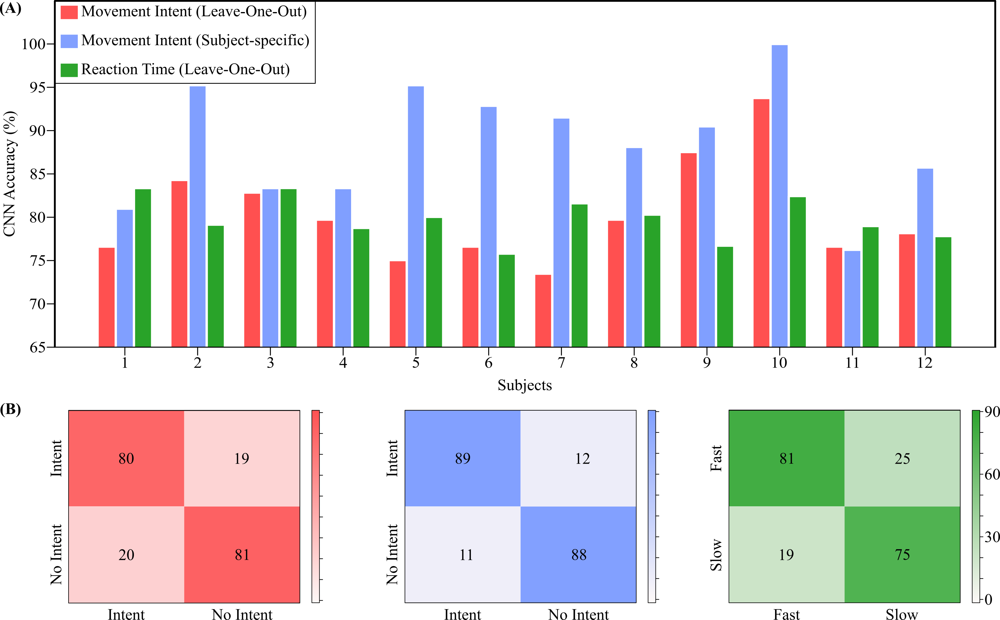
To measure the generalization performance of our CNN, we evaluated it using the leave-one-out technique. Our CNN achieved an average accuracy of for movement intent classification, and for RT. Subject-specific results are shown in (Figure 3A: red and green bars). While the CNN’s prediction of movement intent was most accurate for subject 10 (93.75%), the RT CNN had the best performance for subject 3 (83.34%). In general, the CNN performed well enough for all subjects.
III-B Subject-specific Training
We trained and evaluated our CNN for each subject separately, to test its ability to infer each individual’s intent when data from only that person were available (Figure 3A: blue bar). In this case, our CNN had an average accuracy of for movement intent. Given that the network was trained for each subject separately, its increase in accuracy over leave-one-out was expected because of the reduced variability in the EEG.
III-C Training on All Data
Next, we evaluated our CNN on data from all the subjects, partitioned randomly into train and test in the ratio 4:1. This is a good indication of how our model can perform when data from a large pool of patients is available before-hand. The results, averaged over 10 random partitions, are shown in Table III. We notice that there is an improvement in the performance of the CNNs, when compared to leave-one-out evaluation, since the CNNs were trained on a more representative set of trials that were collected from all the subjects participated in the experiment.
| Task | Mean Accuracy |
|---|---|
| Movement Intent | 87.34% 2.83% |
| Reaction Time | 84.68% 3.68% |
III-D Confusion Matrices
The averaged confusion matrices shown in Figure 3B demonstrate that the network can predict both classes of the two classification tasks equally well. This is further revealed in the F1 scores, defined as the harmonic mean of precision and recall. Specifically, the F1 scores were 0.80 (leave-one-out) and 0.88 (subject-specific) for classifying the movement intent, and 0.80 for classifying RT (leave-one-out).
IV Discussion
Here, we presented a deep learning framework to predict two movement components implicated in explicit and implicit motor learning, which are applicable in adapting rehabilitation robots: i) movement intent and ii) movement RT. Our framework consists of a CNN that associates the EEG activity with these components. The CNN achieved high test accuracies for all the evaluation cases, thus demonstrating its practical effectiveness in our ongoing effort to develop an adaptive robot based on individual components of movement.
Here, we targeted the movement intent from the EEG activity because of the role of voluntary movements in improving motor performance [3]. Neurological deficits, particularly in the supplementary motor area (SMA), have been found to directly affect the ability of patients to initiate movement [32]. Further, [33] suggested a possible compensatory recovery mechanism through recruitment of healthy medial and lateral premotor circuitry. In the clinical setting, ours [9] and other’s [34] studies have revealed significant improvements in the number of self-initiated movements executed by the patients between admission and discharge.
We also targeted the prediction of RT from the EEG, because RT is one of the most well-studied behavioral indicators of neurological integrity. Interestingly, RT has not only been found to be related to neurological deficits, but also it is responsive to intervention [35], including exercise and practice [11, 7]. In light of the fact that the sensorimotor control of the upper extremities is driven by cortical and subcortical areas, our finding that RT behavior is similar in lower and upper extremities [12, 30] expands the role of a presumptive supraspinal pathway from coordinating to controlling discrete lower extremity movements. In fact, when we incorporated RT in the clinical evaluation of the MIT-pediAnklebot [9], we found statistically significant improvements in RT between admission and discharge, when children with cerebral palsy received therapy for 3 weeks.
Taken collectively, the EEG-derived movement components are promising in complementing the kinematics-based adaptation, further assessing and treating the neurological impairments. A hybrid set of kinematics- and EEG-based measures of motor learning could help robots to adapt to one’s cognitive and motor efforts that will no longer be masked by the inherent variability of sensorimotor impairments. For example, we have previously shown how cognitive components of a serious game can be adapted to promote patient engagement during sensorimotor therapy [36]. In addition to opening up the possibilities of adapting gamification of rehabilitation when only gross kinematics measurements can be acquired, a number of robots have also been developed that encourage voluntary movements by remaining passive unless the patient initiates the movement [7]. Further, by giving new knowledge on how EEG-features are related to distinct movement components, our deep learning approach can also be used to assess the level of neurological deficit, and how well such deficit responds to a particular treatment.
Adding to the mounting studies demonstrating the effectiveness of deep neural networks in predicting cognitive functions from EEG [37], this paper shows how CNN can use its two-dimensional structure to extract task-specific spatiotemporal features from the inherently noisy EEG signals, to assess the movement components. The leave-one-out evaluation results suggest that our CNN generalizes well across subjects, which is promising in being used for new patients, with minimal to no training.
Since a successful motor recovery requires both repetitive movements [38] and active participation [39], an adaptation based on specific movement components could further tailor rehabilitation and promote plasticity. For example, an adaptive robotic therapy could target each one of the two movement components: for movement intent, the robot’s assistance would adapt to encourage voluntary movements; for RT, the robot would use the percentage of fast RT as an indicator of functional motor recovery or even relate it to motor skill consolidation. In addition, while we focused here on rehabilitation of upper extremity, it can be argued that the two adaptation criteria apply to lower extremity robots as well. We have previously shown the similarity between the distribution of RTs in the upper and lower extremities [30]. Hence, our work can potentially target a variety of rehabilitation tasks. In that sense, our work can complement the current “assist-as-needed” controllers that rely on robot-derived metrics of motor performance for adaptation [4, 40, 9], by adding an EEG-based measurement that may not be affected by the sensorimotor impairment.
V Conclusion
Overall, the paper discusses our ongoing efforts to decompose motor recovery into its fundamental components and, therefore, it has the potential to shape the design of new rehabilitation robots with a “built-in” adaptability. The integration of the two movement components, namely the intent to move and RT, into the adaptation of assistive devices will result in a) rehabilitation robots that deliver more personalized therapy, by targeting separate attributes of the activity-dependent neural plasticity, and b) an increase of the knowledge of how the brain learns. Overall, this work aims to help in the transition from the evidence-based to science-based rehabilitation, and optimize the collaborative interaction between humans in need and robots in service.
References
- [1] H. I. Krebs, N. Hogan, M. L. Aisen, and B. T. Volpe, “Robot-aided neurorehabilitation,” IEEE transactions on rehabilitation engineering, vol. 6, no. 1, pp. 75–87, 1998.
- [2] C. J. Winstein, J. Stein, R. Arena, B. Bates, L. R. Cherney, S. C. Cramer, F. Deruyter, J. J. Eng, B. Fisher, R. L. Harvey et al., “Guidelines for adult stroke rehabilitation and recovery: a guideline for healthcare professionals from the american heart association/american stroke association,” Stroke, vol. 47, no. 6, pp. e98–e169, 2016.
- [3] M. Lotze, C. Braun, N. Birbaumer, S. Anders, and L. G. Cohen, “Motor learning elicited by voluntary drive,” Brain, vol. 126, no. 4, pp. 866–872, 2003.
- [4] H. I. Krebs, J. J. Palazzolo, L. Dipietro, M. Ferraro, J. Krol, K. Rannekleiv, B. T. Volpe, and N. Hogan, “Rehabilitation robotics: Performance-based progressive robot-assisted therapy,” Autonomous robots, vol. 15, no. 1, pp. 7–20, 2003.
- [5] J. L. Patton and F. A. Mussa-Ivaldi, “Robot-assisted adaptive training: custom force fields for teaching movement patterns,” IEEE Transactions on Biomedical Engineering, vol. 51, no. 4, pp. 636–646, 2004.
- [6] J. L. Patton, M. E. Stoykov, M. Kovic, and F. A. Mussa-Ivaldi, “Evaluation of robotic training forces that either enhance or reduce error in chronic hemiparetic stroke survivors,” Experimental brain research, vol. 168, no. 3, pp. 368–383, 2006.
- [7] L. Marchal-Crespo and D. J. Reinkensmeyer, “Review of control strategies for robotic movement training after neurologic injury,” Journal of neuroengineering and rehabilitation, vol. 6, no. 1, p. 20, 2009.
- [8] H. I. Krebs, B. Volpe, and N. Hogan, “A working model of stroke recovery from rehabilitation robotics practitioners,” Journal of neuroengineering and rehabilitation, vol. 6, no. 1, p. 6, 2009.
- [9] K. P. Michmizos, S. Rossi, E. Castelli, P. Cappa, and H. I. Krebs, “Robot-aided neurorehabilitation: a pediatric robot for ankle rehabilitation,” IEEE Transactions on Neural Systems and Rehabilitation Engineering, vol. 23, no. 6, pp. 1056–1067, 2015.
- [10] K. P. Michmizos and H. I. Krebs, “Pediatric robotic rehabilitation: Current knowledge and future trends in treating children with sensorimotor impairments,” NeuroRehabilitation, vol. 41, no. 1, pp. 69–76, 2017.
- [11] K. E. Light, M. A. Reilly, A. L. Behrman, and W. W. Spirduso, “Reaction times and movement times: Benefits of practice to younger and older adults,” Journal of Aging and Physical Activity, vol. 4, no. 1, pp. 27–41, 1996.
- [12] K. P. Michmizos and H. I. Krebs, “Reaction time in ankle movements: a diffusion model analysis,” Experimental brain research, vol. 232, no. 11, pp. 3475–3488, 2014.
- [13] M. W. Rogers and C. W. Chan, “Motor planning is impaired in parkinson’s disease,” Brain research, vol. 438, no. 1-2, pp. 271–276, 1988.
- [14] C. Marsden, “The mysterious motor function of the basal ganglia: the robert wartenberg lecture.” Neurology, 1982.
- [15] A. P. Georgopoulos, A. B. Schwartz, and R. E. Kettner, “Neuronal population coding of movement direction,” Science, vol. 233, no. 4771, pp. 1416–1419, 1986.
- [16] J. A. Pruszynski, I. Kurtzer, and S. H. Scott, “Rapid motor responses are appropriately tuned to the metrics of a visuospatial task,” Journal of neurophysiology, vol. 100, no. 1, pp. 224–238, 2008.
- [17] E. V. Evarts, “Relation of pyramidal tract activity to force exerted during voluntary movement.” Journal of neurophysiology, vol. 31, no. 1, pp. 14–27, 1968.
- [18] A. Tankus, Y. Yeshurun, T. Flash, and I. Fried, “Encoding of speed and direction of movement in the human supplementary motor area,” Journal of Neurosurgery, vol. 110, no. 6, pp. 1304–1316, 2009.
- [19] M. Gaidica, A. Hurst, C. Cyr, and D. K. Leventhal, “Distinct populations of motor thalamic neurons encode action initiation, action selection, and movement vigor,” Journal of Neuroscience, vol. 38, no. 29, pp. 6563–6573, 2018.
- [20] G. Tacchino, M. Gandolla, S. Coelli, R. Barbieri, A. Pedrocchi, and A. M. Bianchi, “Eeg analysis during active and assisted repetitive movements: evidence for differences in neural engagement,” IEEE Transactions on Neural Systems and Rehabilitation Engineering, vol. 25, no. 6, pp. 761–771, 2016.
- [21] Y. Jin, J. P. O’HALLORAN, L. Plon, C. A. Sandman, and S. G. Potkin, “Alpha eeg predicts visual reaction time,” International Journal of Neuroscience, vol. 116, no. 9, pp. 1035–1044, 2006.
- [22] A. Muralidharan, J. Chae, and D. Taylor, “Extracting attempted hand movements from eegs in people with complete hand paralysis following stroke,” Frontiers in neuroscience, vol. 5, p. 39, 2011.
- [23] W. Park, G. H. Kwon, Y.-H. Kim, J.-H. Lee, and L. Kim, “Eeg response varies with lesion location in patients with chronic stroke,” Journal of neuroengineering and rehabilitation, vol. 13, no. 1, p. 21, 2016.
- [24] K. K. Ang, Z. Y. Chin, H. Zhang, and C. Guan, “Filter bank common spatial pattern (fbcsp) in brain-computer interface,” in 2008 IEEE International Joint Conference on Neural Networks (IEEE World Congress on Computational Intelligence). IEEE, 2008, pp. 2390–2397.
- [25] F. Lotte and C. Guan, “Regularizing common spatial patterns to improve bci designs: unified theory and new algorithms,” IEEE Transactions on biomedical Engineering, vol. 58, no. 2, pp. 355–362, 2010.
- [26] Y. LeCun, Y. Bengio, and G. Hinton, “Deep learning,” nature, vol. 521, no. 7553, pp. 436–444, 2015.
- [27] C. Zhang, S. Bengio, M. Hardt, B. Recht, and O. Vinyals, “Understanding deep learning requires rethinking generalization,” arXiv preprint arXiv:1611.03530, 2016.
- [28] R. T. Schirrmeister, J. T. Springenberg, L. D. J. Fiederer, M. Glasstetter, K. Eggensperger, M. Tangermann, F. Hutter, W. Burgard, and T. Ball, “Deep learning with convolutional neural networks for EEG decoding and visualization,” Human brain mapping, vol. 38, no. 11, pp. 5391–5420, 2017.
- [29] K. P. Michmizos, L. Vaisman, and H. I. Krebs, “A comparative analysis of speed profile models for ankle pointing movements: Evidence that lower and upper extremity discrete movements are controlled by a single invariant strategy,” Frontiers in Human Neuroscience, vol. 8, p. 962, 2014. [Online]. Available: https://www.frontiersin.org/article/10.3389/fnhum.2014.00962
- [30] K. P. Michmizos and H. I. Krebs, “Modeling reaction time in the ankle,” in 5th IEEE RAS/EMBS International Conference on Biomedical Robotics and Biomechatronics. IEEE, 2014, pp. 542–547.
- [31] S. Ioffe and C. Szegedy, “Batch normalization: Accelerating deep network training by reducing internal covariate shift,” arXiv preprint arXiv:1502.03167, 2015.
- [32] A. Krainik, S. Lehéricy, H. Duffau, M. Vlaicu, F. Poupon, L. Capelle, P. Cornu, S. Clemenceau, M. Sahel, C.-A. Valery et al., “Role of the supplementary motor area in motor deficit following medial frontal lobe surgery,” Neurology, vol. 57, no. 5, pp. 871–878, 2001.
- [33] A. Krainik, H. Duffau, L. Capelle, P. Cornu, A.-L. Boch, J.-F. Mangin, D. Le Bihan, C. Marsault, J. Chiras, and S. Lehéricy, “Role of the healthy hemisphere in recovery after resection of the supplementary motor area,” Neurology, vol. 62, no. 8, pp. 1323–1332, 2004.
- [34] D. J. Reinkensmeyer, L. E. Kahn, M. Averbuch, A. McKenna-Cole, B. D. Schmit, and W. Z. Rymer, “Understanding and treating arm movement impairment after chronic brain injury: progress with the arm guide.” 2014.
- [35] G. Mirabella, S. Iaconelli, N. Modugno, G. Giannini, F. Lena, and G. Cantore, “Stimulation of subthalamic nuclei restores a near normal planning strategy in parkinson’s patients,” PloS one, vol. 8, no. 5, 2013.
- [36] R. Kommalapati and K. P. Michmizos, “Virtual reality for pediatric neuro-rehabilitation: adaptive visual feedback of movement to engage the mirror neuron system,” in 2016 38th Annual International Conference of the IEEE Engineering in Medicine and Biology Society (EMBC). IEEE, 2016, pp. 5849–5852.
- [37] A. Craik, Y. He, and J. L. Contreras-Vidal, “Deep learning for electroencephalogram (eeg) classification tasks: a review,” Journal of neural engineering, vol. 16, no. 3, p. 031001, 2019.
- [38] H. I. Krebs, K. Michmizos, T. Susko, H. Lee, A. Roy, and N. Hogan, “Beyond human or robot administered treadmill training,” in Neurorehabilitation Technology. Springer, 2016, pp. 409–433.
- [39] N. Bernstein, “The co-ordination and regulation of movements,” The co-ordination and regulation of movements, 1966.
- [40] K. P. Michmizos and H. I. Krebs, “Assist-as-needed in lower extremity robotic therapy for children with cerebral palsy,” in 2012 4th IEEE RAS & EMBS international conference on biomedical robotics and biomechatronics (BioRob). IEEE, 2012, pp. 1081–1086.