Observation of Magnetic Proximity Effect Using Resonant Optical Spectroscopy of an Electrically Tunable MoSe2/CrBr3 Heterostructure
Abstract
Van der Waals heterostructures combining two-dimensional magnetic and semiconducting layers constitute a promising platform for interfacing magnetism, electronics, and optics. Here, we use resonant optical reflection spectroscopy to observe the magnetic proximity effect in a gate-tunable MoSe2/CrBr3 heterostructure. High quality of the interface leads to a giant zero-field splitting of the K and K’ valley excitons in MoSe2, equivalent to an external magnetic field of , with a weak but distinct electric field dependence that hints at potential for electrical control of magnetization. The magnetic proximity effect allows us to use resonant optical spectroscopy to fully characterize the CrBr3 magnet, determining the easy-axis coercive field, the magnetic anisotropy energy, and critical exponents associated with spin susceptibility and magnetization.
Two-dimensional (2D) magnetic materials have attracted considerable attention due to their potential applications in spintronic devices [1, 2]. Since the first demonstration that magnetism persists down to the monolayer limit in chromium trihalides (CrX3, X = Cl, Br, I)[3, 4], much progress has been made, both in understanding fundamental properties of these materials [5, 6, 7, 8, 9, 10, 11, 12, 13, 14] and investigation of crucial steps towards applications [15, 16, 17, 18, 19, 20, 21, 22, 23]. Concurrently, transition metal dichalcogenides (TMDs) have established themselves as 2D semiconductors with remarkable optical properties [24, 25, 26] and possible applications in photonics and valleytronics [27, 28, 29]. Van der Waals heterostructures composed of different 2D materials have the potential to realize atomically smooth interfaces that are not affected by lattice structure mismatch between the layers, allowing in principle arbitrary combinations of materials [30]. Magnetic proximity effect in such structures on the one hand leads to transfer of magnetization to otherwise non-magnetic layers, and on the other hand may allow for controlling magnetization using electrical or optical excitation.
In this Letter, we use resonant optical spectroscopy to unequivocally demonstrate the magnetic proximity effect in a MoSe2/CrBr3 heterostructure, where we observe a large zero-field splitting of the K and K’ exciton resonances in MoSe2. We find that the magnetization of MoSe2 is exclusively induced by exchange coupling of conduction band electrons. We use the shift of MoSe2 excitonic resonances to study the magnetic properties of CrBr3, and determine the magnetic anisotropy as well as the critical exponents associated with magnetization and susceptibility. Our work establishes resonant optical measurements in heterostructures incorporating TMD monolayers and 2D magnetic materials as a powerful spectroscopic tool that could be invaluable for studying magnetic materials with weak optical transitions without requiring high power laser excitation.
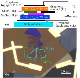
The sample we studied consists of a monolayer MoSe2 in direct contact with a bilayer CrBr3 encapsulated in hexagonal boron nitride (hBN) on a SiO2 substrate, as shown schematically in Fig. 1(a). In contrast to the layer-antiferromagnets CrI3 and CrCl3, the interlayer exchange in bilayer CrBr3 has been shown to be ferromagnetic [4]. Monolayer graphene gates and a graphene contact allow for independent tuning of the charge carrier density and out-of-plane electric field in the sample. In the optical micrograph in Fig. 1(b), the regions of bare MoSe2, bare CrBr3, and the overlapping region can be seen. Details on the sample fabrication, optical setup, and data analysis are given in the Supplemental Material [31]. All measurements were performed at approximately unless stated otherwise.
Normalized polarization-resolved reflection spectra of the bare MoSe2 and the MoSe2/CrBr3 heterostructure are shown in Fig. 2(a) and (b), for a choice of gate voltages that ensure charge neutrality of MoSe2. In the absence of an external magnetic field, the K and K’ valley excitons are degenerate in bare MoSe2 and the spectrum shows no polarization dependence. In contrast, a valley splitting of emerges in the MoSe2/CrBr3 heterostructure region, equivalent to an external magnetic field of , assuming the electronic g-factor to be . This splitting can be attributed to an exchange coupling between electronic states in MoSe2 and spin-polarized states in CrBr3 that leads to different energy shifts for the MoSe2 K and K’ valley excitons. Due to strain and disorder in the heterostructure, the splitting varies spatially by approximately .
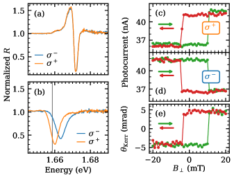
To demonstrate that the MoSe2 exciton valley splitting originates from the magnetization of CrBr3, we compare hysteresis measurements of the MoSe2 reflectance as a function of an external out-of-plane magnetic field to the established method of measuring CrBr3 magnetization along the easy axis using the magneto-optical Kerr effect (MOKE) [4]. Figures 2(c) and (d) show the reflectance of a right- and left-hand circularly polarized (/) laser ( CW) tuned to the low-frequency tail of the exciton resonance, as indicated by the vertical line in Fig. 2(b), as a function of . For this choice of laser detuning, the valley splitting gives rise to maximal contrast in magnetic circular dichroism (MCD). The MOKE depicted in Fig. 2(e) is measured on the same spot with a linearly polarized laser at (, CW). The one-to-one correspondence between the measurements confirms that the valley splitting is directly linked to the magnetization of CrBr3. We verified that the MOKE signal is not altered by the presence of MoSe2 by comparing measurements on the heterostructure and bare CrBr3 (see Supplemental Material [31]). Using resonant spectroscopy on MoSe2 instead of the MOKE to access the magnetization of CrBr3 is advantageous, since it allows us to perform the same measurement with a simpler technique and lower illumination power. Avoiding measurements requiring high laser intensity is particularly important for chromium trihalides where sizeable MOKE signals are only obtained using above-band-gap lasers that could cause heating. Moreover, identifying peak positions instead of measuring laser intensities after a polarizing beam splitter makes our spectroscopic method less sensitive to imperfections in the polarization selection than traditional techniques.
An exchange splitting of similar magnitude has been reported in the pioneering work on photoluminescence (PL) measurements of WSe2/CrI3 heterostructures [5, 20, 14]. We were not able to observe the splitting in PL measurements (see Supplemental Material [31]). Instead, we see broad emission lines with an integrated intensity that is smaller by a factor twenty compared to bare MoSe2, which suggests that tunneling to CrBr3 provides a fast non-radiative relaxation channel for conduction band electrons and excitons in MoSe2. Because the exchange coupling responsible for the splitting relies on second-order virtual tunnel coupling, the PL splitting is expected to be large where the tunnel coupling is large, leading to a short exciton lifetime. Since PL primarily originates from long-lived states, we would expect it to be dominated by low-oscillator-strength localized excitations in parts of the heterostructure where the tunnel coupling is small. Consequently, disorder-induced spatial variations of the tunnel coupling could lead to a PL signal that shows small or possibly vanishing exciton valley splitting. This is in contrast to resonant reflection/absorption measurements which probe extended states with high oscillator strength within the optical excitation spot.
To explore the nature of the exchange coupling between CrBr3 and MoSe2 we measure the gate voltage dependence of the reflection spectra in polarization, shown in Fig. 3(a) and (b). The voltages indicated on the vertical axis were applied to both top and bottom gate. The bare MoSe2 flake can be charged with electrons or holes, evidenced by the appearance of attractive polaron lines at both positive and negative gate voltages [32]. In the presence of CrBr3, only holes can be injected into MoSe2, consistent with the type-II band alignment schematically shown in Fig. 3(c) and predicted from ab initio calculations [33, 34]. Injected electrons accumulate in the lower-lying conduction band of CrBr3, leaving the MoSe2 undoped and leading to screening of the top gate (see Supplemental Material [31]).
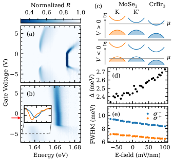
The attractive polaron line on the p-doped side exhibits the same valley splitting as the neutral exciton, as shown in the inset of Fig. 3(b). If the itinerant holes in MoSe2 were subject to a sizeable exchange interaction, we would observe a strong valley polarization of holes, leading to a single circularly polarized attractive polaron resonance [35, 36]. The observation of polaron resonances with equal strength for the two polarizations demonstrates that electron exchange is predominantly responsible for the exciton and polaron valley splitting. A detailed understanding of the underlying coupling mechanism is beyond the scope of this Letter and requires additional theoretical work.
The presence of top and bottom gates allows us to probe the electric field dependence of the reflectance for constant chemical potential. In the absence of mobile charges in the heterostructure, the electric field is approximately given by and the chemical potential by , where and are the the thicknesses of the top and bottom hBN flakes; the actual electric field may deviate by a constant factor due to the dielectric constants and finite thickness of MoSe2 and CrBr3 (see Supplemental Material [31]). To keep the chemical potential constant while varying the electric field, we tune the gate voltages with a fixed ratio , where determines the chemical potential; we determined this ratio experimentally from 2D gate sweeps (see Supplemental Material [31]).
Figures 3(d) and (e) show the valley splitting and the reflection peak widths, respectively, for the neutral exciton as a function of the applied electric field. The choice of gate voltages, indicated by the red arrow in Fig. 3(b), ensures the charge neutrality of the heterostructure. Clear dependence of both the splitting and the linewidth on the electric field suggests that the tunnel coupling strength is modified. Such an approximately linear electric field dependence of the splitting was predicted in theoretical works [33, 34] and may have implications for future gate-tunable spintronic devices. Additionally, the higher-energy exciton line (here ) is consistently broader than the lower-energy line, presumably due to the spin-dependent charge transfer between MoSe2 and CrBr3; similar observations were previously reported in heterostructures composed of different 2D magnetic layers [20, 14].
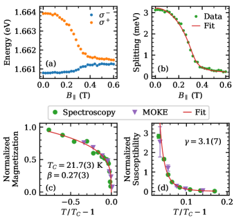
Having demonstrated the magnetic proximity effect in MoSe2, we use resonant spectroscopy of the MoSe2 exciton resonance to determine the magnetic properties of CrBr3. Figure 4(a) shows fitted positions of the split exciton peaks as function of an applied in-plane magnetic field . We observe that for , the splitting gradually decreases and saturates at a value of for . The reduction of the splitting is a consequence of the canting of the CrBr3 spins into the plane. The small remaining splitting at high magnetic fields is due to a tilt of the magnetic field axis with respect to the sample plane that leads to an out-of-plane component of the magnetization and consequently a non-zero exchange field. A striking feature of the data in Fig. 4(a) is the asymmetry in the -induced change in resonance energy between the low- and high-energy exciton peaks: we speculate that this asymmetry could arise from an energy splitting between the spin-polarized conduction bands of CrBr3 that play a prominent role in determining the exchange coupling to MoSe2 electrons in different valleys.
The observed dependence of the splitting vs. allows us to estimate the anisotropy energy of CrBr3. To this end, we assume that the CrBr3 flake has a uniform magnetization and numerically minimize its potential energy , where is the anisotropy energy along the easy axis, is the angle of the magnetic moment with respect to the easy axis, is the external magnetic field, and is the angle of with respect to the easy axis. We set to the magnetization per Cr atom [37, 38] and fit the model to the experimental data to obtain an anisotropy energy per Cr atom and . Although this simple model does not tell us anything about the type of anisotropy, previous calculations have shown that it is expected to originate from anisotropic exchange coupling rather than on-site anisotropy [39]. By using the previously reported values of the isotropic intralayer exchange of bulk CrBr3 () [40, 41], we find that the exchange interaction is weakly anisotropic, . The small coercive field we measure (Fig. 2(c)–(e)) is consistent with the weak anisotropy of the intralayer exchange interaction. Remarkably, the exciton valley splitting in MoSe2 is comparable to the CrBr3 intralayer exchange , even though the exchange between two CrBr3 layers is expected to be significantly smaller [40, 41, 42].
Next, we measure the critical temperature and critical exponents of the second-order magnetic phase transition of CrBr3 through resonant MoSe2 exciton spectroscopy. To this end, we measure the hysteresis curves of circularly polarized reflection spectra vs. as a function of temperature (see Supplemental Material [31]). For , shown in Fig. 4(c), the splitting at provides a measure for the remnant magnetization , which is the order parameter for the phase transition. For , shown in Fig. 4(d), the slope of the splitting vs. is proportional to the magnetic susceptibility . By fitting the functional forms and simultaneously to the experimental data, we find the Curie temperature as well as the critical exponents and . We also perform similar measurements using the MOKE and find that the data points fall onto the same curves when normalized with respect to the peak values of and . The values we obtain for and are consistent with the 2D-Heisenberg model with weak anisotropy [43].
In conclusion, we use resonant exciton reflection measurements of valley splitting in MoSe2 to demonstrate a strong magnetic proximity effect due to ferromagnetic CrBr3. From the absence of itinerant hole valley polarization in the reflection spectra we infer that the resulting valley Zeeman effect is predominantly due to exchange coupling between conduction band electrons in MoSe2 and CrBr3: remarkably, the strength of this interlayer exchange coupling is comparable to the intralayer exchange coupling in CrBr3. Our investigation of the magnetic properties of CrBr3 using resonant optical spectroscopy reveals several features such as an electric field dependence of the proximity effect, weak anisotropy of the exchange interaction, and the critical exponents associated with the magnetic phase transition.
The data that supports the findings of this Letter is available in the ETH Research Collection [44].
I Acknowledgments
We gratefully acknowledge the contributions of Alberto Morpurgo and Zhe Wang who introduced us to the field of 2D magnets. Y. Shimazaki and A. Popert contributed to the fabrication of the heterostructure. We also express our gratitutde to A. Vindigni and P. Maletinsky for insightful discussions. This work was supported by the Swiss National Science Foundation (SNSF) under Grant No. 200021-178909/1. K. W. and T. T. acknowledge support from the Elemental Strategy Initiative conducted by the MEXT, Japan, and the CREST (JPMJCR15F3), JST.
References
- Gong and Zhang [2019] C. Gong and X. Zhang, Science 363, eaav4450 (2019).
- Cortie et al. [2019] D. L. Cortie, G. L. Causer, K. C. Rule, H. Fritzsche, W. Kreuzpaintner, and F. Klose, Advanced Functional Materials , 1901414 (2019).
- Huang et al. [2017] B. Huang, G. Clark, E. Navarro-Moratalla, D. R. Klein, R. Cheng, K. L. Seyler, D. Zhong, E. Schmidgall, M. A. McGuire, D. H. Cobden, W. Yao, D. Xiao, P. Jarillo-Herrero, and X. Xu, Nature 546, 270 (2017).
- Zhang et al. [2019] Z. Zhang, J. Shang, C. Jiang, A. Rasmita, W. Gao, and T. Yu, Nano Letters 19, 3138 (2019).
- Zhong et al. [2017] D. Zhong, K. L. Seyler, X. Linpeng, R. Cheng, N. Sivadas, B. Huang, E. Schmidgall, T. Taniguchi, K. Watanabe, M. A. McGuire, W. Yao, D. Xiao, K.-M. C. Fu, and X. Xu, Science Advances 3, e1603113 (2017).
- Chen et al. [2018] L. Chen, J.-H. Chung, B. Gao, T. Chen, M. B. Stone, A. I. Kolesnikov, Q. Huang, and P. Dai, Physical Review X 8, 041028 (2018).
- Jin et al. [2018] W. Jin, H. H. Kim, Z. Ye, S. Li, P. Rezaie, F. Diaz, S. Siddiq, E. Wauer, B. Yang, C. Li, S. Tian, K. Sun, H. Lei, A. W. Tsen, L. Zhao, and R. He, Nature Communications 9, 5122 (2018).
- Thiel et al. [2019] L. Thiel, Z. Wang, M. A. Tschudin, D. Rohner, I. Gutiérrez-Lezama, N. Ubrig, M. Gibertini, E. Giannini, A. F. Morpurgo, and P. Maletinsky, Science 364, 973 (2019).
- Jin et al. [2019] C. Jin, Z. Tao, K. Kang, K. Watanabe, T. Taniguchi, K. F. Mak, and J. Shan, (2019), arXiv:1910.13023 .
- Song et al. [2019] T. Song, Z. Fei, M. Yankowitz, Z. Lin, Q. Jiang, K. Hwangbo, Q. Zhang, B. Sun, T. Taniguchi, K. Watanabe, M. A. McGuire, D. Graf, T. Cao, J.-H. Chu, D. H. Cobden, C. R. Dean, D. Xiao, and X. Xu, Nature Materials 18, 1298 (2019).
- Cai et al. [2019] X. Cai, T. Song, N. P. Wilson, G. Clark, M. He, X. Zhang, T. Taniguchi, K. Watanabe, W. Yao, D. Xiao, M. A. McGuire, D. H. Cobden, and X. Xu, Nano Letters 19, 3993 (2019).
- Chen et al. [2019] W. Chen, Z. Sun, Z. Wang, L. Gu, X. Xu, S. Wu, and C. Gao, Science 366, 983 (2019).
- Sun et al. [2019] Z. Sun, Y. Yi, T. Song, G. Clark, B. Huang, Y. Shan, S. Wu, D. Huang, C. Gao, Z. Chen, M. McGuire, T. Cao, D. Xiao, W.-T. Liu, W. Yao, X. Xu, and S. Wu, Nature 572, 497 (2019).
- Zhong et al. [2020] D. Zhong, K. L. Seyler, X. Linpeng, N. P. Wilson, T. Taniguchi, K. Watanabe, M. A. McGuire, K.-M. C. Fu, D. Xiao, W. Yao, and X. Xu, Nature Nanotechnology 10.1038/s41565-019-0629-1 (2020).
- Wang et al. [2018] Z. Wang, I. Gutiérrez-Lezama, N. Ubrig, M. Kroner, M. Gibertini, T. Taniguchi, K. Watanabe, A. Imamoğlu, E. Giannini, and A. F. Morpurgo, Nature Communications 9, 2516 (2018).
- Song et al. [2018] T. Song, X. Cai, M. W.-Y. Tu, X. Zhang, B. Huang, N. P. Wilson, K. L. Seyler, L. Zhu, T. Taniguchi, K. Watanabe, M. A. McGuire, D. H. Cobden, D. Xiao, W. Yao, and X. Xu, Science 360, 1214 (2018).
- Ghazaryan et al. [2018] D. Ghazaryan, M. T. Greenaway, Z. Wang, V. H. Guarochico-Moreira, I. J. Vera-Marun, J. Yin, Y. Liao, S. V. Morozov, O. Kristanovski, A. I. Lichtenstein, M. I. Katsnelson, F. Withers, A. Mishchenko, L. Eaves, A. K. Geim, K. S. Novoselov, and A. Misra, Nature Electronics 1, 344 (2018).
- Klein et al. [2018] D. R. Klein, D. MacNeill, J. L. Lado, D. Soriano, E. Navarro-Moratalla, K. Watanabe, T. Taniguchi, S. Manni, P. Canfield, J. Fernández-Rossier, and P. Jarillo-Herrero, Science 10.1126/science.aar3617 (2018).
- Jiang et al. [2018a] S. Jiang, L. Li, Z. Wang, K. F. Mak, and J. Shan, Nature Nanotechnology 13, 549 (2018a).
- Seyler et al. [2018] K. L. Seyler, D. Zhong, B. Huang, X. Linpeng, N. P. Wilson, T. Taniguchi, K. Watanabe, W. Yao, D. Xiao, M. A. McGuire, K.-M. C. Fu, and X. Xu, Nano Letters 18, 3823 (2018).
- Huang et al. [2018] B. Huang, G. Clark, D. R. Klein, D. MacNeill, E. Navarro-Moratalla, K. L. Seyler, N. Wilson, M. A. McGuire, D. H. Cobden, D. Xiao, W. Yao, P. Jarillo-Herrero, and X. Xu, Nature Nanotechnology 13, 544 (2018).
- Jiang et al. [2018b] S. Jiang, J. Shan, and K. F. Mak, Nature Materials 17, 406 (2018b).
- Farooq and Hong [2019] M. U. Farooq and J. Hong, npj 2D Materials and Applications 3, 3 (2019).
- Xu et al. [2014] X. Xu, W. Yao, D. Xiao, and T. F. Heinz, Nature Physics 10, 343 (2014).
- Mak et al. [2018] K. F. Mak, D. Xiao, and J. Shan, Nature Photonics 12, 451 (2018).
- Onga et al. [2017] M. Onga, Y. Zhang, T. Ideue, and Y. Iwasa, Nature Materials 16, 1193 (2017).
- Mak et al. [2010] K. F. Mak, C. Lee, J. Hone, J. Shan, and T. F. Heinz, Physical Review Letters 105, 136805 (2010).
- Manzeli et al. [2017] S. Manzeli, D. Ovchinnikov, D. Pasquier, O. V. Yazyev, and A. Kis, Nature Reviews Materials 2, 17033 (2017).
- Deng et al. [2018] Y. Deng, Y. Yu, Y. Song, J. Zhang, N. Z. Wang, Z. Sun, Y. Yi, Y. Z. Wu, S. Wu, J. Zhu, J. Wang, X. H. Chen, and Y. Zhang, Nature 563, 94 (2018).
- Geim and Grigorieva [2013] A. K. Geim and I. V. Grigorieva, Nature 499, 419 (2013).
- [31] See Supplemental Material at URL for information on the sample fabrication, experimental details, data analysis, and extended data on gate dependence, photoluminescence, and optical doping.
- Sidler et al. [2016] M. Sidler, P. Back, O. Cotlet, A. Srivastava, T. Fink, M. Kroner, E. Demler, and A. Imamoglu, Nature Physics 13, 255 (2016).
- Zollner et al. [2019] K. Zollner, P. E. Faria Junior, and J. Fabian, Physical Review B 100, 085128 (2019).
- Xie et al. [2018] J. Xie, L. Jia, H. Shi, D. Yang, and M. Si, Japanese Journal of Applied Physics 58, 010906 (2018).
- Back et al. [2017] P. Back, M. Sidler, O. Cotlet, A. Srivastava, N. Takemura, M. Kroner, and A. Imamoglu, Phys. Rev. Lett. 118, 237404 (2017).
- Smoleński et al. [2019] T. Smoleński, O. Cotlet, A. Popert, P. Back, Y. Shimazaki, P. Knüppel, N. Dietler, T. Taniguchi, K. Watanabe, M. Kroner, and A. Imamoglu, Physical Review Letters 123, 097403 (2019).
- Kotani [1949] M. Kotani, Journal of the Physical Society of Japan 4, 293 (1949).
- Dillon [1962] J. F. Dillon, in Proceedings of the Seventh Conference on Magnetism and Magnetic Materials, edited by J. A. Osborn (Springer US, Boston, MA, 1962) pp. 1191–1192.
- Lado and Fernández-Rossier [2017] J. L. Lado and J. Fernández-Rossier, 2D Materials 4, 035002 (2017).
- Davis and Narath [1964] H. L. Davis and A. Narath, Physical Review 134, A433 (1964).
- Samuelsen et al. [1971] E. J. Samuelsen, R. Silberglitt, G. Shirane, and J. P. Remeika, Physical Review B 3, 157 (1971).
- de Jongh and Miedema [1974] L. de Jongh and A. Miedema, Advances in Physics 23, 1 (1974).
- Fisher [1974] M. E. Fisher, Reviews of Modern Physics 46, 597 (1974).
- [44] See https://doi.org/10.3929/ethz-b-000397581.
- Kopp and Ashworth [1972] F. J. Kopp and T. Ashworth, Review of Scientific Instruments 43, 327 (1972).
- Wang et al. [2013] L. Wang, I. Meric, P. Y. Huang, Q. Gao, Y. Gao, H. Tran, T. Taniguchi, K. Watanabe, L. M. Campos, D. A. Muller, J. Guo, P. Kim, J. Hone, K. L. Shepard, and C. R. Dean, Science 342, 614 (2013).