Modeling supercoiled DNA interacting with an anchored cluster of proteins:
towards a quantitative estimation of chromosomal DNA supercoiling
Abstract
We investigate the measurement of DNA supercoiling density () along chromosomes using interaction frequencies between DNA and DNA-anchored clusters of proteins. Specifically, we show how the physics of DNA supercoiling leads, in bacteria, to the quantitative modeling of binding properties of ParB proteins around their centromere-like site, parS. Using this framework, we provide an upper bound for in the Escherichia coli chromosome, consistent with plasmid values, and offer a proof of concept for a high accuracy measurement. To reach these conclusions, we revisit the problem of the formation of ParB clusters. We predict, in particular, that they result from a non-equilibrium, stationary balance between an influx of produced proteins and an outflux of excess proteins, i.e., they behave like liquid-like protein condensates with unconventional “leaky” boundaries.
In most bacteria, DNA is underwound. Despite its critical role for genome structuring Vologodskii and Cozzarelli (1994) and coordination of gene expression Dorman and Dorman (2016), measurement of the negative supercoiling along chromosomes remains highly challenging, with both biological and physical difficulties. Biological difficulties stem from the complex functioning of cells. For instance, a large part of supercoiling is known to be absorbed by various histone-like proteins Travers and Muskhelishvili (2005). The remaining supercoiling, which is responsible for the formation of branched plectonemic structures Vologodskii et al. (1992), is usually referred to as ”free” or ”effective” Bliska and Cozzarelli (1987). Physical difficulties are inherent in the dual nature of supercoiling. That is, in the absence of topoisomerases, a topologically constrained DNA molecule (such as circular molecules or constrained linear domains Liu and Wang (1987)) is characterized by a constant linking number, Lk, equal to the sum of the twist (Tw), the cumulative helicity of the molecule, and the writhe (Wr), the global intricacy of the molecule White (1969). As a consequence, supercoiling, i.e. the change of Lk with respect to , the value at rest, leads to changes in the mean values of both Tw and Wr. Having access to only Tw, when using e.g. DNA intercaling agents, is thus a priori insufficient to fully characterize the topological status associated with chromosomal loci Lal et al. (2016). This explains why supercoiling density, , has been estimated quantitatively using plasmid reporters only, since their compaction level can be quantitatively assessed in vitro – see Sinden et al. (1980) for an exception, although the chromosomal measurement is global, not local. Note, in this regard, that a genetic recombination-based system sensitive to the tightness of plectonemes has been developed to probe supercoiling density along the chromosome Booker et al. (2010); Rovinskiy et al. (2019). The quantitative estimation of in vivo still remains problematic, because the method can only be calibrated in vitro Booker et al. (2010).
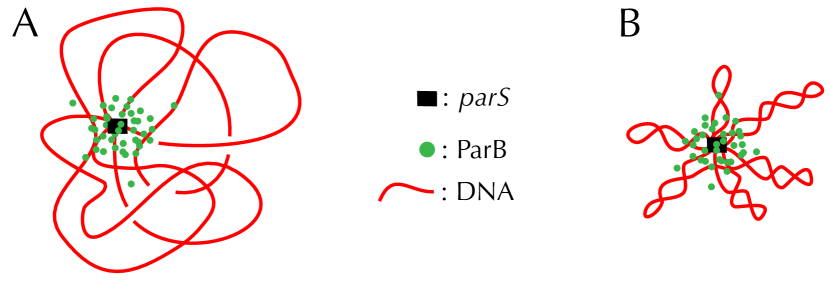
Here, we investigate the possibility of measuring the effective chromosomal supercoiling density using DNA binding properties of the centromere-binding protein ParB, part of the active ParABS system of DNA segregation. Specifically, it has been argued that the capture by chromatin immuno-precipitation sequencing (ChIP-seq) of the binding of ParB onto DNA in the vicinity of its specific binding site (parS) is driven by stochastic binding involving DNA looping properties Sanchez et al. (2015) (Fig. 1). More precisely, ParB proteins cluster around parS Sanchez et al. (2015); Debaugny et al. (2018) through a phase separation-like mechanism David et al. (2018). In this context, it has been shown that only a process of looping, which brings DNA loci inside the cluster, can explain the long range decay of the ParB binding profile as the genomic distance to parS increases (black curves in Fig. 3) Sanchez et al. (2015); Walter et al. (2018). Knowing that supercoiling properties strongly influence DNA looping properties, we thus assess whether a quantitative reproduction of the ParB binding profile in the vicinity of parS is possible using a model with no other free parameter than DNA supercoiling density (). To this end, we consider, on one hand, a realistic model of supercoiled DNA that has been independently calibrated using single-molecule techniques and, on the other hand, an independent estimation of the size of the ParB cluster using high-resolution microscopic experiments.
Compared to the previous stochastic binding model where a very small DNA persistence length (), difficult to justify on physical grounds, was required to match experimental data Sanchez et al. (2015), here we show, using numerical simulations of realistic long (i.e. ) molecules, that DNA supercoiling indeed leads to a quantitative reproduction of ChIP-seq ParB binding profiles. In this context, we provide a bound for the chromosomal supercoiling density, propose new experimental protocols to further fix its exact values and demonstrate, for the first time to our knowledge, the consistency between chromosomal and plasmid measurements. In addition, we provide novel insights into the physical properties of ParB clusters. In particular, we predict a cluster shape that differs from the usual sharp boundaries of liquid droplets. Namely, we show that the cluster density profile display unconventional “leaky” boundaries, which can be explained as a perturbation induced by a source of proteins located at the edge of the cluster core. Altogether, our work thus offers insights into both bacterial DNA organization and liquid-like protein condensates. It also offers a proof of concept for measuring chromosomal supercoiling with high accuracy.
Stochastic Binding model.
ChIP-seq detection of DNA-bound proteins involves sub-nm crosslinking between DNA and proteins Hoffman et al. (2015). Thus we expect that the non-specific ParB binding profile results from ”collisions” between DNA and the ParB proteins located in the parS-anchored cluster (Fig. 1). We therefore suppose that, except at parS, the timescale for ParB to unbind DNA is much shorter than the timescale for DNA to diffuse away from the location where binding occurs (instantaneous unbinding hypothesis). The modeled non-specific ParB binding profile, , thus reads Sanchez et al. (2015):
| (1) |
describes DNA looping properties: it stands for the equilibrium probability distribution function for a DNA locus at a genomic distance from parS to be located at a distance from parS in the three-dimensional space. For simplicity, here we neglect effects coming from the interaction between DNA and the cluster, therefore is computed by considering an isolated DNA chain.
stands for the probability to find a ParB protein at distance from parS. Although its exact shape is not known (see below for predictions), we have by definition of the strong binding of ParB to parS. Next, the full width at half maximum of the cluster, , has been estimated using high-resolution fluorescent microscopy, leading to Guilhas et al. (2020). In this experiment, cell contents were chemically fixed so that refers to the probability to find a ParB protein at a distance from the cluster center, not from parS, with . Considering the positional degrees of freedom of the cluster center with respect to parS, we then have , where stands for the probability density of finding the cluster center at a distance given a point at distance from parS (Supp. Info.).
Self-avoiding rod-like chain model of DNA.
We consider a realistic resolution polymer model of bacterial DNA, namely the self-avoiding rod-like chain (sRLC) model Vologodskii et al. (1992) (detailed simulation procedure in Lepage and Junier (2017)). Specifically, DNA is modeled as a discrete chain of long ( of B-DNA) articulated hard-core cylinders, with radius reflecting the short-range electrostatic repulsions of DNA for in vivo salt conditions Lepage et al. (2015). The chain is iteratively deformed using crankshaft elementary motions with Metropolis-Hastings transition rates, under the condition that it does not cross itself. Each articulating site is associated with bending and torsional energies such that the resulting persistence length ( or, equivalently, ) and torsional length () are typical of B-DNA for in vivo salt conditions Vologodskii et al. (1992); Strick et al. (2003); Lepage et al. (2015).
Here, we discuss results obtained with a long chain by varying from to slowly enough so that chain statistical properties are insensitive to the associated rate of change (see Fig. S1 and simulation details in Supp. Info.). Simulated conformations are thus expected to reflect thermodynamic equilibrium, even at low values of where plectonemes are tight. We further checked that our results did not depend significantly on the length of the chain by performing additional simulations of long chains (Fig. S2). Note, here, that the motivation to work with is both biological and physical: in the worst case of topoisomerase mutants, the total supercoiling density in E. coli has been shown to remain above Bliska and Cozzarelli (1987), while recent work has revealed the existence of a transition toward a hyperbranched regime occurring at Krajina and Spakowitz (2016), which is beyond the scope of our discussion.
Leaky vs quenched cluster.
Having in hand the corresponding for , we now consider the spatial distribution of ParB proteins associated with the parS-anchored clusters. In this regard, high-resolution microscopic measurements Guilhas et al. (2020) suggest that these clusters result from a phase transition-like mechanism. Theoretical models further suggest that this phase transition is unconventional as it implies a framework of a lattice gas on a fluctuating polymer David et al. (2018). Moreover, the physical formation of a cluster is likely to interfere with biological processes like, e.g., the production of ParB close to the cluster, just as membrane proteins are often produced close to the membrane Libby et al. (2012). In other words, the spatial distribution of ParB proteins around parS remains an open question.
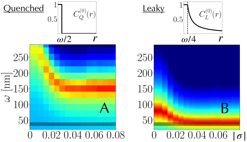
Here, we investigate two extreme cases for the shape of these clusters, referred to as quenched and leaky. A quenched cluster (Fig. 2A) is defined by , with the Heaviside function. It corresponds to the conventional sharp interface of a droplet. A leaky cluster (Fig. 2B) further includes the stationary solution of a diffusion process where ParB proteins are continuously produced at the edge of the cluster core and diluted due to cell growth and division (Supp. Info.). That is, the leaky cluster releases proteins in excess, while for (cluster core) reflects the saturation regime in which experiments are performed Debaugny et al. (2018). As a result, includes a long range decay such that . Note that for both quenched and leaky cases, the full width at half maximum of is equal to .
We computed binding profiles for ranging in and for values of between and . We compared them with profiles obtained for E. coli by inserting parS along the chromosome (black curve in Fig. S3) – only one side of the chromosome is analyzed as the other side is distorted by the presence of strong promoter regions Debaugny et al. (2018). In this experiment, parS sites interspersed by base pairs, as found in the natural parS region, were inserted at xylE locus Debaugny et al. (2018). A careful analysis of the binding properties among these parS sites actually revealed significant variations of the ChIP-seq signal, which was thus normalized with respect to the maximum value. The origin of the curvilinear abscissa was set right at the edge of the most extreme parS site.
We are interested in explaining the global shape of the binding profile as it is expected to reflect generic polymer physics principles. To that end, we quantify the explanatory power of each model by reporting the root mean square deviation with respect to the experimental binding profile for . Both the lower and upper bounds at and , respectively, are used to avoid specific, reproducible distortions of the signal associated with the presence of gene promoters and sites for regulatory DNA proteins Debaugny et al. (2018).
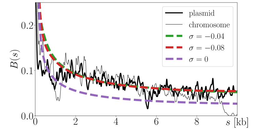
We find that both quenched and leaky clusters can accurately capture experimental data (Fig. S3). However, the best quenched models are found at (Fig. 2A), which is much larger than . In contrast, the best leaky models are found at when (Fig. 2B). That is, they explain data in the physiologically relevant plectonemic regime of bacterial DNA. They also solve the small DNA persistence length issue associated with the previous version of the stochastic binding model where DNA supercoiling was neglected Sanchez et al. (2015) – a small persistence length was indeed needed to “mimic” compaction due to plectonemes. Interestingly, compared to chromosomal parS data, ParB binding profiles in the vicinity of a parS located on a plasmid ( long F-plasmid Debaugny et al. (2018)) show less distortion (Fig. 3) – just as for the chromosome, only one side of the plasmid is analyzed as the other side is distorted by binding sites for a replication initiator Sanchez et al. (2015). In this context, the best leaky models lead to similar model parameters ( when ), while providing an even better match with the data (Fig. 3). Compared to the chromosomal situation where the gene parB is located away from parS, this better match might reflect a phenomenology of the plasmid fitting particularly well the leaky situation, with parB located only away from parS Bouet et al. (2006). The hypothesis of a source located on the edge of the cluster core is indeed even more relevant since the production of proteins in bacteria often occurs close to their gene Llopis et al. (2010).
-sensitive probes for strong supercoiling.
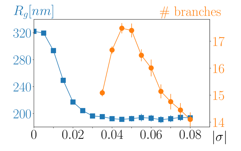
While leaky models with experimentally relevant capture experimental data rather well, resulting binding profiles are almost indistinguishable for (Fig. 3). This lack of sensitivity is concomitant with a poor variation of the radius of gyration (blue curve in Fig. 4) in the plectonemic regime. Note that, in contrast, branching properties can vary significantly in this regime Vologodskii and Cozzarelli (1994); Krajina and Spakowitz (2016). For instance, we find that the number of plectonemic branches reaches a maximum at (orange curve in Fig. 4), in accord with previous analyses with smaller molecules Vologodskii and Cozzarelli (1994) and with a minimum value of the hydrodynamic radius for long plasmids Wang (1974); Vologodskii and Cozzarelli (1994); Krajina and Spakowitz (2016).
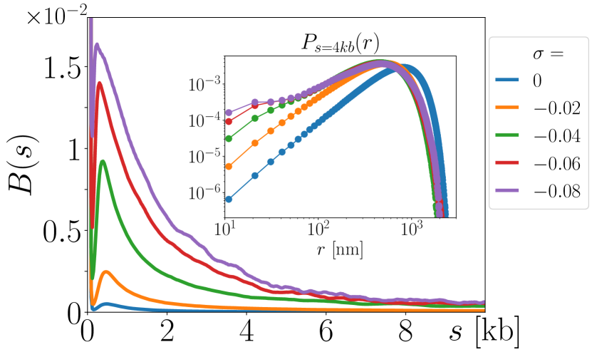
A natural question, then, is whether it is possible to build a probe that is sensitive to variations of for strong supercoiling. Interestingly, we have found a possible solution consisting of a system that senses intertwining properties of plectonemes, in the spirit of the recombination system Booker et al. (2010). In that respect, one would need a quenched (instead of a leaky) cluster that is small enough such that the binding properties of proteins is sensitive to the diameter and pitch of plectonemes Marko and Siggia (1995); Barde et al. (2018). For instance, our simulations reveal a strong sensitivity of , at the kb genomic scale for , with respect to all values of for spatial distances on the order of (inset of Fig. 5). One can verify, then, that a quenched cluster with provides well-distinct binding profiles for (Fig. 5). Notice the much smaller values of in this case, compared e.g. to results in Fig. 3. ParB ChIP-seq experiments can nevertheless report very low binding frequencies as demonstrated by titration assays Debaugny et al. (2018).
Discussion and perspectives.
We have shown that the binding profile of ParB proteins in the vicinity of parS can be quantitatively explained considering a stochastic binding process between a supercoiled DNA and proteins that are issued from a saturated parS-anchored core cluster. To this end, we had to consider clusters from a non-equilibrium, stationary perspective, with the presence of a spatially localized source and sink. Biologically, the sink reflects protein dilution due to cell growth and division, while the source may arise from two effects: the continuous activity of genes producing new proteins in a saturated cluster and the effect of an unconventional liquid-like nature of the cluster. Namely, we predict the cluster core to result from a balance between an influx of continuously produced proteins and an outflux of proteins in excess. In the plasmid, the situation may even be more prototypical with the production of ParB occurring close to parS.
In this context, and for the first time to the best of our knowledge, we provide an upper bound () for the in vivo supercoiling density at a chromosomal location of a bacterium (E. coli during its exponential growth) and we show that it also holds for plasmids. Interestingly, this value corresponds to the onset of the plectonemic regime characterized by a poor variation of the radius of gyration, on one hand, and a significant variation of branching properties, on the other hand. Importantly, we also offer a proof of concept to obtain a finer estimate of the supercoiling density. Specifically, in the spirit of existing genetic recombination-based probes Booker et al. (2010); Rovinskiy et al. (2019), we demonstrate that a small quenched cluster provides a supercoiling-sensitive probe as it ”senses” physical properties of plectonemes.
Compared to ”biological” genetic recombination-based probes, our ”physical” ChIP-seq-based probe is expected to be much less invasive. It should also be less sensitive to molecular environment as it is based on generic (polymer) physics properties – for instance, recombination-based systems depend on (slow) enzymatic recombinase reactions, whose quantitative modeling has, to the best of our knowledge, remained elusive. In practice, while genetic design of quenched clusters of ParB proteins might be tricky, transcription factors could provide an efficient system. These proteins have indeed the capacity of binding both cognate DNA sites strongly and other DNA sites non-specifically with (short) millisecond residence times Elf et al. (2007). They could also be used in conjunction with a DNA methyltransferase to generate methylation (instead of binding) profiles without the need of crosslinking stages Redolfi et al. . Finally, a sensitive system would require having the designed artificial DNA devoid as much as possible of interfering biological elements, such as gene promoters, which distort the utilizable physical signal. Along this line, one would like to have an explicit description of ParB nucleation and diffusion properties to develop a detailed model of the interactions between ParB and DNA using e.g. molecular dynamics approaches. In particular, the discrepancy between experimental and modeling profiles below (Fig. 3) might be the result of our approximation of neglecting hard-core interactions between ParB proteins and DNA. At large scales, cellular confinement of DNA should also be included in the model. We note, nevertheless, that a complete picture would require studying the melting of a plectonemic tree-like structure at the chromosome scale, which is currently beyond the capacities of numerical simulations.
Acknowledgements.
We thank Daniel Jost for useful suggestions. JCW was supported by a “Modélisation pour le Vivant” CNRS Grant (CoilChrom). J.Y.B. is supported by an AO80Prime (Numacoiled) CNRS grant. I.J. was supported by an ATIP-Avenir grant (Centre National de la Recherche Scientifique).References
- Vologodskii and Cozzarelli (1994) A. Vologodskii and N. Cozzarelli, Annual Review of Biophysics and Biomolecular Structure 23, 609 (1994).
- Dorman and Dorman (2016) C. J. Dorman and M. J. Dorman, Biophysical reviews 8, 89 (2016).
- Travers and Muskhelishvili (2005) A. Travers and G. Muskhelishvili, Current Opinion in Genetics & Development 15, 507 (2005).
- Vologodskii et al. (1992) A. V. Vologodskii, S. D. Levene, K. V. Klenin, M. Frank-Kamenetskii, and N. R. Cozzarelli, Journal of Molecular Biology 227, 1224 (1992).
- Bliska and Cozzarelli (1987) J. B. Bliska and N. R. Cozzarelli, Journal of molecular biology 194, 205 (1987).
- Liu and Wang (1987) L. F. Liu and J. C. Wang, PNAS 84, 7024 (1987).
- White (1969) J. H. White, American Journal of Mathematics 91, 693 (1969).
- Lal et al. (2016) A. Lal, A. Dhar, A. Trostel, F. Kouzine, A. S. N. Seshasayee, and S. Adhya, Nature communications 7, 11055 (2016).
- Sinden et al. (1980) R. R. Sinden, J. O. Carlson, and D. E. Pettijohn, Cell 21, 773 (1980).
- Booker et al. (2010) B. M. Booker, S. Deng, and N. P. Higgins, Molecular Microbiology 78, 1348 (2010).
- Rovinskiy et al. (2019) N. S. Rovinskiy, A. A. Agbleke, O. N. Chesnokova, and N. P. Higgins, Microorganisms 7 (2019).
- Sanchez et al. (2015) A. Sanchez, D. I. Cattoni, J.-C. Walter, J. Rech, A. Parmeggiani, M. Nollmann, and J.-Y. Bouet, Cell Systems 1, 163 (2015).
- Debaugny et al. (2018) R. E. Debaugny, A. Sanchez, J. Rech, D. Labourdette, J. Dorignac, F. Geniet, J. Palmeri, A. Parmeggiani, F. Boudsocq, V. Anton Leberre, J.-C. Walter, and J.-Y. Bouet, Molecular Systems Biology 14, e8516 (2018).
- David et al. (2018) G. David, J. C. Walter, C. P. Broedersz, J. Dorignac, F. Geniet, A. Parmeggiani, N. O. Walliser, and J. Palmeri, arXiv.org , arXiv:1811.09234 (2018).
- Walter et al. (2018) J.-C. Walter, N.-O. Walliser, G. David, J. Dorignac, F. Geniet, J. Palmeri, A. Parmeggiani, N. S. Wingreen, and C. P. Broedersz, New Journal of Physics 20, 035002 (2018).
- Hoffman et al. (2015) E. A. Hoffman, B. L. Frey, L. M. Smith, and D. T. Auble, The Journal of biological chemistry 290, 26404 (2015).
- Guilhas et al. (2020) B. Guilhas, J. C. Walter, J. Rech, G. David, N. O. Walliser, J. Palmeri, C. Mathieu-Demaziere, A. Parmeggiani, J. Y. Bouet, A. L. Gall, and M. Nöllmann, bioRxiv (2020), http://dx.doi.org/10.1101/791368.
- Lepage and Junier (2017) T. Lepage and I. Junier, Methods in molecular biology (Clifton, N.J.) 1624, 323 (2017).
- Lepage et al. (2015) T. Lepage, F. Képès, and I. Junier, Biophysical Journal 109, 135 (2015).
- Strick et al. (2003) T. R. Strick, M.-N. Dessinges, G. Charvin, N. H. Dekker, J.-F. Allemand, D. Bensimon, and V. Croquette, Reports on Progress in Physics 66 (2003).
- Krajina and Spakowitz (2016) B. A. Krajina and A. J. Spakowitz, Biophysical Journal 111, 1339 (2016).
- Libby et al. (2012) E. A. Libby, M. Roggiani, and M. Goulian, Proceedings of the National Academy of Sciences 109, 7445 (2012).
- Bouet et al. (2006) J.-Y. Bouet, M. Bouvier, and D. Lane, Molecular Microbiology 62, 1447 (2006).
- Llopis et al. (2010) P. M. Llopis, A. F. Jackson, O. Sliusarenko, I. Surovtsev, J. Heinritz, T. Emonet, and C. Jacobs-Wagner, Nature 466, 77 (2010).
- Wang (1974) J. C. Wang, Journal of molecular biology 87, 797 (1974).
- Marko and Siggia (1995) J. Marko and E. Siggia, Physical Review E 52, 2912 (1995).
- Barde et al. (2018) C. Barde, N. Destainville, and M. Manghi, Physical Review E 97, 032412 (2018).
- Elf et al. (2007) J. Elf, G. Li, and X. S. Xie, Science 316, 1191 (2007).
- (29) J. Redolfi, Y. Zhan, C. Valdes, M. Kryzhanovska, I. M. Guerreiro, V. Iesmantavicius, G. Tiana, T. Pollex, J. Kind, S. Smallwood, W. d. Laat, and L. Giorgetti, bioRxiv .