Feeding the zombies: Synthesizing brain volumes
using a 3D progressive growing GAN
Abstract
Deep learning requires large datasets for training (convolutional) networks with millions of parameters. In neuroimaging, there are few open datasets with more than 100 subjects, which makes it difficult to, for example, train a classifier to discriminate controls from diseased persons. Generative adversarial networks (GANs) can be used to synthesize data, but virtually all research is focused on 2D images. In medical imaging, and especially in neuroimaging, most datasets are 3D or 4D. Here we therefore present preliminary results showing that a 3D progressive growing GAN can be used to synthesize MR brain volumes.
1 Introduction
Generative adversarial networks (GANs) [1] can today produce very realistic synthetic images [2]. GANs use adversarial training, where a generator creates an image from noise and a discriminator classifies each image as synthetic or real. During the training, the generator becomes better at generating realistic images, and the discriminator becomes better at discriminating images as synthetic or real. GANs can broadly be divided into noise-to-image GANs [1, 2], which produce an image from a noise vector, and image-to-image GANs, which produce an image from another image (image to image translation) [3, 4].
GANs can be particularly useful in the medical imaging field [5], where large datasets (e.g. data from more than 100, 1,000 or 10,000 subjects) are uncommon [6]. However, medical imaging data are normally not 2D, but 3D or 4D, and therefore a 3D GAN should be used to synthesize realistic volumes. Compared to 2D GANs, the current progress on 3D noise-to-image GANs is rather limited, with a few exceptions [7, 8].
3D CycleGAN [4] has been used for image-to-image translation of medical volumes [9, 10, 11, 12] and 2D noise-to-image GANs have been used to synthesize medical images [13, 6, 14, 15, 16], but the work on 3D noise-to-image GANs in the medical imaging domain is very limited. [17] used a 3D GAN to synthesize MR patches of 32 x 32 x 32 voxels, but did not generate full size volumes. [18] used a 3D GAN to synthesize full size volumes of 64 x 64 x 64 voxels. It is difficult to judge the image quality due to the small images, but the volumes appear to lack detail.
In this work, we evaluate if a 3D progressive growing GAN [2] can be used to synthesize realistic T1-weighted MR volumes.
2 Data
We used T1-weighted MR volumes from the Human Connectome Project (HCP) for training our 3D GAN111Data collection and sharing for this project was provided by the Human Connectome Project (U01-MH93765) (HCP; Principal Investigators: Bruce Rosen, M.D., Ph.D., Arthur W. Toga, Ph.D., Van J.Weeden, MD). HCP fund- ing was provided by the National Institute of Dental and Craniofacial Research (NIDCR), the National Institute of Mental Health (NIMH), and the National Institute of Neurological Disorders and Stroke (NINDS). HCP data are disseminated by the Laboratory of Neuro Imaging at the University of Southern California. [19, 20]. Out of the 1,113 subjects we used the first 900 subjects for training, and reserved the remaining 213 subjects for evaluations. The T1-weighted volumes were acquired with a mm isotropic voxel size, giving volumes of 260 x 311 x 260 voxels. We used preprocessed [20] structural images, which have been distortion corrected and registered to MNI space. Each volume was downsampled a factor 2 and then cropped to obtain volumes of 128 x 128 x 128 voxels.
3 Methods
3.1 Data augmentation
To increase the number of training volumes we performed data augmentation by applying 10 random 3D rotations to each of the 900 volumes, to achieve a total of 9000 training volumes. The random rotations were generated from a normal distribution with a mean of 0 and a standard deviation of 10 degrees.
3.2 3D PGAN
Noise-to-image GANs are rather difficult to train, especially for high resolutions, and a common pitfall is the mode collapse problem, where the GAN synthesizes a single image. A progressive growing GAN (PGAN) [2] is trained progressively, i.e. by first generating images of 4 x 4 pixels, then images of 8 x 8 pixels, all the way to the desired resolution. Here we investigate if the same idea can be used to synthesize high resolution volumes, by starting the training on 4 x 4 x 4 volumes, and then continue the training on 8 x 8 x 8 volumes etc.
We based our 3D PGAN on the 2D PGAN Tensorflow implementation provided in [2]. We replaced all 2D convolutions with 3D convolutions, and added an extra dimension to all relevant Tensorflow calls. We also added reading and writing of nifti files using the nibabel Python package [21]. Our 3D PGAN is available on Github222https://github.com/wanderine/ProgressiveGAN3D to facilitate replication of our results [22]. Compared to the original 2D PGAN implementation, we lowered the number of filters from 512 to 128. Table 2 states the used parameters for each resolution level, in general the learning rate is lower compared to the original 2D PGAN. The number of real volumes to show before doubling the resolution was increased from 600,000 to 1,000,000.
| Resolution | Minibatch size | Learning rate |
|---|---|---|
| 4 x 4 x 4 | 512 | 0.0003 |
| 8 x 8 x 8 | 256 | 0.0003 |
| 16 x 16 x 16 | 16 | 0.0006 |
| 32 x 32 x 32 | 4 | 0.0006 |
| 64 x 64 x 64 | 4 | 0.0007 |
Table 2 states the training times for the 3D PGAN, using an Nvidia DGX station (containing 4 Nvidia Tesla V100 graphics cards with 32 GB of memory each).
| Resolution | Training time |
|---|---|
| 4 x 4 x 4 | 8 min |
| 8 x 8 x 8 | 35 min |
| 16 x 16 x 16 | 4 h |
| 32 x 32 x 32 | 23 h |
| 64 x 64 x 64 | 81 h |
4 Results
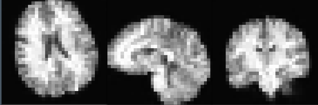
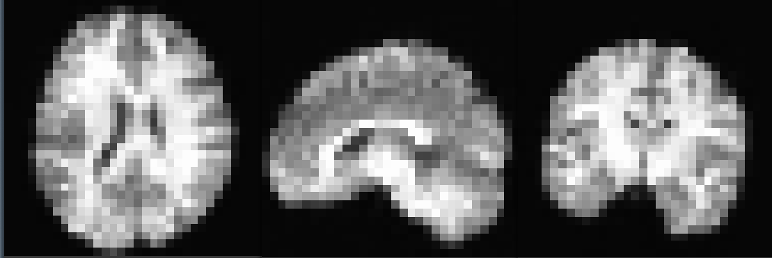
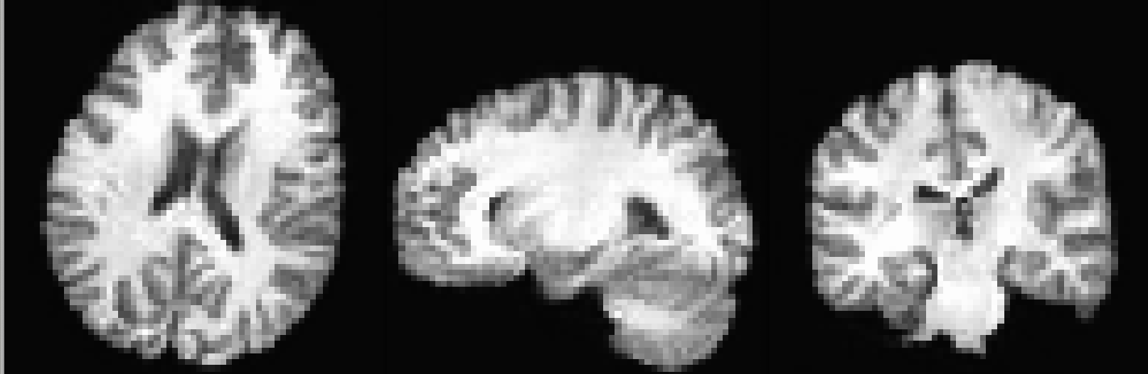
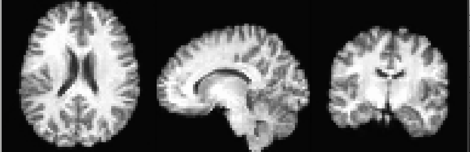
Figure 1 shows some synthetic brains with a resolution of 32 x 32 x 32 voxels generated by our 3D PGAN, while Figure 2 shows some synthetic brains with a resolution of 64 x 64 x 64 voxels.
5 Conclusion
We have demonstrated that a 3D progressive growing GAN can be used to synthesize T1-weighted volumes of 64 x 64 x 64 voxels. The synthetic volumes can be used for training (convolutional) networks that perform classification or segmentation. Furthermore, synthetic volumes can be shared freely, as they do not belong to a specific person, and can therefore facilitate data sharing [23]. In future work we will synthesize volumes of higher resolution.
Acknowledgements
This study was supported by Swedish research council grant 2017-04889. Funding was also provided by VINNOVA Analytic Imaging Diagnostics Arena (AIDA) and the ITEA3 / VINNOVA funded project ”Intelligence based iMprovement of Personalized treatment And Clinical workflow supporT” (IMPACT).
References
- [1] Ian Goodfellow, Jean Pouget-Abadie, Mehdi Mirza, Bing Xu, David Warde-Farley, Sherjil Ozair, Aaron Courville, and Yoshua Bengio, “Generative adversarial nets,” in Advances in neural information processing systems, 2014, pp. 2672–2680.
- [2] Tero Karras, Timo Aila, Samuli Laine, and Jaakko Lehtinen, “Progressive growing of GANs for improved quality, stability, and variation,” arXiv preprint arXiv:1710.10196, 2017.
- [3] Phillip Isola, Jun-Yan Zhu, Tinghui Zhou, and Alexei A Efros, “Image-to-image translation with conditional adversarial networks,” in Proceedings of the IEEE conference on computer vision and pattern recognition, 2017, pp. 1125–1134.
- [4] Jun-Yan Zhu, Taesung Park, Phillip Isola, and Alexei A Efros, “Unpaired image-to-image translation using cycle-consistent adversarial networks,” in Proceedings of the IEEE international conference on computer vision, 2017, pp. 2223–2232.
- [5] Xin Yi, Ekta Walia, and Paul Babyn, “Generative adversarial network in medical imaging: A review,” Medical image analysis, p. 101552, 2019.
- [6] Christopher Bowles, Liang Chen, Ricardo Guerrero, Paul Bentley, Roger Gunn, Alexander Hammers, David Alexander Dickie, Maria Valdés Hernández, Joanna Wardlaw, and Daniel Rueckert, “Gan augmentation: augmenting training data using generative adversarial networks,” arXiv preprint arXiv:1810.10863, 2018.
- [7] Jiajun Wu, Chengkai Zhang, Tianfan Xue, Bill Freeman, and Josh Tenenbaum, “Learning a probabilistic latent space of object shapes via 3D generative-adversarial modeling,” in Advances in neural information processing systems, 2016, pp. 82–90.
- [8] Weiyue Wang, Qiangui Huang, Suya You, Chao Yang, and Ulrich Neumann, “Shape inpainting using 3D generative adversarial network and recurrent convolutional networks,” in Proceedings of the IEEE International Conference on Computer Vision, 2017, pp. 2298–2306.
- [9] Dong Nie, Roger Trullo, Jun Lian, Caroline Petitjean, Su Ruan, Qian Wang, and Dinggang Shen, “Medical image synthesis with context-aware generative adversarial networks,” in International Conference on Medical Image Computing and Computer-Assisted Intervention, 2017, pp. 417–425.
- [10] Janne J Näppi and Hiroyuki Yoshida, “Cycle-consistent 3D-generative adversarial network for virtual bowel cleansing in CT colonography,” in Medical Imaging 2019: Image Processing, 2019, vol. 10949, p. 109492Z.
- [11] Biting Yu, Luping Zhou, Lei Wang, Jurgen Fripp, and Pierrick Bourgeat, “3D cGAN based cross-modality MR image synthesis for brain tumor segmentation,” in IEEE International Symposium on Biomedical Imaging (ISBI), 2018, pp. 626–630.
- [12] David Abramian and Anders Eklund, “Generating fMRI volumes from T1-weighted volumes using 3D CycleGAN,” arXiv preprint arXiv:1907.08533, 2019.
- [13] Maria JM Chuquicusma, Sarfaraz Hussein, Jeremy Burt, and Ulas Bagci, “How to fool radiologists with generative adversarial networks? a visual turing test for lung cancer diagnosis,” in 2018 IEEE 15th international symposium on biomedical imaging (ISBI 2018). IEEE, 2018, pp. 240–244.
- [14] Maayan Frid-Adar, Idit Diamant, Eyal Klang, Michal Amitai, Jacob Goldberger, and Hayit Greenspan, “GAN-based synthetic medical image augmentation for increased CNN performance in liver lesion classification,” Neurocomputing, vol. 321, pp. 321–331, 2018.
- [15] Andrew Beers, James Brown, Ken Chang, J Peter Campbell, Susan Ostmo, Michael F Chiang, and Jayashree Kalpathy-Cramer, “High-resolution medical image synthesis using progressively grown generative adversarial networks,” arXiv preprint arXiv:1805.03144, 2018.
- [16] Changhee Han, Hideaki Hayashi, Leonardo Rundo, Ryosuke Araki, Wataru Shimoda, Shinichi Muramatsu, Yujiro Furukawa, Giancarlo Mauri, and Hideki Nakayama, “GAN-based synthetic brain MR image generation,” in 2018 IEEE 15th International Symposium on Biomedical Imaging (ISBI 2018). IEEE, 2018, pp. 734–738.
- [17] Arnab Kumar Mondal, Jose Dolz, and Christian Desrosiers, “Few-shot 3D multi-modal medical image segmentation using generative adversarial learning,” arXiv preprint arXiv:1810.12241, 2018.
- [18] Gihyun Kwon, Chihye Han, and Dae-shik Kim, “Generation of 3D brain MRI using auto-encoding generative adversarial networks,” in International Conference on Medical Image Computing and Computer-Assisted Intervention, 2019, pp. 118–126.
- [19] David C Van Essen, Stephen M Smith, Deanna M Barch, Timothy EJ Behrens, Essa Yacoub, Kamil Ugurbil, Wu-Minn HCP Consortium, et al., “The WU-Minn human connectome project: an overview,” Neuroimage, vol. 80, pp. 62–79, 2013.
- [20] Matthew F Glasser, Stamatios N Sotiropoulos, J Anthony Wilson, Timothy S Coalson, Bruce Fischl, Jesper L Andersson, Junqian Xu, Saad Jbabdi, Matthew Webster, Jonathan R Polimeni, et al., “The minimal preprocessing pipelines for the Human Connectome Project,” Neuroimage, vol. 80, pp. 105–124, 2013.
- [21] Matthew Brett, Michael Hanke, Ben Cipollini, Marc-Alexandre Côté, Chris Markiewicz, Stephan Gerhard, Eric Larson, Gregory R Lee, Yaroslav Halchenko, Erik Kastman, et al., “nibabel: 2.1. 0,” Zenodo, 2016.
- [22] Anders Eklund, Thomas E Nichols, and Hans Knutsson, “Reply to Brown and Behrmann, Cox, et al., and Kessler et al.: Data and code sharing is the way forward for fMRI,” Proceedings of the National Academy of Sciences, vol. 114, no. 17, pp. E3374–E3375, 2017.
- [23] Russell A Poldrack and Krzysztof J Gorgolewski, “Making big data open: data sharing in neuroimaging,” Nature neuroscience, vol. 17, no. 11, pp. 1510, 2014.