Magnetic structure in square cupola compound Ba(TiO)Cu4(PO4)4: a 31P NMR Study
Abstract
The magnetic structure of the antiferromagnetic square cupola compound Ba(TiO)Cu4(PO4)4 with the tetragonal structure is studied with 31P nuclear magnetic resonance techniques. The magnetic hyperfine shift shows a clear splitting at the Néel temperature TN = 9.5 K, where the resonance splits into two lines when an external magnetic field is oriented along the axis and into four lines when the field is along the axis. In the paramagnetic region follows temperature dependence of the magnetic susceptibility . From vs plot we determined nearly isotropic hyperfine field values H = 765 mT/ and H = 740 mT/ for the magnetic field oriented along and , respectively. From the rotation of the single crystal in the external magnetic field we determined eight different orientations of -tensor in the paramagnetic region. In the antiferromagnetic state at = 6 K we found that the local field at phosphorus is mainly due to dipolar field of coppers. Here the rotation of the single crystal shows eight different orientations of the local field mT. The orientations correspond to the calculation of dipolar fields at phosphorus assuming magnetic quadrupolar configuration of magnetic moments described previously [Nat. Commun. 7, 13039 (2016); Phys. Rev. B 96, 214436 (2017)].
pacs:
76.60.-k, 75.47.Lx, 75.50.EeI Introduction

Recently, an interesting class of compounds,(TiO)Cu4(PO4)4 (TCPO; = Ba, Sr, or Pb), was newly synthesized Kimura2016magneto ; Kimura2018a . The structure of TCPO is tetragonal with the space group 4212 and consists of the layers of up and down square cupola Cu4O12 (FIG. 1). Each square cupola is made of four corner sharing CuO4 plaquets. The layers are separated by ions and TiO5 pyramids. These compounds undergo an antiferromagnetic (AF) transition at low temperatures. For example, Ba(TiO)Cu4(PO4)4 (BaTCPO) exhibits an AF ordered state at temperatures below the Néel temperature TN = 9.5 K. Below TN the arrangement of magnetic moments of coppers shows an antiferroic quadrupolar order. Because of the antiferroic order, BaTCPO does not exhibit a magnetoelectric effect where an electric (magnetic) field causes magnetization (electric polarization). Instead, this compound exhibits a remarkable magnetodielectric effect. Sr(TiO)Cu4(PO4)4 (SrTCPO) also shows similar magnetic and magnetodielectric properties below TN = 6.2 K SrPRRB2019 , while Pb(TiO)Cu4(PO4)4 (PbTCPO) exhibits a ferroic quadrupolar order resulting in a linear magnetoelectric effect below TN = 6.5 K PbPRMat2018 ; PbJPSJ2019 .
Magnetic structure of BaTCPO was carefully studied by neutron scattering experiments Kimura2016magneto . These studies yielded two possible arrangements of the spins: one, denoted , where the moments are confined approximately in the CuO4 planes; and the other, , where the moments are approximately perpendicular to the CuO4 planes forming two-in-two-out-type structure (FIG. 1). The latter arrangement had smaller R-factor, thus being the most probable arrangement. Later Babkevich et al. Babkevich2017 confirmed the two possible structures using spherical neutron polarimetry, inclining by a discernible, albeit small advantage towards the spin structure.
In the present report we will use 31P NMR techniques to study the local magnetic fields in a single crystal of BaTCPO. Phosphorus ions in PO4 tetrahedra see four coppers in two cupolas as next-nearest-neighbours. Therefore, the local field at phosphorus is definitely influenced by the magnetic arrangement of coppers. Previous NMR studies of SrTPCO Islam2018 and BaTPCO Kumar2019 have used powder samples where detailed information about the magnetic structure is difficult to get.
II Experimental details
Single crystals of Ba(TiO)Cu4(PO4)4 were grown with the flux method by Kimura et al. Kimura2018a ; Kimura2016inorganic . The sample crystal used in the experiments sized 1.9 x 2.0 x 3.9 mm and weighed 61.84 mg. NMR measurements were conducted using the spectrometer MAGRes2000 attached to a B = 4.7 T superconducting magnet, 31P resonance frequency 80.97 MHz. He-flow cryostat (JANIS Research Inc.) allowed measurements in the temperature range of 4.5 to 300 K. The NMR probe was our own made and had a single-axis goniometer. LakeShore CERNOX calibrated sensors and Model 332 temperature controller were used for temperature regulation. Spin-lattice relaxation T1 was measured using inversion-recovery method. The magic angle spinning (MAS) room temperature spectrum was recorded using a home-built probe with the mm O.D. rotor spinning kHz. The magnetic shifts are given relative to the resonance of H3PO4.
III Results and analysis
III.1 Magnetization
The temperature dependence of magnetic susceptibility was measured with the applied magnetic field B = 4.7 T along the [001] and [100] directions as shown in FIG. 2. At temperature region T 17 K there is a broad maximum of vs T curve, which indicates onset of short range order.

At high temperature followed the Curie-Weiss law:
| (1) |
Here is temperature-independent susceptibility. At T 100 K the fitting gives the following parameters: for the magnetic field along the [001] direction: cm3/mole per Cu, cm3 K per mole Cu, K; and for the field along the [100] direction: cmmole-Cu, cm3 K mole-Cu, and K. From the Curie constant one gets the effective copper magnetic moment as , where is the Boltzmann factor, and is Avogadro’s number. We get and 1.911 for [001] and [100] directions, respectively. Here, is the Bohr magneton. Using we get almost equal Lande -factor values as and for [001] and [100], respectively, which are common for Cu2+ ions.
Here we note, that in regular, collinear antiferromagnets the vanishes in the direction where the local magnetic moments are aligned parallel or antiparallel to an external magnetic field and stays constant in the directions where the external field is perpendicular to the local magnetic moments Singer1956 . Thus the local moments here may prefer the [001] direction, but the non-vanishing could also just be a signature of the non-collinear order.
III.2 31P Knight shift

Temperature dependence of 31P Knight shift is presented in FIG. 3. Above the Néel temperature TN = 9.5 K, the Knight shift (T) follows perfectly the magnetic susceptibility curve (T), as given Clogston1961 by Eq.(2):
| (2) |
Here, is the temperature-independent shift, the chemical shift, and H is the hyperfine coupling constant. From the vs plots (insets of FIG. 3) we found the hyperfine field values as H = 7.65(5) kOe/ for direction and slightly smaller value, H = 7.40(5) kOe/ for direction .
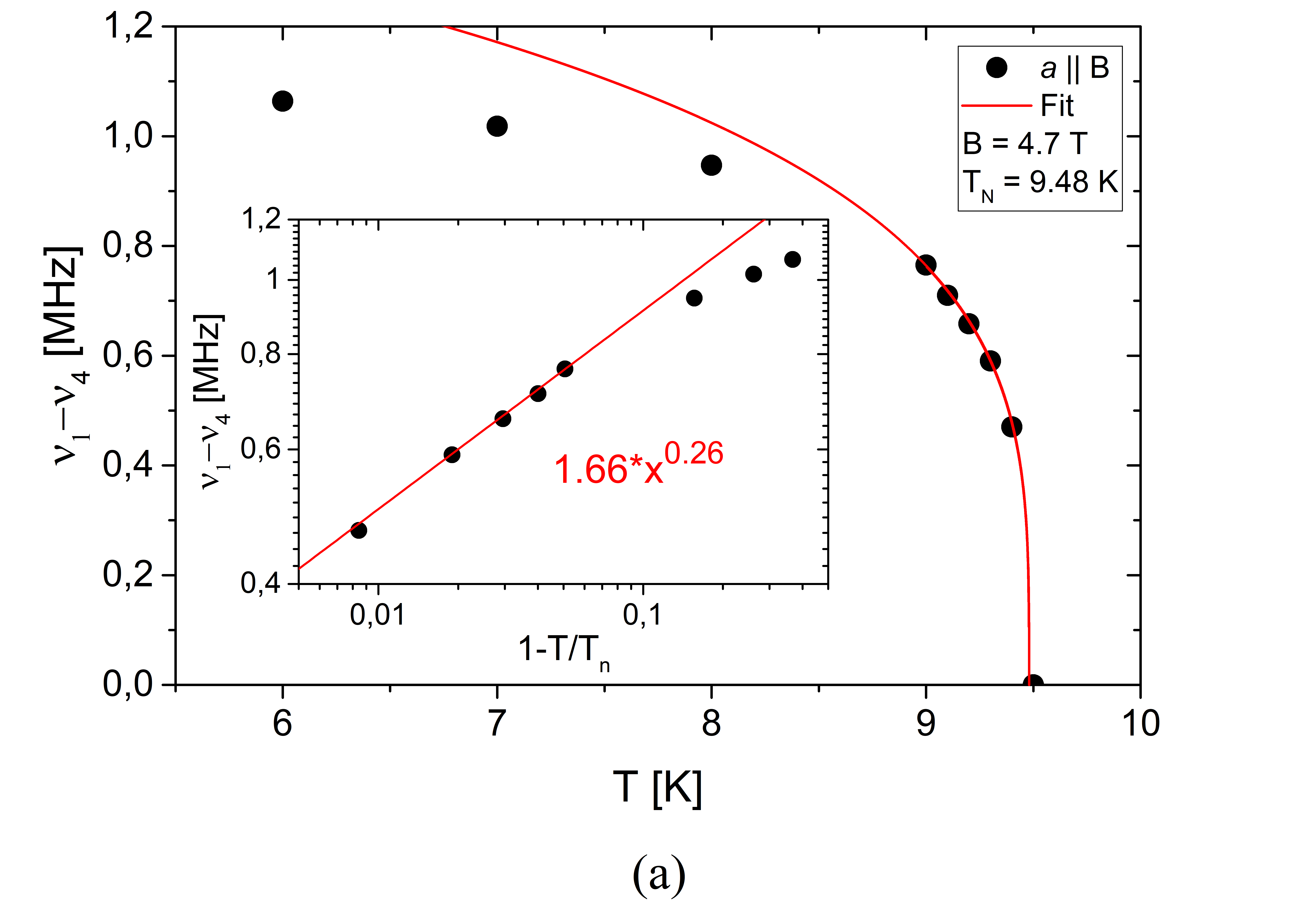
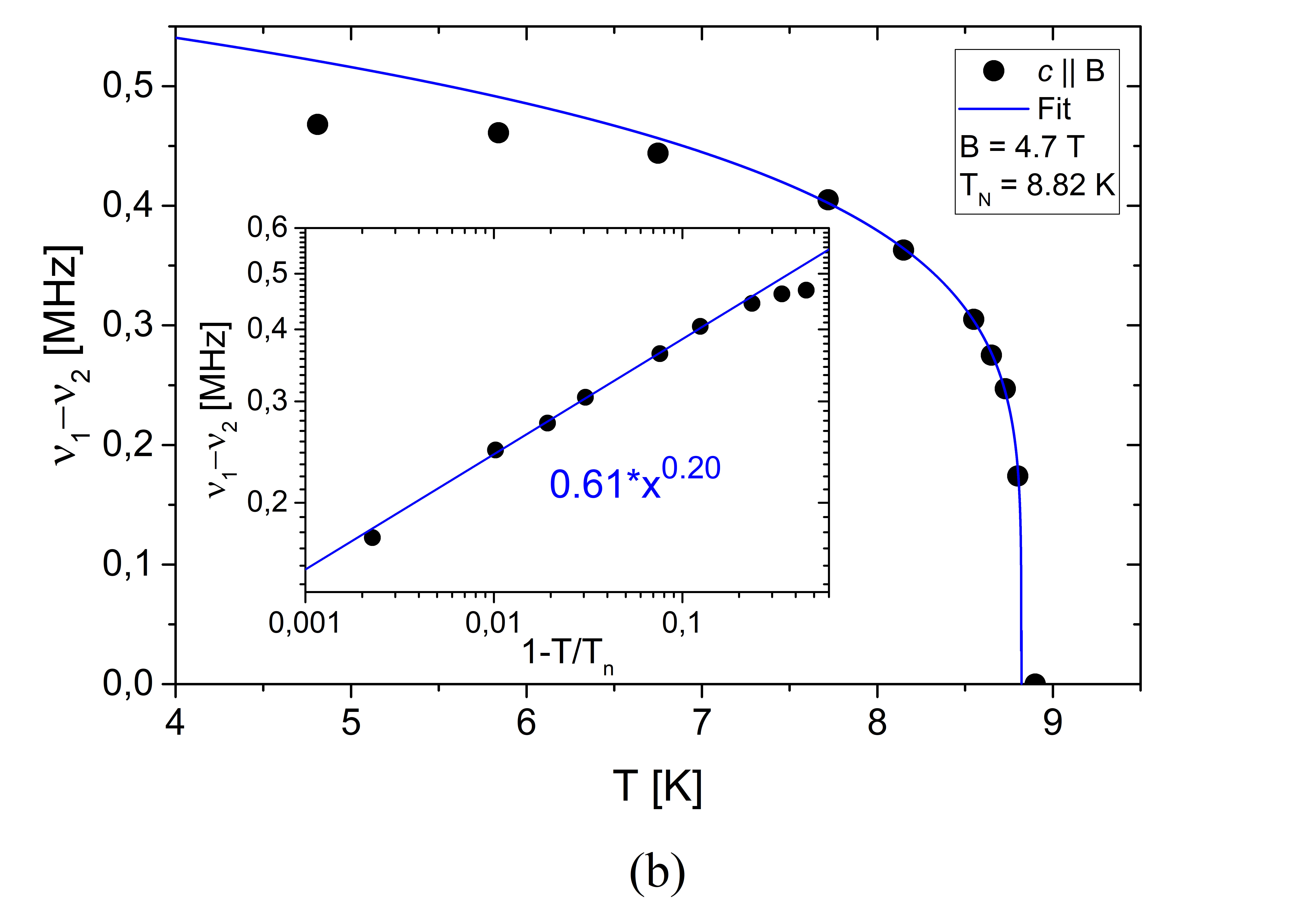
Below TN, due to the onset of local magnetic fields, the resonance line splits into four lines in the orientation and into two lines when the crystal is oriented with . Following the approach given in Ref. Islam2018 we will estimate the critical order parameter of the phase transition from the growth of the internal field in the vicinity of the ordering temperature. As a measure of the internal field we use the frequency difference between the most shifted resonance lines instead of the internal ones, which is proportional to the internal field by gyromagnetic ratio = 17.237 MHz/T. The temperature dependences of the frequency differences in the two orientations of the single crystal are given in FIG. 4.
The temperature dependence of the frequency difference is fitted by the formula
| (3) |
The best fit was obtained with TN = 9.48 K, = 0.26 for the orientation , and TN = 8.82 K, = 0.20 for . The critical exponent values, if compared to some theoretical values (as given e. g. in Ref. Nath2009 ), indicate that the ordering scheme is the closest to the 2D XY case.
III.3 31P NMR of powder sample

Before starting 31P analysis of single crystal we recorded the spectrum of a powder sample. FIG. 5(a) shows 31P NMR spectrum recorded with magic angle spinning MAS-NMR_reference of the sample. The spectrum shows a single sharp line at isotropic magnetic shift. The spectrum of a static sample (FIG. 5(b)) shows a typical powder line shape with singularities at the principal values of the Knight shift tensor:
| (4) |
with K %, K %, K %. These values give us the reference for the interpretation of the rotation patterns of the single crystal.
III.4 Orientation of the 31P Knight shift tensor
Determination of tensor orientation in a single crystal is not very easy task Mehring1976 . For that, in general case, one needs to record resonance frequencies rotating the sample around three different axes. Depending on symmetry or having some principal values of the shift tensor pre-determined, the number of necessary rotation patterns may be smaller. After obtaining those rotation patterns, one needs to find unitary transformation that will transform the principal axis system (PAS) into the crystal frame. As usual, the Hamiltonian of spin- nucleus consists of the Zeeman and the Knight shift interaction
| (5) |
where , , and are Pauli matrices:
| (6) |
Three successive rotations, each characterized by three Euler angles, transform the Hamiltonian from the principal axes frame (PAS) into the laboratory frame:
There are four frames of reference: PAS with diagonal tensor (), crystal frame (), goniometer frame (), and laboratory frame (K). The transformations between them are , , with the corresponding Euler angles. We used the conventional Euler ZYZ rotation of the Hamiltonian easyspin .
In BaTCPO there are eight different positions of the phosphorus ions in the unit cell, each giving the resonance corresponding to different transformation . The rotation patterns around the and axis at temperatures T = 295 K and T = 18 K are shown in FIG. 6. The angles , , and , , are unique for each experiment; , , are desired eight sets of the Euler angles transforming the tensor in PAS to the crystal frame for each phosporus site. It turns out that the principal axis of the Knight shift tensor is tilted by 45 degrees from the crystal axis. Schematics of the Knight tensor’s eight orientations are presented in Fig. 7.


| (a) T=295K | T | (c) 295K | (d) 18K | T=295K | ||||||||||||
| nr | ||||||||||||||||
| czg | (-12:372) | 90 | 0 | 85 | 0 | 0 | 1 | 30 | 45 | -45 | 25 | 45 | -40 | |||
| azg | (-12:372) | 90 | 0 | 0 | 90 | 0 | 2 | -30 | 45 | 45 | -25 | 45 | 50 | |||
| 3 | 30 | 45 | 135 | 25 | 45 | 140 | ||||||||||
| (b) T=18K | 4 | -30 | 45 | -135 | -25 | 45 | -130 | T=18K | ||||||||
| 5 | 30 | 135 | 45 | 25 | 135 | 50 | ||||||||||
| czg | (-12:372) | 90 | 0 | 80 | 0 | 0 | 6 | -30 | 135 | -45 | -25 | 135 | -40 | |||
| azg | (-12:372) | 90 | 0 | 0 | 90 | 0 | 7 | 30 | 135 | -135 | -25 | 135 | 140 | |||
| 8 | -30 | 135 | 135 | 25 | 135 | -130 | ||||||||||
III.5 Spin-lattice relaxation results
Spin-lattice relaxation was measured with inversion-recovery pulse sequence at magnetic fields along [001] and [100] directions (Fig. 8). The magnetization recovery was exponential throughout all the measurements:
| (8) |
where is the magnetization at delay after inversion, is the equilibrium magnetization, and 2 is a constant depending on the accuracy of the inversion.

The relaxation rate at T 60 K is almost constant which is typical for a paramagnetic material, where the relaxation is caused by the fluctuation of the magnetic moments. Before the phase transition at TN, a sharp spike occurs in the relaxation speed which is connected to the rapid slowing of the fluctuations. Below TN, relaxation speed decreases sharply proportional to T7. In case of , the relaxation rate seems to have a discontinuity in close vicinity of TN. Here we note, that 1/T1 T7 was also observed for SrTCPO Islam2018 .
In paramagnetic temperature region we can use the Moriya’s theory of relaxation Moriya , where:
| (9) |
Here is the nuclear gyromagnetic ratio, is the nuclear spin, is the hyperfine field as in Eq. 2, is the number of nearest Cu2+ neighbors for the nucleus and is the Heisenberg exchange frequency (in units rad-1), where = 2 is the number of Cu2+ ions as nearest neighbors to 31P, = 1/2 is the electronic spin, and is the exchange interaction.
With the hyperfine field value of = 7.650 kOe/ and the relaxation rate for the paramagnetic region 1/T1 = 1410 s-1 we get an estimate for the exchange interaction inside the square cupola = 35 K with an exchange frequency of = 4.5 rad/s. This value is in coherence with previous results of = 3.0 meV = 34.8 K Kimura2016magneto .
III.6 Local magnetic structure in the ordered state
The resonance frequencies by rotation of the single crystal around the [001] and [100] at T = 6 K are given in Fig. 9. The results are in coherence with the Knight shift temperature dependence (FIG. 3). Once the magnetic field is turned around the axis, one can see that when B, there are four different magnetic field projections in the AFM region as given in FIG. 3(a). When the crystal is oriented B, there are two different local field projections to the external field direction as given in FIG. 3(b). Rotating the sample around and gives eight different rotation patterns for eight phosphorus ions in the unit cell. Each rotation pattern can be described by the equation:
| (10) |
where the constant term is the Larmor frequency plus average chemical shift; the second term describes the angle dependence of the local field projection to the external field direction. The third term describes the angle dependence of the resonance frequency due to turning of the chemical shift tensor.
The rotation patterns around the [001] axis show two sets of lines (blue and red). The phase shift within the set is 90 degrees. The red lines are shifted from B by +16 degrees, while the blue lines are shifted -16 degrees. We can assign the blue lines to the phosphorus ions in “up” cupola and the red lines to the ions in “down” cupola. The rotation patterns of the crystal around the [100] (FIG. 9(b), Table 2b) are not so well resolved. Here, the approximation of the frequencies by Eq. 10 is not particularly good. A possible reason might be that the magnetic structure in 4.7 T magnetic field at B direction is not yet well settled at temperature 6 K. We found above (see FIG. 4) that the ordering temperature in the direction B was TN = 8.8 K, while in case of B we had TN = 9.5 K.
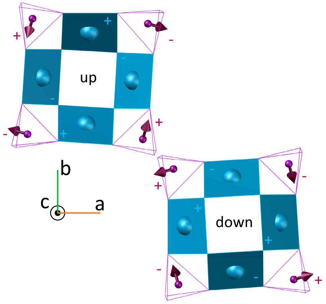
Despite of that, one can clearly see the rotation patterns with two different amplitudes as expected for two different local field projections along the axis. The assignment of the resonances to “up” and “down” cupola is not unique. For example, we cannot distinguish the cases, where all the local field directions of one cupola have positive projection to the axis and the moments of the other cupola have negative projection, from the case, where the ions of one cupola have two positive and two negative projections and the field on ions of the other cupola have, respectively, two negative and two positive projections to the axis. The assignment in FIG. 9(b) corresponds to the local field configuration as given below in FIG. 11.

| nr | |||||
|---|---|---|---|---|---|
| 1 | 81.59 | 0.56 | 347 | 0.075 | 155 |
| 2 | 81.59 | 0.56 | 20 | 0.075 | 25 |
| 3 | 81.58 | 0.56 | 80 | 0.075 | 50 |
| 4 | 81.59 | 0.56 | 110 | 0.075 | 125 |
| 5 | 81.59 | 0.55 | 170 | 0.075 | 155 |
| 6 | 81.595 | 0.56 | 198 | 0.065 | 30 |
| 7 | 81.59 | 0.575 | 258 | 0.078 | 50 |
| 8 | 81.60 | 0.56 | 288 | 0.065 | 130 |
| nr | |||||
|---|---|---|---|---|---|
| 1 | 81.47 | 0.62 | 294 | 0.17 | 87 |
| 2 | 81.47 | 0.62 | 246 | 0.17 | 97 |
| 3 | 81.45 | 0.29 | 212 | 0.17 | 72 |
| 4 | 81.44 | 0.29 | 148 | 0.17 | 115 |
| 5 | 81.47 | 0.62 | 114 | 0.17 | 84 |
| 6 | 81.47 | 0.62 | 66 | 0.17 | 95 |
| 7 | 81.45 | 0.29 | 32 | 0.17 | 69 |
| 8 | 81.45 | 0.29 | 328 | 0.17 | 111 |
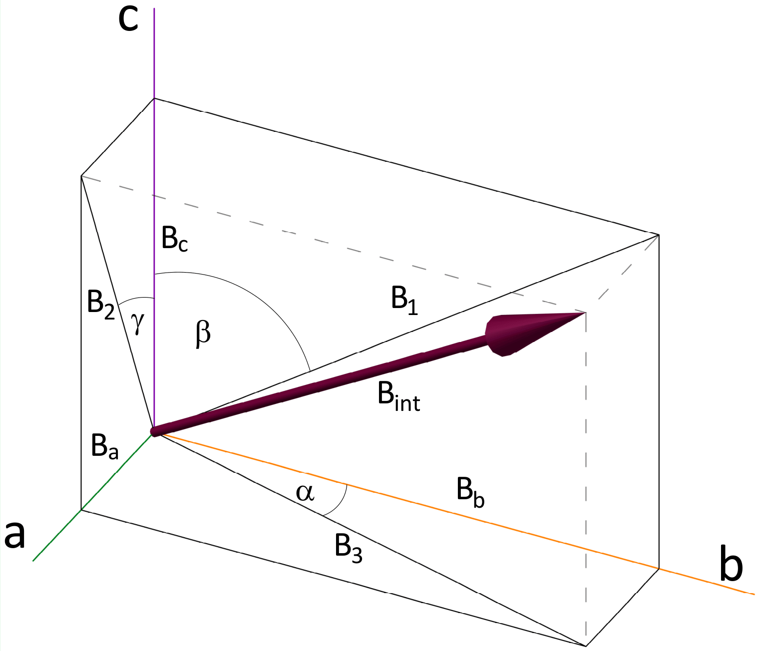
The analysis of the data given in Table 2 can be carried out using the scheme of the local field direction as given in FIG. 10. Three cosine amplitudes in Table 2 correspond to the local field projections B1, B2, and B3 in FIG. 10. Using the gyromagnetic ratio of 31P = 17.237 MHz/T, we find B1 = 36 mT, B2 = 32.5 mT, and B3 = 16.8 mT. The angles = 16, = 66, and = 32 degrees. It is not difficult to calculate the projections Ba = 8.9 mT, Bb = 31.2 mT, and Bc = 14.6 mT, as well as the module of the internal field B = 35.6 mT.
As noted above, unique assignment of the resonances to certain phosphorus in the unit cell is not possible. One possible local field configuration is given in FIG. 11.
The internal field at phosphorus ions consists of two components:
| (11) |
where is transferred hyperfine field, and is the dipolar field from the magnetic moments of Cu2+ ions. In the following we assume that is very well cancelled in AF ordered state, and the local magnetic field at phosphorus is due to dipolar field of Cu2+ ions.
We did calculate the dipolar field at each phosphorus ion of the unit cell assuming the two magnetic structures proposed in Refs. Kimura2016magneto ; Babkevich2017 . In the calculation we did sum the dipolar field from every Cu2+ inside a sphere of 50 Å around given phosphorus. At that we took into account that the unit cell of magnetic structure is doubled along direction, i. e. the magnetic moments of every other layer along the axis were reversed. As a result, we obtain the following dipolar field projections along the , , and axis
| a) | |||
for the structure noted , and
| b) | |||
for the structure noted .
In comparison, our experiments above give the values:
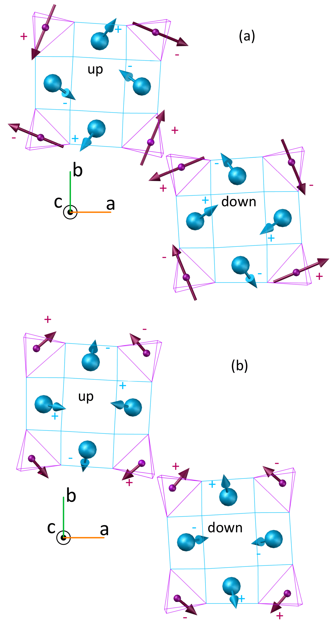
Calculated dipolar field directions at the phosphorus ions are given in FIG. 12.
Comparison of the experimental field pattern to the calculated dipolar fields gives remarkable similarity to the case calculated for structure - the calculated dipolar field directions are close to the experimental values in FIG. 11, although the calculated Bc value is relatively small and the calculated local field is 2.7 times larger than the experimental value. Three times larger value of the calculated dipolar field compared to the experimentally determined local field was reported earlier for dipolar field at Ba site in antiferromagnetic YBa2Cu3O6.05 Ref. Lombardi1996 . The authors ascribed this controversy to the possible effect of delocalization of the copper - electron. Dipolar field calculated for structure is quite different. It is almost confined to the plane, with nearly equal Ba and Bb components. Therefore, we can conclude that the NMR data are better consistent with the magnetic structure .
IV Conclusion
We performed 31P NMR of the antiferromagnetic square cupola compound Ba(TiO)Cu4(PO4)4 in the applied magnetic field B = 4.7 T and provided an in-depth overview of the local magnetic environment around the Cu2+ cupolas. From the 31P NMR frequency dependence of the single crystal orientation we successfully determined the principal values of the 31P magnetic shift tensor and the orientation of eight magnetic tensors in the unit-cell at room temperature and at temperature T = 6 K. The Knight shift temperature dependence in comparison of that of the bulk magnetic susceptibility enabled to determine the hyperfine field on 31P nuclei H = 7.65(5) kOe/ for and H = 7.40(5) kOe/ for . The temperature dependence of 31P spin-lattice relaxation resulted in an approximation for the exchange interaction constant between Cu2+ ions . 31P NMR frequency dependence on the single crystal orientation in the antiferromagnetic state gave a clear picture of local magnetic fields at 31P ions. The static magnetic field at every phosphorus was determined as mT. Experimental configuration of the local field was compared to the calculated dipolar field for several magnetic arrangement of the copper magnetic moments. We found that the magnetic structure determined by the previous neutron diffraction studies Kimura2016magneto ; Babkevich2017 is most consistent with the NMR data.
Acknowledgemnts
This research was supported by the Estonian Science Council Grants IUT23-7 and PRG4, the European Regional Development Fund Grant TK134, JSPS KAKENHI Grant Numbers JP17H01143, JP19H05823 and JP19H01847, and by the MEXT Leading Initiative for Excellent Young Researchers (LEADER).
References
- (1) K. Kimura, P. Babkevich, M. Sera, M. Toyoda, K. Yamauchi, G.S. Tucker, J. Martius, T. Fennell, P. Manuel, D.D. Khalyavin, R.D. Johnson, T. Nakano, Y. Nozue, H.M. Rønnow, and T. Kimura, Nat. Commun. 7, 13039 (2016).
- (2) K. Kimura, M. Toyoda, P. Babkevich, K. Yamauchi, M. Sera, V. Nassif, H.M. Rønnow, and T. Kimura, Phys. Rev. B 97, 134418 (2018).
- (3) Y. Kato, K. Kimura, A. Miyake, M. Tokunaga, A. Matsuo, K. Kindo, M. Akaki, M. Hagiwara, S. Kimura, T. Kimura, and Y. Motome, Phys. Rev. B 99, 024415 (2019).
- (4) K. Kimura, Y. Kato, K. Yamauchi, A. Miyake, M. Tokunaga, A. Matsuo, K. Kindo, M. Akaki, M. Hagiwara, S. Kimura, M. Toyoda, Y. Motome, and T. Kimura, Phys. Rev. Materials 2, 104415 (2018).
- (5) K. Kimura, S. Kimura, and T. Kimura, J. Phys. Soc. Jpn. 88, 093707 (2019).
- (6) P. Babkevich, L. Testa, K. Kimura, T. Kimura, G.S. Tucker, B. Roessli, and H.M. Rønnow, Phys. Rev. B 96, 214436 (2017).
- (7) S.S. Islam, K.M. Ranjith, M. Baenitz, Y. Skourski, A.A. Tsirlin, and R. Nath, Phys. Rev. B 97, 174432 (2018).
- (8) V. Kumar, A. Shahee, S. Kundu, M. Baenitz, and A.V. Mahajan, J. Magn. Magn. Mater. 492, 165600 (2019).
- (9) K. Kimura, M. Sera, and T. Kimura, Inorg. Chem. 55, 1002 (2016).
- (10) J.R. Singer, Phys. Rev. 121, 1357 (1961).
- (11) A.M. Clogston and V. Jaccarino, Phys. Rev. 104, 929 (1956).
- (12) R. Nath, Y. Furukawa, F. Borsa, E.E. Kaul, M. Baenitz, C. Geibel, and D.C. Johnston, Phys. Rev. B 80, 214430 (2009).
- (13) Low-temperature NMR: Techniques and Applications, D. Arčon, I. Heinmaa, and R. Stern, Edited by: P. Hodgkinson, in Modern Methods in Solid-state NMR: A Practitioner’s Guide, Book Series: New Developments in NMR Vol. 15, Pages: 231-261 (2018).
- (14) High Resolution NMR Spectroscopy in Solids, M. Mehring, Book Series: NMR Basic Principles and Progress Vol. 11, (1976).
- (15) Easyspin.org: Rotations and Euler angles.
- (16) T. Moriya, Prog. Theor. Phys. 16, 23 (1956); J. Phys. Soc. Jpn. 18, 516 (1963).
- (17) A. Lombardi, M. Mali, J. Roos, and D. Brinkmann, Phys. Rev. B 53, 14269 (1996).