Epitaxial stabilization of ultra thin films of high entropy perovskite
Abstract
High entropy oxides (HEOs) are a class of materials, containing equimolar portions of five or more transition metal and/or rare-earth elements. We report here about the layer-by-layer growth of HEO [(La0.2Pr0.2Nd0.2Sm0.2Eu0.2)NiO3] thin films on NdGaO3 substrates by pulsed laser deposition. The combined characterizations with in-situ reflection high energy electron diffraction, atomic force microscopy, and X-ray diffraction affirm the single crystalline nature of the film with smooth surface morphology. The desired +3 oxidation of Ni has been confirmed by an element sensitive X-ray absorption spectroscopy measurement. Temperature dependent electrical transport measurements revealed a first order metal-insulator transition with the transition temperature very similar to the undoped NdNiO3. Since both of these systems have a comparable tolerance factor, this work demonstrates that the electronic behaviors of -site disordered perovskite-HEOs are primarily controlled by the average tolerance factor.
Finding new materials and new ways to tune material’s properties are essential to fulfill the demand of the constantly evolving modern technology. Transition metal oxides show various fascinating electronic and magnetic phenomena such as metal-insulator transition, superconductivity, colossal magnetoresistance, multiferroicity, skyrmions, etc., which have lots of prospect for technological applications Imada, Fujimori, and Tokura (1998); Tokura (2006); Yang, Ko, and Ramanathan (2011); Catalan et al. (2012); Lorenz et al. (2016); Matsuno et al. (2018). Furthermore, transition metal (TM) based high entropy oxides (HEOs) are being explored in recent years to achieve tunable properties in unexplored parts of complex phase diagram Rost et al. (2015); Bérardan et al. (2016); Rost et al. (2017); Sarkar et al. (2017); Djenadic et al. (2017); Jiang et al. (2018); Anand et al. (2018); Sarkar et al. (2018); Sharma et al. (2018); Dkabrowa et al. (2018); Witte et al. (2019); Sarkar et al. (2019); Zhang et al. (2019); Meisenheimer, Kratofil, and Heron (2017); Sharma et al. (2018, 2019). In general, the configurational entropy of a multi-component solid solution can be enhanced by mixing a large number of cations in equiatomic proportions and a single structural phase is formed if the entropy contribution overcomes enthalpy driven phase separation (=-; , , are Gibbs free energy, enthalpy and entropy of mixing, respectively) Rost et al. (2015); Sarkar et al. (2019). After the report of the first HEO [Mg0.2Ni0.2Co0.2Cu0.2Zn0.2O with rocksalt structure] by Rost et al. Rost et al. (2015), HEOs with other structural symmetry such as perovskite Sharma et al. (2018); Witte et al. (2019), spinel Dkabrowa et al. (2018) have been also synthesized. However, this promising field of HEO is at a very early stage and most of the aspects of HEOs are yet to be explored experimentally. For example, it is still unknown whether the strong disorder or the average tolerance factor () determines the electronic and magnetic behaviors of perovskite-HEOs.
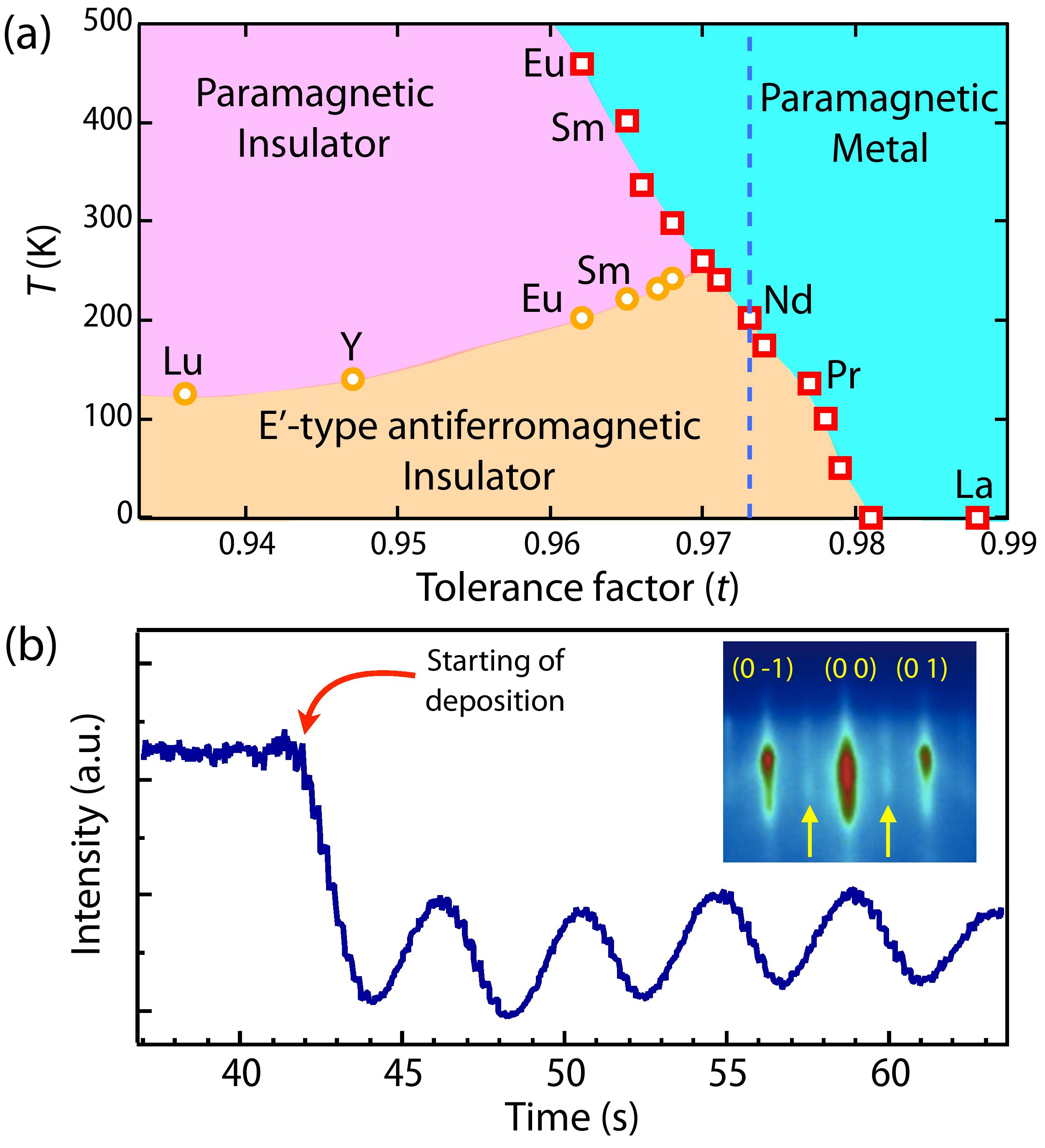
As a prototypical example of perovskite (O3) series, NiO3 (= La, Pr, Nd, Sm, Eu…Lu) exhibits an interesting phase diagram as a function of tolerance factor (=, where , , are radii of , Ni and O, respectively) Medarde (1997); Catalan (2008). LaNiO3, the least distorted member of this series remains metallic and paramagnetic down to the lowest temperature. Bulk PrNiO3 and NdNiO3 (NNO) show temperature driven simultaneous transitions from an orthorhombic, paramagnetic, metallic phase to a monoclinic, antiferromagnetic, insulating phase respectively (see Fig. 1(a)). The insulating phase is also characterized by a checkerboard type charge ordering Staub et al. (2002). In case of the more distorted members, such as SmNiO3, EuNiO3, etc, the magnetic transition gets decoupled from the other three simultaneous transitions, resulting in an intermediate paramagnetic, insulating, charge ordered phase. The quest to understand the origin of these transitions have led to remarkable progress in epitaxial stabilization of NiO3 family (see Refs. Middey et al., 2016; Catalano et al., 2018, and literature cited therein), and thin films of NiO3 with =La, Pr, Nd, Sm, and Eu have been stabilized so far Middey et al. (2016); Catalano et al. (2018); Ha et al. (2012); Liu et al. (2013); Feigl et al. (2013); Meyers et al. (2013); Mikheev et al. (2015); Hepting et al. (2014); Scherwitzl et al. (2011); Bruno et al. (2013). This further provides a unique opportunity to verify the role of disorder vs. , in determining the electronic and magnetic properties of perovskite HEO with a strong disorder at the site.
| Compound | (Å) | (Å) | (Å) | = (Å) | (Å) | Referrence |
|---|---|---|---|---|---|---|
| LaNiO3 | 5.457 | 5.457 | 13.146 | 3.838 | 3.838 | García-Muñoz et al.,1992 |
| PrNiO3 | 5.419 | 5.380 | 7.626 | 3.818 | 3.813 | García-Muñoz et al.,1992 |
| NdNiO3 | 5.389 | 5.382 | 7.610 | 3.808 | 3.805 | García-Muñoz et al.,1992 |
| SmNiO3 | 5.327 | 5.432 | 7.565 | 3.804 | 3.782 | Alonso et al.,1999 |
| EuNiO3 | 5.294 | 5.458 | 7.537 | 3.802 | 3.769 | Alonso et al.,1999 |
In this letter, we report successful layer-by-layer epitaxial growth of (La0.2Pr0.2Nd0.2Sm0.2Eu0.2)NiO3 [(LPNSE)NO] thin films on a single crystalline NdGaO3 (NGO) substrate by pulsed laser deposition (PLD). The variation in bulk lattice constants together with the pseudo-cubic lattice constants of several members of the NiO3 series have been listed in Table-I. Since the average of the of NiO3 with =La, Pr, Nd, Sm, Eu (indicated by a vertical line in Fig. 1(a)) is comparable to NNO, the electronic behavior of [(LPNSE)NO] thin films have been also compared with NNO films. Several characterization techniques including in-situ RHEED (reflection high energy electron diffraction) and ex-situ atomic force microscopy (AFM), X-ray diffraction (XRD), X-ray absorption spectroscopy (XAS) confirmed high structural quality of these (LPNSE)NO] thin films with proper oxidation state of Ni. Transport measurements and XAS experiments further revealed that in-spite of having a strong structural disorder, the electronic behaviors of (LPNSE)NO sample are very similar to a single site cation NNO film.
(LPNSE)NO films with thickness 15 uc, 30 uc and 45 uc (uc=unit cell in pseudocubic notation) and NNO film with 15 uc were grown on NGO (1 1 0)or [(0 0 1)pc] substrates (here or and pc denote orthorhombic and pseudocubic setting) by a PLD system at 735∘C under a dynamic oxygen pressure of 100-150 millitorr. A KrF excimer laser, operating with 4 Hz and energy density 1.5 J/cm2 was used for the deposition. The layer by layer growth was monitored by a high pressure RHEED system. The films were post-annealed at the growth temperature under an oxygen pressure of 500 torr for 30 minutes. A Park system AFM was used to check the morphology of these films. X-ray diffraction patterns were recorded using a Rigaku Smartlab X-ray diffractometer. Temperature dependent resistivity was measured by using the Van der Pauw geometry in a Quantum Design PPMS (physical property measurement system). XAS spectra at Ni- and O- edges were collected in bulk-sensitive TFY (total fluorescence yield) mode at the 4-ID-C beamline of Advanced Photon Source, Argonne National Laboratory.
The time dependent intensity of the specular reflection of RHEED pattern (Fig. 1(b)), recorded during the deposition shows very prominent oscillations, confirming the layer-by-layer growth of (LPNSE)NO film. The inset of Fig. 1(b) shows a RHEED image of a (LPNSE)NO film, taken after cooling to the room temperature. The streaky pattern of specular (0 0) and off-specular (0 1), (0 -1) Bragg reflections is a characteristic of smooth surface morphology. Akin to the RHEED pattern of NNO film on NGO substrate Ojha et al. (2019), (LPNSE)NO films also have half-order spots: (0 1/2) and (0 -1/2) (denoted by the arrows), implying orthorhombic/monoclinic symmetry at room temperature. Inset of Fig. 2(a) shows AFM image of the 45 uc (LPNSE)NO film and the roughness is found to be 1.8Å well below the , further testifying excellent surface morphology of the film.
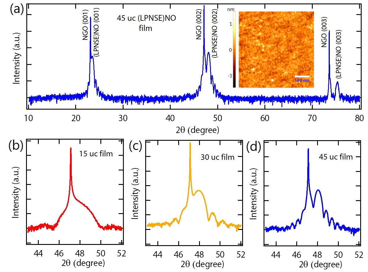
In order to check the structural quality of the samples and to detect the presence of any impurity phase, we have recorded 2- diffraction scan for (LPNSE)NO films using Cu radiation. Such a long scan XRD for the 45 uc (LPNSE)NO film (Fig. 2(a)) consists of broad film peaks in the vicinity of sharp substrate peaks, confirming the single crystalline nature of the film. Most importantly, the absence of any impurity peaks (within the detection limit of XRD) infers the growth conditions, used in this work is able to stabilize the multicomponent system into a single phase. XRD patterns around the (0 0 2)pc substrate peak for 15 uc, 30 uc, and 45 uc (LPNSE)NO films are shown in Fig. 2(b), (c), and (d), respectively. The very close proximity between the film peak and the substrate peak (Fig. 2(b)) in case of the 15 uc LPNSE)NO film prohibits a reliable estimation of out-of-plane lattice constant (). for 30 uc and 45 uc film are found to be 3.792Å and 3.784Å. The presence of thickness fringes in the vicinity of the film peaks further supports the excellent flatness of the film-substrate interface. The thickness of the films calculated from the position of the fringes (e.g. 17.3 nm for 45 uc film) are very close to the value expected from the RHEED oscillations. of the 15 uc NNO film is found to be 3.845Å (XRD pattern not shown).
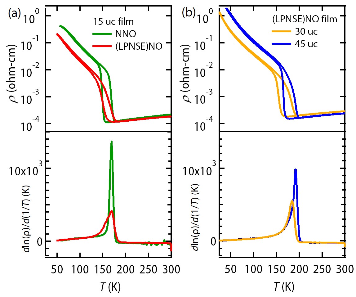
After confirming the high structural and morphological quality, we have investigated the electrical transport of the films. As reported earlier Liu et al. (2013); Mikheev et al. (2015); Ojha et al. (2019), 15 uc NNO thin film on NGO substrate undergoes first order MIT (upper panel of Fig. 3(a)). The transition temperature in the cooling run ( 160 K) and heating ( 180 K) is lower compared to the bulk NNO and is related to the epitaxial strain and finite thickness Liu et al. (2013). Surprisingly, the resistivity () of 15 uc (LPNSE)NO film at 300 K is very similar to that of 15 uc NNO film in-spite of having a strong disorder on the site. It further exhibits a MIT with strong thermal hysteresis ( 175 K, 185 K). However, the transition is more sluggish and of the insulating phase is also lower than that of 15 uc NNO film. With the increase of the film thickness, becomes approximately 200 K ((upper panel of Fig. 3(b)), which is very close to the transition temperature expected for the corresponding of (LPNSE)NO phase from the bulk phase diagram (Fig. 1(a)). This finding clearly establishes that the average tolerance factor controls the for this HEO, rather than the disorder at -site.
All NiO3 with an insulating phase also host -type antiferromagnetic ordering García-Muñoz, Rodríguez-Carvajal, and Lacorre (1994); Scagnoli et al. (2006); Liu et al. (2013); Hepting et al. (2014); Middey et al. (2018a, b). The magnetic transition temperature () can be approximately estimated from (ln)/(1/) vs. plot, as demonstrated recently for NNO films and EuNiO3/LaNiO3 superlattices Middey et al. (2018b, c); Ojha et al. (2019). Such resistivity analysis of the heating run data (lower panel of Fig. 3(a), (b)) provides a of 170 K for both 15 uc NNO and 15 uc (LPNSE)NO sample, 185 and 190 K for 30 uc and 45 uc (LPNSE)NO film, respectively. This suggests a simultaneous electronic and magnetic transitions in these (LPNSE)NO films. Soft X-ray resonant scattering experiments can further confirm this Scagnoli et al. (2006); Liu et al. (2013); Hepting et al. (2014); Middey et al. (2018a, b).
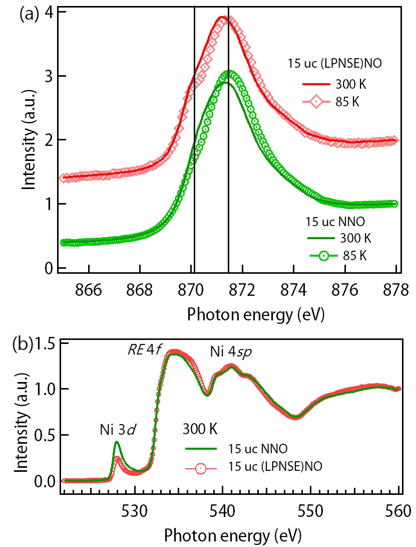
The required high +3 oxidation state of Ni makes NiO3 based systems very susceptible to the oxygen nonstoichiometry. In order to further understand the electronic and chemical structure of these films, we have measured XAS spectra on Ni edge and O- edge at 300 K and 85 K, i.e. much below . Due to strong overlap of La edge with Ni for (LPNSE)NO film, we discuss here only edge. First of all, the XAS line shape of both 15 uc NNO and 15 uc (LPNSE)NO film at 300 K (Fig. 4(a)) is consistent with Ni3+ in the metallic phase of nickelates Medarde et al. (1992); Liu et al. (2010); Wu et al. (2015); Middey et al. (2014), affirming the stabilization of desired oxidation state of Ni. Further, the appearance of strong multiple structures (around 870.05 eV) in the insulating phase of the (LPNSE)NO film is also consistent with the observation of NNO film (Fig. 4(a)) and the insulating phase of other nickelates Medarde et al. (1992); Liu et al. (2010); Meyers et al. (2013); Wu et al. (2015). Similar to the high cuprates, NiO3 also contains ligand holes, which can be observed as a pre-peak around 528 eV in O edge XAS spectrum due to the ( denotes hole in the oxygen 1 core state and corresponds to a hole in O 2 state) Medarde et al. (1992); Liu et al. (2013); Middey et al. (2014, 2018c); Freeland, van Veenendaal, and Chakhalian (2016). While the intensity of the pre peak is reduced in (LPNSE)NO film at 300 K, the position and FWHM (full width at half maxima) are very similar for both samples. The measurements at 85 K have found lowering of peak width in both samples (not shown), which is expected due to the band narrowing across the MIT Meyers et al. (2016); Freeland, van Veenendaal, and Chakhalian (2016). Thus, transport and XAS measurements conclude that the overall electronic structure effect across the MIT of (LPNSE)NO film is very similar to that of NNO film.
To summarize, we have successfully grown high quality epitaxial films of multicomponent (LPNSE)NO in a layer-by-layer fashion by pulsed laser deposition. RHEED, XRD, AFM, XAS and transport measurements have been carried out to investigate the structure and the electronic behavior of these films. In spite of having multi elements and strong disorder at the site, the average tolerance factor determines the electronic transition temperature. However, the microscopic details e.g. nucleation of insulating/metallic phase around the transition temperature Post et al. (2018), charge transfer between Ni and sites Upton et al. (2015), conductivity noise Daptary et al. (2019), etc. may depend on the on the details of composition and needs to be explored further. Stabilization of such multi system in a single crystalline form would further allow to investigate the complexity of phase transitions around the triple point of NiO3 phase diagram Park et al. (2013). Due to the chemical diversity of the perovskite family, huge numbers of HEO with a strong disorder at either site or site or both sites can be studied in the future to explore disorder driven physics in strongly correlated systems.
This work is funded by a DST Nanomission grant (DST/NM/NS/2018/246) and a SERB Early Career Research Award (ECR/2018/001512). The authors acknowledge AFM and XRD facilities at the Department of Physics, IISc Bangalore. RKP and SM thank the Department of Science and Technology, India (SR/NM/Z-07/2015) for the financial support to conduct synchrotron experiment at Advanced Photon Source and Jawaharlal Nehru Centre for Advanced Scientific Research (JNCASR) for managing the project. This research used resources of the Advanced Photon Source, a U.S. Department of Energy Office of Science User Facility operated by Argonne National Laboratory under Contract No. DE-AC02-06CH11357.
References
- Imada, Fujimori, and Tokura (1998) M. Imada, A. Fujimori, and Y. Tokura, Rev. Mod. Phys. 70, 1039 (1998).
- Tokura (2006) Y. Tokura, Reports on Progress in Physics 69, 797 (2006).
- Yang, Ko, and Ramanathan (2011) Z. Yang, C. Ko, and S. Ramanathan, Annual Review of Materials Research 41, 337 (2011).
- Catalan et al. (2012) G. Catalan, J. Seidel, R. Ramesh, and J. F. Scott, Reviews of Modern Physics 84, 119 (2012).
- Lorenz et al. (2016) M. Lorenz, M. R. Rao, T. Venkatesan, E. Fortunato, P. Barquinha, R. Branquinho, D. Salgueiro, R. Martins, E. Carlos, A. Liu, et al., Journal of Physics D: Applied Physics 49, 433001 (2016).
- Matsuno et al. (2018) J. Matsuno, J. Fujioka, T. Okuda, K. Ueno, T. Mizokawa, and T. Katsufuji, Science and technology of advanced materials 19, 899 (2018).
- Rost et al. (2015) C. M. Rost, E. Sachet, T. Borman, A. Moballegh, E. C. Dickey, D. Hou, J. L. Jones, S. Curtarolo, and J.-P. Maria, Nature communications 6, 8485 (2015).
- Bérardan et al. (2016) D. Bérardan, S. Franger, A. Meena, and N. Dragoe, Journal of Materials Chemistry A 4, 9536 (2016).
- Rost et al. (2017) C. M. Rost, Z. Rak, D. W. Brenner, and J.-P. Maria, Journal of the American Ceramic Society 100, 2732 (2017).
- Sarkar et al. (2017) A. Sarkar, R. Djenadic, N. J. Usharani, K. P. Sanghvi, V. S. Chakravadhanula, A. S. Gandhi, H. Hahn, and S. S. Bhattacharya, Journal of the European Ceramic Society 37, 747 (2017).
- Djenadic et al. (2017) R. Djenadic, A. Sarkar, O. Clemens, C. Loho, M. Botros, V. S. Chakravadhanula, C. Kübel, S. S. Bhattacharya, A. S. Gandhi, and H. Hahn, Materials Research Letters 5, 102 (2017).
- Jiang et al. (2018) S. Jiang, T. Hu, J. Gild, N. Zhou, J. Nie, M. Qin, T. Harrington, K. Vecchio, and J. Luo, Scripta Materialia 142, 116 (2018).
- Anand et al. (2018) G. Anand, A. P. Wynn, C. M. Handley, and C. L. Freeman, Acta Materialia 146, 119 (2018).
- Sarkar et al. (2018) A. Sarkar, L. Velasco, D. Wang, Q. Wang, G. Talasila, L. de Biasi, C. Kübel, T. Brezesinski, S. S. Bhattacharya, H. Hahn, et al., Nature communications 9, 3400 (2018).
- Sharma et al. (2018) Y. Sharma, B. L. Musico, X. Gao, C. Hua, A. F. May, A. Herklotz, A. Rastogi, D. Mandrus, J. Yan, H. N. Lee, et al., Physical Review Materials 2, 060404 (2018).
- Dkabrowa et al. (2018) J. Dkabrowa, M. Stygar, A. Mikua, A. Knapik, K. Mroczka, W. Tejchman, M. Danielewski, and M. Martin, Materials Letters 216, 32 (2018).
- Witte et al. (2019) R. Witte, A. Sarkar, R. Kruk, B. Eggert, R. A. Brand, H. Wende, and H. Hahn, Physical Review Materials 3, 034406 (2019).
- Sarkar et al. (2019) A. Sarkar, Q. Wang, A. Schiele, M. R. Chellali, S. S. Bhattacharya, D. Wang, T. Brezesinski, H. Hahn, L. Velasco, and B. Breitung, Advanced Materials 31, 1806236 (2019).
- Zhang et al. (2019) J. Zhang, J. Yan, S. Calder, Q. Zheng, M. A. McGuire, D. L. Abernathy, Y. Ren, S. H. Lapidus, K. Page, H. Zheng, et al., Chemistry of Materials (2019).
- Meisenheimer, Kratofil, and Heron (2017) P. Meisenheimer, T. Kratofil, and J. Heron, Scientific reports 7, 13344 (2017).
- Sharma et al. (2019) Y. Sharma, Q. Zheng, A. R. Mazza, E. Skoropata, T. Heitmann, Z. Gai, B. Musico, P. F. Miceli, B. C. Sales, V. Keppens, et al., arXiv preprint arXiv:1909.05019 (2019).
- Medarde (1997) M. L. Medarde, Journal of Physics: Condensed Matter 9, 1679 (1997).
- Catalan (2008) G. Catalan, Phase Transitions 81, 729 (2008).
- Staub et al. (2002) U. Staub, G. I. Meijer, F. Fauth, R. Allenspach, J. G. Bednorz, J. Karpinski, S. M. Kazakov, L. Paolasini, and F. d’Acapito, Phys. Rev. Lett. 88, 126402 (2002).
- Middey et al. (2016) S. Middey, J. Chakhalian, P. Mahadevan, J. W. Freeland, A. J. Millis, and D. D. Sarma, Annual Review of Materials Research 46, 305 (2016).
- Catalano et al. (2018) S. Catalano, M. Gibert, J. Fowlie, J. Íñiguez, J.-M. Triscone, and J. Kreisel, Reports on Progress in Physics 81, 046501 (2018).
- Ha et al. (2012) S. D. Ha, M. Otaki, R. Jaramillo, A. Podpirka, and S. Ramanathan, Journal of Solid State Chemistry 190, 233 (2012).
- Liu et al. (2013) J. Liu, M. Kargarian, M. Kareev, B. Gray, P. J. Ryan, A. Cruz, N. Tahir, Y.-D. Chuang, J. Guo, J. M. Rondinelli, J. W. Freeland, G. A. Fiete, and J. Chakhalian, Nat Commun 4, 2714 (2013).
- Feigl et al. (2013) L. Feigl, B. Schultz, S. Ohya, D. Ouellette, A. Kozhanov, and C. Palmstram, Journal of Crystal Growth 366, 51 (2013).
- Meyers et al. (2013) D. Meyers, S. Middey, M. Kareev, M. van Veenendaal, E. J. Moon, B. A. Gray, J. Liu, J. W. Freeland, and J. Chakhalian, Phys. Rev. B 88, 075116 (2013).
- Mikheev et al. (2015) E. Mikheev, A. J. Hauser, B. Himmetoglu, N. E. Moreno, A. Janotti, C. G. Van de Walle, and S. Stemmer, Science Advances 1, e1500797 (2015), http://advances.sciencemag.org/content/1/10/e1500797.full.pdf .
- Hepting et al. (2014) M. Hepting, M. Minola, A. Frano, G. Cristiani, G. Logvenov, E. Schierle, M. Wu, M. Bluschke, E. Weschke, H.-U. Habermeier, E. Benckiser, M. Le Tacon, and B. Keimer, Phys. Rev. Lett. 113, 227206 (2014).
- Scherwitzl et al. (2011) R. Scherwitzl, S. Gariglio, M. Gabay, P. Zubko, M. Gibert, and J.-M. Triscone, Phys. Rev. Lett. 106, 246403 (2011).
- Bruno et al. (2013) F. Y. Bruno, K. Z. Rushchanskii, S. Valencia, Y. Dumont, C. Carrétéro, E. Jacquet, R. Abrudan, S. Blügel, M. Ležaić, M. Bibes, and A. Barthélémy, Phys. Rev. B 88, 195108 (2013).
- García-Muñoz et al. (1992) J. L. García-Muñoz, J. Rodríguez-Carvajal, P. Lacorre, and J. B. Torrance, Phys. Rev. B 46, 4414 (1992).
- Alonso et al. (1999) J. A. Alonso, M. J. Martínez-Lope, M. T. Casais, M. A. G. Aranda, and M. T. Fernández-Díaz, Journal of the American Chemical Society 121, 4754 (1999), http://dx.doi.org/10.1021/ja984015x .
- Zhou, Goodenough, and Dabrowski (2003) J.-S. Zhou, J. Goodenough, and B. Dabrowski, Physical Review B 67, 020404 (2003).
- Ojha et al. (2019) S. K. Ojha, S. Ray, T. Das, S. Middey, S. Sarkar, P. Mahadevan, Z. Wang, Y. Zhu, X. Liu, M. Kareev, and J. Chakhalian, Phys. Rev. B 99, 235153 (2019).
- García-Muñoz, Rodríguez-Carvajal, and Lacorre (1994) J. L. García-Muñoz, J. Rodríguez-Carvajal, and P. Lacorre, Phys. Rev. B 50, 978 (1994).
- Scagnoli et al. (2006) V. Scagnoli, U. Staub, A. M. Mulders, M. Janousch, G. I. Meijer, G. Hammerl, J. M. Tonnerre, and N. Stojic, Phys. Rev. B 73, 100409 (2006).
- Middey et al. (2018a) S. Middey, D. Meyers, R. Kumar Patel, X. Liu, M. Kareev, P. Shafer, J.-W. Kim, P. J. Ryan, and J. Chakhalian, Applied Physics Letters 113, 081602 (2018a).
- Middey et al. (2018b) S. Middey, D. Meyers, M. Kareev, Y. Cao, X. Liu, P. Shafer, J. W. Freeland, J.-W. Kim, P. J. Ryan, and J. Chakhalian, Phys. Rev. Lett. 120, 156801 (2018b).
- Middey et al. (2018c) S. Middey, D. Meyers, M. Kareev, X. Liu, Y. Cao, J. W. Freeland, and J. Chakhalian, Phys. Rev. B 98, 045115 (2018c).
- Medarde et al. (1992) M. Medarde, A. Fontaine, J. L. García-Muñoz, J. Rodríguez-Carvajal, M. de Santis, M. Sacchi, G. Rossi, and P. Lacorre, Phys. Rev. B 46, 14975 (1992).
- Liu et al. (2010) J. Liu, M. Kareev, B. Gray, J. W. Kim, P. Ryan, B. Dabrowski, J. W. Freeland, and J. Chakhalian, Applied Physics Letters 96, 233110 (2010).
- Wu et al. (2015) M. Wu, E. Benckiser, P. Audehm, E. Goering, P. Wochner, G. Christiani, G. Logvenov, H.-U. Habermeier, and B. Keimer, Phys. Rev. B 91, 195130 (2015).
- Middey et al. (2014) S. Middey, P. Rivero, D. Meyers, M. Kareev, X. Liu, Y. Cao, J. W. Freeland, S. Barraza-Lopez, and J. Chakhalian, Sci. Rep. 4, 6819 (2014).
- Freeland, van Veenendaal, and Chakhalian (2016) J. W. Freeland, M. van Veenendaal, and J. Chakhalian, Journal of Electron Spectroscopy and Related Phenomena 208, 56 (2016), special Issue: Electronic structure and function from state-of-the-art spectroscopy and theory.
- Meyers et al. (2016) D. Meyers, J. Liu, J. W. Freeland, S. Middey, M. Kareev, J. Kwon, J. M. Zuo, Y.-D. Chuang, J. W. Kim, P. J. Ryan, and et al., Scientific Reports 6, 27934 (2016).
- Post et al. (2018) K. Post, A. McLeod, M. Hepting, M. Bluschke, Y. Wang, G. Cristiani, G. Logvenov, A. Charnukha, G. Ni, P. Radhakrishnan, et al., Nature Physics 14, 1056 (2018).
- Upton et al. (2015) M. H. Upton, Y. Choi, H. Park, J. Liu, D. Meyers, J. Chakhalian, S. Middey, J.-W. Kim, and P. J. Ryan, Phys. Rev. Lett. 115, 036401 (2015).
- Daptary et al. (2019) G. N. Daptary, S. Kumar, M. Kareev, J. Chakhalian, A. Bid, and S. Middey, Phys. Rev. B 100, 125105 (2019).
- Park et al. (2013) J. H. Park, J. M. Coy, T. S. Kasirga, C. Huang, Z. Fei, S. Hunter, and D. H. Cobden, Nature 500, 431 (2013).