Multiphoton-excited DUV photolithography for 3D nanofabrication
Abstract
Light-matter interactions in the deep ultraviolet (DUV) wavelength region exhibits a variety of optical effects such as luminescence, photoisomerization, and polymerization in many materials. Despite the rich photochemistry and high spatial resolution due to the short wavelength, the notorious lack of DUV-compatible optical components and devices precludes use of DUV light in microscopy and lithography as a routine laboratory tool. Here, we present the use of two-photon excitation with visible laser light to realizes photo-polymerization of molecules with an excitation energy equivalent to DUV light. Using standard optics for visible light, methacrylate oligomers were polymerized with 400 nm femtosecond pulses without any addition of photo-initiators and sensitizers. By scanning the laser focus in 3D, a series of fine 3D structures were created with the smallest resolved line-space features of 80 nm. We found DUV polymerizations induced by two-photon absorption is surprisingly efficient and requires laser intensity only on the order of 100 kW/cm2. With the variety of successful demonstrations including organic- and inorganic-material-made-structures presented, our direct nano-3D-printing method would be a valuable tool for nanofabrication in 3D.
The spatial resolution in optical microscopy and lithography is determined by the wave nature of light, namely, the half the wavelength. As a result of the successive shortening of the operating wavelength, the advanced deep UV (DUV) lithography machines is operated with the 193 nm wavelength of argon fluoride laser Wagner and Harned (2010); Totzeck et al. (2007) and electron beam lithography (EBL) makes use of even shorter de Broglie wave of electrons to resolve nanoscale features. The major limitation facing when using such short wavelengths is the lack of optically transparent materials and compatible devices in the short wavelength region below 340 nm.
As a solution, we present the use of two-photon excitation with visible wavelength (400 nm) to realize energetically equivalent DUV excitation (200 nm) for photo-polymerization. Previous studies in fluorescence microscopy demonstrate two- Yamanaka et al. (2015) and three-photon Maiti et al. (1997) excitation is capable to access high energy molecular transition at DUV using standard optics for visible. Thanks to the strong nonlinearly involved in two-photon polymerization process Kawata et al. (2001), a superior spatial resolution is achievable beyond the diffraction limit of the light, which can be comparable to DUV lithography and EBL Gan et al. (2013). The nonlinearity also offers 3D fabrication capability Maruo et al. (1997); Kawata et al. (2001), the feature not accessible by DUV lithography technology based on one-photon excitation.
Traditionally, TPP technique employs a Ti:sapphire-based femtosecond oscillator operating at NIR wavelength (800 nm) as an excitation laser. The use of short excitation wavelength at 400 nm in TPP, as presented here, changes the picture of photochemistry. To explain this, we illustrated energy diagrams for photo-excitation and radical generations in Figs. 1(a)–(c). As illustrated in Fig. 1(a), excitation energy given by two-photon absorption of NIR pulses is too low to directly excite monomers to the electronic excited state.
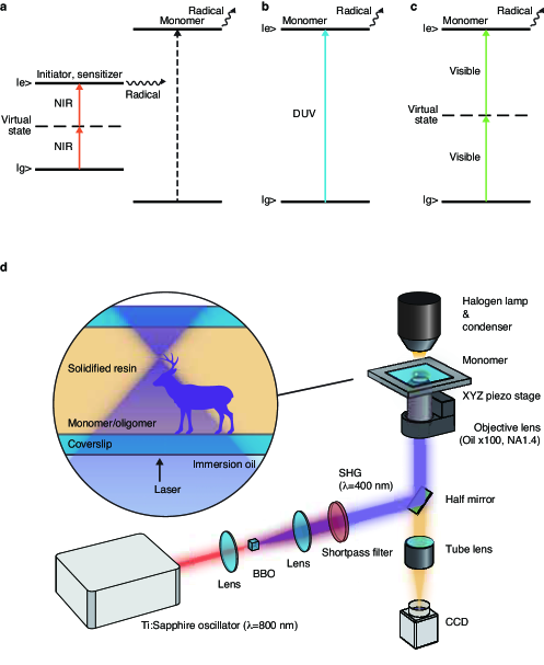
To trigger polymerization chain reactions, photo-initiators and sensitizers that have a transition energy corresponding to the two-photon energy of NIR light are added in monomer solution. However, the addition of initiators, for example, in fabricating gelatin hydrogel scaffolds for tissue engineering shows problematic cytotoxicity during cell culturing Li et al. (2013); Williams et al. (2005), suggesting the addition of initiators can affect properties of the fabricated devices. On the other hand, the wavelength shorter than 220 nm covers absorption bands of double bonds including C=C, C=O, benzene ring, and so on (Supplementary Table S1). As shown in Fig. 1(b), DUV wavelength as short as 200 nm allows direct excitation of the chemical bonds natively existing in organic molecules, which enables polymerization without using initiators. Excited with energetically equivalent two-photon absorption (Fig. 1(c)), initiator-free DUV polymerization can be realized with visible excitation wavelength. To the best of our knowledge, the present work is the first to use natively existing double bonds in organic molecules for TPP nanofabrications.
Figure 1(d) illustrates the optical setup (details in the Methods). Briefly, femtosecond pulse laser from Ti:sapphire oscillator operated at NIR wavelength (800 nm) is frequency-doubled to produce visible femtosecond pulse (400 nm) and focused by a high NA objective (NA1.4) into a sample cell containing liquid monomer/oligomer. The position of the sample cell is scanned three-dimensionally in with respect to the position of the laser focus spot to pile up voxels to form a structure.
As a material for proof-of-concept, we used acrylate oligomer, di-pentaerythritol hexaacrylate (DPHA) (Fig. 2(a), inset), which is a popular cross-linker material Kawata et al. (2001); Maruo et al. (1997).
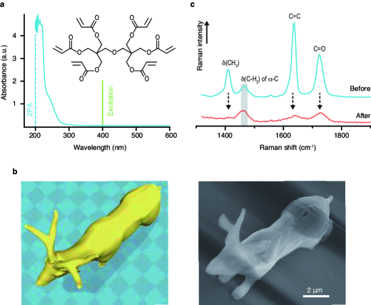
DPHA is viscous liquid in room temperature and has six conjugated acryloyl moieties which become radical sites upon the cleavage of the C=C and C=O double bonds. The absorption spectrum of DPHA is shown in Fig. 2(a). A pronounced absorption is seen in the DUV wavelength region from 200 to 230 nm, attributed to C=C and C=O functional groups in DPHA, while no absorption is seen at a wavelength of 400 nm where our femtosecond laser for TPP-excitation is tuned.
We focused 400 nm-femotosecond laser into the liquid oligomer and scan the focus spot along with the surface of a deer model (Fig. 2(b), left panel). After the scan is completed, the sample was taken out of the residual liquid oligomer and observed by SEM. The result is shown in the right panel in Fig. 2(b). The deer sculpture stands with the shape well reproducing the CAD model (left panel). The length of body is about 10 m. The horns and the branches are free-standing with a diameter of about 300 nm, demonstrating the capability to build high-aspect-ratio structures.
To investigate how DPHA undergoes chemical structural changes upon the laser irradiation, we measured Raman spectra of DPHA before and after the laser irradiation (details in the Methods). The measured Raman spectra are shown in Fig. 2(c) with the lines in blue and red indicate the spectra before and after laser irradiation, respectively. The Raman spectrum of DPHA shows four distinctive peaks (blue spectrum). The peaks at 1402 cm-1 is assigned to the in-plane deformation mode of methylene group (CH2); 1460 cm-1, the deformation mode of (C–H2) in –C; 1630 cm-1 and 1720 cm-1, the stretching modes of C=C and C=O, respectively Edwards et al. (2006); Willis et al. (1969). Assuming C–H2 in –C is not involved in polymerization reaction due to the transparency (no absorption) against DUV light, we used the peak intensity of (C–H2) in –C at 1460 cm-1 as an internal reference to normalize Raman intensities. As seen from Fig. 2(c), we observed a drastic decrease in the peak intensity of C=C at 1630 cm-1, indicating the consumption of C=C bond to form C–C networks. The C=C consumption is accompanied by cleavage of C=O bond that is conjugated with the C=C bond Baldacchini et al. (2009), which manifests itself as a simultaneous decreasing of C=O peak at 1720 cm-1. The decreasing intensity of CH2 band at 1402 cm-1 is explained by the cleavage of C=C bond connecting the CH2 group.
The applied laser intensity to obtain the deer sculpture was 300 kW/cm2 with the exposure time of 4 ms at each scanning step. The laser intensity required for initiating TPP process was found to be seven times smaller than the value used for previous NIR-excited-TPP for the same DPHA but added with photo-initiator and a photo-sensitizer (2.1 MW/cm2) Ushiba et al. (2014). In case of NIR excitation, the photo-initiator occupies only 1 wt% of monomer/cross-liner solution Ushiba et al. (2014), whereas in case of DUV-TPP, all oligomer molecules inside the laser focus work as initiator. The highly concentrated radicals generated by DUV absorption explains the unexpectedly high efficiency of DUV-TPP.
Previously, Parkatzidis et al. reported TPP of pre-synthesized gelatin methacrylamide without addition of initiators using an excitation wavelength of 520 nm Parkatzidis et al. (2018). In the previous work using 520 nm, the reported applied laser intensity at the focus was on the order of tens of TW/cm2 Parkatzidis et al. (2018), which is more than eight orders of magnitudes higher than that we applied for polymerizing methacrylate oligomers with 400 nm excitation. In the former case, the two-photon energy of 520 nm laser (i.e. 260 nm) is hardly overlaps with the molecular absorption Parkatzidis et al. (2018), while in our case, two-photon energy of 400 nm (i.e. 200 nm) falls at the peak position of absorption (Fig. 2(a)). The huge difference in required laser intensity indicates that the efficiency of two-photon polymerization is highly enhanced by using two-photon excitation energy consistent with one-photon absorption energy of molecules.
To rationalize laser intensity and resolution in DUV-TPP, we systematically measure the voxel size at different irradiation intensities and times. The SEM images of voxels are shown in Fig. 3(a) for the exposure power of 380, 500 and 630 kW/cm2 and time of 512, 256, 128, 64, and 32 ms.
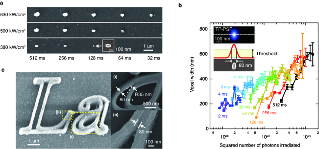
The polarization of the excitation laser was linear and horizontally oriented, resulting in slightly elongated shape of voxels along with the polarized direction. It is clearly seen that the size of voxel is smaller for lower laser intensity and shorter exposure time. The smallest voxel was 80 nm in width, which was obtained for the laser intensity of 380 kW/cm2 and the exposure time of 128 ms. Below 380 kW/cm2, we were unable to find a voxel on the substrate, partly because smaller voxels would have been rinsed off during the cleaning process before SEM observation. For laser intensity of 630 kW/cm2 and the exposure time of 512 ms, the voxel size increased as much as 600 nm, which is larger than the size of laser spot given by the wavelength of 400 nm and NA1.4. Such large voxel was produced plausibly as a result of diffusion of radicals out of the laser focus Gleeson and Sheridan (2009); Zhou et al. (2015).
In Fig. 3(b), we plotted the voxel widths as a function of the squared number of photons irradiated to sample for different exposure time and intensities. The voxel width phenomenologically lies linear-proportional to the logarithm of the squared number of photons, indicating a strong non-linearity. For the fixed exposure time, the voxel width increases with increasing numbers of photon dose. We numerically calculated two-photon point spread function (TP-PSF) with the condition of wavelength of 400 nm and objective NA of 1.4. The resulting image of TP-PSF and the sectional line profile at the center are shown in the inset in Fig. 3(b). The TP-PSF has a full-width at half-maximum of 139 nm and 99 nm in parallel and perpendicular to the polarization direction, respectively. The observed smallest voxel width of 80 nm falls to a fraction of the calculated TP-PSF width of 99 nm. Considering the thresholding effect in photo-polymerization Kawata et al. (2001); Zhou et al. (2015) (also illustrated in the inset), the experimentally obtained voxel size of 80 nm reasonably agreed with the value numerically predicted from PSF.
To evaluate the spatial resolution of DUV-TPP, we show in Fig. 3(c) the SEM images of “La” letters drawn by DUV-TPP. The edge of the fabricated structure is remarkably sharp. Regions marked by (i) and (ii) are magnified and shown in the right upper and lower panels, respectively. The line width of 80 nm and a curved feature with a radius of curvature of 35 nm were observed in panel (i). In panel (ii), the space resolved between two crossing lines was about 80 nm, reasonably consistent with the smallest voxel size observed in Fig. 3(a). From these results, we concluded the finest resolved space is about 80 nm wide, corresponding to of excitation wavelength. The value of spatial resolution achieved with 400 nm excitation is superior to those previously reported for NIR-excitation Kawata et al. (2001) and 520 nm excitation Malinauskas et al. (2010).
Next, we apply initiator-free DUV-TPP to another class of material, inorganic metal oxides. Zirconium dioxide (ZrO2) exhibits large dielectric constant, known as high- material, and has potentially important applications in optical Liang et al. (2009) and electronic devices Park et al. (2011). Titanium dioxide (TiO2) also has a wide range of applications including photocatalysis Fujishima et al. (2008). There are numbers of techniques to make a planer thin film of metal oxides Liang et al. (2009), but a technique to form 3D nanostructures will be demanded, for example, to integrate metal oxides nanostructures in electronic devices.
Figure 4(a) explains the reaction pathway to synthesize metal oxides.
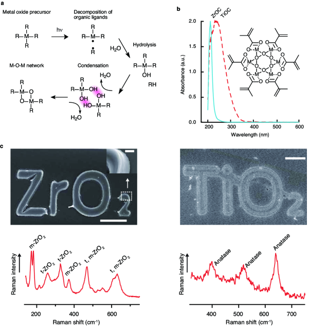
It starts from metal oxo cluster (MOC) as a precursor (Method). The MOC in solution phase has organic ligands surrounding the metal cation. Photo excitation of ligands promotes decomposition of organic ligands. The generated radicals leads hydrolysis and condensation, and metal-oxygen-metal (MOM) network is created. We patterned the MOM structures by DUV-TPP. No photo-initiator and sensitizers were added to MOC solution. The solidified MOM structures were taken out of residual unsolidified MOC solution and finally thermally annealed to crystalize metal oxides.
The absorption spectrum of synthesized MOC solution is shown in Fig. 4(b) for ZrOC and TiOC. ZrOC has an absorption peak at 210 nm, attributed to C=C bond from ligand MMA. TiOC has an absorption originated from the C=C bond from ligand MMA, but the peak wavelength is slightly shifted to 230 nm and the width is broader. In both cases, the absorption band of the MOC overlaps with the two-photon excitation energy with a laser wavelength centered at 400 nm.
Figure 4(c) shows the SEM image of the fabricated structures with letters “ZrO2” and “TiO2” from the respective ZrOC and TiOC solutions. The inset of “ZrO2” image shows the magnified portion in the side wall of the letter “2”. A layered feature in vertical direction is seen on the wall of the letter, demonstrating the 3D capability. The structures were slightly deformed during thermal annealing. Further optimization is needed to avoid deformation and cracks during annealing process Kozuka (2006). To identify the chemical components of the obtained structures, we took Raman spectra from a part of the structures. The result is shown in the bottom panels in Fig. 4(c). The spectrum in the left panel shows characteristic Raman peaks. Comparing these peaks to the literatures Li et al. (2001), we found the peaks at 269 and 310 cm-1 are assigned to the Raman modes of tetragonal phase t-ZrO2, Eg, and B1g, respectively Li et al. (2001). The other peaks at 178, 189, and 380 cm-1 are assigned to the monoclinic phase m-ZrO2 Li et al. (2001). Consequently, the fabricated “ZrO2” structure is composed of polycrystalline ZrO2 that has both tetragonal and monoclinic phases. The remaining peaks at 469 and 640 cm-1 are reported for both t- and m-ZrO2 Li et al. (2001). In the measured spectrum, the Raman intensity at 640 cm-1 is stronger than that at 469 cm-1, which is an indication that the obtained structure is t-ZrO2-phase rich. The dominant phase is affected by the annealing temperature of the crystallization process Li et al. (2001). No remaining peak of ligand MMA was observed in the spectrum. The Raman spectrum from “TiO2” structure shows three distinct peaks, 4008, 517, and 639 cm-1. These peaks are all consistent with the reported Raman modes in TiO2 anatase crystal Berger et al. (1993). The results of Raman spectroscopy show the obtained structures are indeed ZrO2 and TiO2 crystals. To the best of our knowledge, this is the first to build structures made of inorganic metal compound by direct laser writing.
In order to gain further insight about applicability and limitation of DUV-TPP, we have tested polymerization of MMA monomers with the experimental conditions similar to the one used for MMA oligomers. As a result, however, we were unable to find any structure remaining on the substrate after laser irradiations. Unlike acrylate oligomer having six acryloyl moieties within a molecule, MMA monomer has only a single set of C=C and C=O. Since there are a limited number of double bonds available to form crosslinks with each other, MMA monomer has less chance to form polymer matrix during a given lifetime of radicals than MMA oligomer does. Applying DUV-TPP to simpler molecules having a small numbers of radical sites would become possible by increasing monomer concentrations or reducing a solution viscosity, which will be a future work.
Another limitation of DUV-TPP from material point of view is the molecules with conjugated system. It is known that the electronic transition energy of conjugated system becomes lower when the number of conjugated double bond increases. When polymerized molecules absorbs visible light, one-photon polymerization dominates over two-photon polymerization, which spoils 3D fabrication capability. As a solution, single-pulse polymerization Mills et al. (2013) may work.
There are reports of cytotoxicity during cell culturing on gelatin hydrogel scaffolds fabricated with photo-initiators Li et al. (2013); Williams et al. (2005). While chosing appropriate initiator is a crucial issue in NIR-excited TPP, initiator-free DUV-TPP is a promising alternative approach for applications that require high degree of bio-compatibility and nontoxicity, such as cell culturing scaffolds, regenerative medicines, drag delivery containers Xing et al. (2015); Lee et al. (2019). Experiments are ongoing to fabricate structures with collagen type I molecules using DUV-TPP, and obtained solidified structures after irradiation of 400 nm laser (Supplementary Information and Fig. S1).
As demonstrated with metal oxides structures, DUV-TPP widens the choice of materials for 3D nanofabrications. Using synthesized ligand MMA as cross-linker, DUV-TPP is potentially applicable to 3D nanofabrications of wide variety of inorganic materials including metals Tanaka et al. (2006) and SiO2 Kotz et al. (2017). With the material versatility, an interesting extension is fabrication of multi-material nanodevices Lind et al. (2017); Kwon et al. (2018); Dietrich et al. (2018)
Finally, multi-photon excitation with visible laser offers facile and convenient way to realize maskless 3D printing with equivalent operating wavelength in DUV. This enables rapid prototyping or testing of arbitrary macro- and microstructures and devices for many applications in both industry and fundamental researches. Since laser intensity required is relatively small in DUV-TPP, writing throughput can be improved by parallelization using multi-spots scheme. Strong nonlinearity involved in polymerization process would be utilized to further improve the spatial resolution to the level comparable to the state-of-the-art DUV lithography and EBL Gan et al. (2013); Xing et al. (2007).
In summary, we have described DUV photo-polymerization with two-photon excitation at visible wavelength. Without using photo-initiators and sensitizers, direct multi-photon excitation of molecular native absorption has shown to induce photo-polymerizations. This expands the scope of TPP-polymerizable materials from traditional organic materials to inorganic materials as demonstrated by fabrication of both acrylate and metal oxides nanostrucutures in 3D.
I acknowledgments
This work was supported partly by the JSPS KAKENHI Grant Numbers JP17K05076, and JSPS Core-to-Core Program on Advanced Nanophotonics.
II Author contributions
A.T. and K.F. conceived the project and designed the experiments. A.N. performed the experiments and R.O. provided the calculation data. S.K. participated in planning the experiments and provided conceptual input. The manuscript was written by A.T., A.N. and K.F.
References
- Wagner and Harned (2010) C. Wagner and N. Harned, Nat. Photonics 4, 24 (2010).
- Totzeck et al. (2007) M. Totzeck, W. Ulrich, A. Goehnermeier, and W. Kaiser, Nat. Photonics 1, 629 (2007).
- Yamanaka et al. (2015) M. Yamanaka, K. Saito, N. I. Smith, Y. Arai, K. Uegaki, Y. Yonemaru, K. Mochizuki, S. Kawata, T. Nagai, and K. Fujita, J. Biomed. Opt. 20, 101202 (2015).
- Maiti et al. (1997) S. Maiti, J. Shear, R. Williams, W. Zipfel, and W. Webb, Science 275, 530 (1997).
- Kawata et al. (2001) S. Kawata, H. B. Sun, T. Tanaka, and K. Takada, Nature 412, 697 (2001).
- Gan et al. (2013) Z. Gan, Y. Cao, R. A. Evans, and M. Gu, Nat. Commun. 4, 2061 (2013).
- Maruo et al. (1997) S. Maruo, O. Nakamura, and S. Kawata, Opt. Lett. 22, 132 (1997).
- Li et al. (2013) Z. Li, J. Torgersen, A. Ajami, S. Muehleder, X. Qin, W. Husinsky, W. Holnthoner, A. Ovsianikov, J. Stampfl, and R. Liska, RSC Advances 3, 15939 (2013).
- Williams et al. (2005) C. G. Williams, A. N. Malik, T. K. Kim, P. N. Manson, and J. H. Elisseeff, Biomaterials 26, 1211 (2005).
- Edwards et al. (2006) H. G. M. Edwards, K. S. Johal, and A. F. Johnson, Vibrational Spectroscopy 41, 160 (2006).
- Willis et al. (1969) H. A. Willis, V. J. I. Zichy, and P. J. Hendra, Polymer 10, 737 (1969).
- Baldacchini et al. (2009) T. Baldacchini, M. Zimmerley, C.-H. Kuo, E. O. Potma, and R. Zadoyan, J. Phys. Chem. B 113, 12663 (2009).
- Ushiba et al. (2014) S. Ushiba, K. Masui, N. Taguchi, T. Hamano, S. Kawata, and S. Shoji, Sci. Rep. 5, 17152 (2014).
- Parkatzidis et al. (2018) K. Parkatzidis, E. Kabouraki, A. Selimis, M. Kaliva, A. Ranella, M. Farsari, and M. Vamvakaki, Macromol. Mater. Eng. 34, 1800458 (2018).
- Gleeson and Sheridan (2009) M. R. Gleeson and J. T. Sheridan, J. Opt. Soc. Am. B 26, 1736 (2009).
- Zhou et al. (2015) X. Zhou, Y. Hou, and J. Lin, AIP Advances 5, 030701 (2015).
- Malinauskas et al. (2010) M. Malinauskas, V. Purlys, M. Rutkauskas, A. Gaidukevičiūtė, and R. Gadonas, Lithuanian Journal of Physics 50, 201 (2010).
- Liang et al. (2009) L. Liang, Y. Xu, D. Wu, and Y. Sun, Materials Chemistry and Physics 114, 252 (2009).
- Park et al. (2011) Y. M. Park, J. Daniel, M. Heeney, and A. Salleo, Adv. Mater. 23, 971 (2011).
- Fujishima et al. (2008) A. Fujishima, X. Zhang, and D. A. Tryk, Surf Sci Rep 63, 515 (2008).
- Kozuka (2006) H. Kozuka, Journal of Sol-Gel Science and Technology 40, 287 (2006).
- Li et al. (2001) M. J. Li, Z. H. Feng, G. Xiong, P. L. Ying, Q. Xin, and C. Li, J. Phys. Chem. B 105, 8107 (2001).
- Berger et al. (1993) H. Berger, H. TANG, and F. Lévy, Journal of Crystal Growth 130, 108 (1993).
- Mills et al. (2013) B. Mills, J. A. Grant-Jacob, M. Feinaeugle, and R. W. Eason, Opt. Express 21, 14853 (2013).
- Xing et al. (2015) J.-F. Xing, M.-L. Zheng, and X.-M. Duan, Chem. Soc. Rev. 44, 5031 (2015).
- Lee et al. (2019) A. Lee, A. R. Hudson, D. J. Shiwarski, J. W. Tashman, T. J. Hinton, S. Yerneni, J. M. Bliley, P. G. Campbell, and A. W. Feinberg, Science 365, 482 (2019).
- Tanaka et al. (2006) T. Tanaka, A. Ishikawa, and S. Kawata, Appl. Phys. Lett. 88, 081107 (2006).
- Kotz et al. (2017) F. Kotz, K. Arnold, W. Bauer, D. Schild, N. Keller, K. Sachsenheimer, T. M. Nargang, C. Richter, D. Helmer, and B. E. Rapp, Nature 544, 337 (2017).
- Lind et al. (2017) J. U. Lind, T. A. Busbee, A. D. Valentine, F. S. Pasqualini, H. Yuan, M. Yadid, S.-J. Park, A. Kotikian, A. P. Nesmith, P. H. Campbell, et al., Nat. Mater. 16, 303 (2017).
- Kwon et al. (2018) J. Kwon, Y. Takeda, R. Shiwaku, S. Tokito, K. Cho, and S. Jung, Nat. Commun. 10, 54 (2018).
- Dietrich et al. (2018) P. I. Dietrich, M. Blaicher, I. Reuter, M. Billah, T. Hoose, A. Hofmann, C. Caer, R. Dangel, B. Offrein, U. Troppenz, et al., Nat. Photonics 12, 241 (2018).
- Xing et al. (2007) J.-F. Xing, X.-Z. Dong, W.-Q. Chen, X.-M. Duan, N. Takeyasu, T. Tanaka, and S. Kawata, Appl. Phys. Lett. 90, 131106 (2007).
- Stehlin et al. (2014) F. Stehlin, F. Wieder, A. Spangenberg, J.-M. Le Meins, and O. Soppera, J. Mater. Chem. C 2, 277 (2014).
- Maruo et al. (2009) S. Maruo, T. Hasegawa, and N. Yoshimura, Opt. Express 17, 20945 (2009).
III Methods
Optical configuration of two-photon polymerizations
An experimental setup is shown in Fig. 1(d). A Ti:sapphire oscillator system (Tsunami, Spectra Physics) was used as a light source. The fundamental 800 nm NIR laser beam (80 fs pulse width, 82 MHz repetition rate, 10 nm full-width at half- maximum (FWHM) bandwidth) was focused to a -BaB2O4 (BBO) crystal using a lens (f=50 mm) to provide a visible laser pulse having a center wavelength at 400 nm. The frequency-doubled laser beam was expanded and collimated by a lens (f=100 mm). The laser was passed through a short-pass filter to cut the fundamental 800 nm beam, and the visible laser coupled into an Olympus IX71 inverted microscope on which a sample cell is set. A 100 oil immersion objective (NA 1.4, Zeiss) was used to focus the visible light into sample solution. The sample stage moves three-dimensionally in using a piezo stage (P-517, Physik Instrument) which was controlled by home-written programs.
Experimental procedures for two-photon polymerizations
Acrylate sample preparation
For the two-photon polymerizations of acrylate oligomer shown in Fig. 2, we used di-pentaerythritol hexaacrylate (DPHA) (Kyoeisha Chemical Co., Ltd.), which is a popular acrylate oligomer. The stock solution of DPHA was used as received. The absorption spectra was measured using UV-Vis-NIR spectrometer (UV-3600, Shimadzu). A droplet of DPHA solution was sealed between a pair of two coverslips (Matsunami glass) with a 50 m-thick silicone sheet sandwiched as a spacer.
Metal oxio-cluster (MOC) preparation
Zr oxio-cluster (ZrOC) was prepared following the procedures described in the literatureStehlin et al. (2014). Briefly, Zr alkoxide (zirconium(IV) propoxide (ca. 70% in 1-Propanol, Tokyo Chemical Industry Co., Ltd.) precursor was mixed with methyl methacrylate (MMA) (Tokyo Chemical Industry Co., Ltd.) at the molar ratio of Zr:MMA=1:8. After stirring of five minutes, a volume of 20 L of 1-propanol (Tokyo Chemical Industry Co., Ltd.) was added. After ten minutes of stirring, de-ionized water was added at a molar ratio of Zr:H2O=1:20. Finally, an additional volume of 20 L 1-propanol was added to adjust the viscosity of solution.
Ti oxio-cluster (TiOC) was prepared with the same procedure except for using Ti alkoxide (titanium (IV) isopropoxide, 97%, Aldrich) in place of Zr alkoxide.
Fabrications
To produce a three dimensional structure, we first generate a computer model using a computer-aided design (CAD) software. The CAD model is then converted to a batch of G-codes using a slicer software (Cura), and fed into a scan controller to move the sample stage. After the scan is completed, residual liquid oligomer is rinsed off using ethanol followed by a supercritical cleaning that uses CO2 supercritical fluid having extremely small viscosity to reduce a risk of collapsing nanostructures during cleaning process Maruo et al. (2009).
For fabrication of metal oxides structures, the experimental conditions used for DUV-TPP were the same as the one used for the fabrication of acrylic polymer except for the laser intensity 130 kW/cm2 and exposure time of 4ṁs per voxel. As a solvent for rinsing the residual liquid MOC, cyclohexanone (Tokyo Chemical Industry) was used. After cleaning, the MOM structure was thermally annealed at a temperature of 600C∘ for two hours to completely decompose the organic ligands and to crystalize the metal oxide structure.
SEM observation
To observe the fabricated structures, the sample was coated with a thin platinum layer of 2 nm thick. A field emission scanning electron microscope (S-4800, Hitachi) was used.
Raman microscopy characterization
Raman spectra were taken using a Raman microscope (Raman-11, Nanophoton). The excitation laser wavelength was 532 nm and a 100 objective lens (NA0.9, Nikon) was used. The laser power was 100 mW and the exposure time was 1 s.