Integrating Transposable Elements in the 3D Genome
Abstract
Chromosome organisation is increasingly recognised as an essential component of genome regulation, cell fate and cell health. Within the realm of transposable elements (TEs) however, the spatial information of how genomes are folded is still only rarely integrated in experimental studies or accounted for in modelling. Here, we propose a new predictive modelling framework for the study of the integration patterns of TEs based on extensions of widely employed polymer models for genome organisation. Whilst polymer physics is recognised as an important tool to understand the mechanisms of genome folding, we now show that it can also offer orthogonal and generic insights into the integration and distribution profiles (or “topography”) of TEs across organisms. Here, we present polymer physics arguments and molecular dynamics simulations on TEs inserting into heterogeneously flexible polymers and show with a simple model that polymer folding and local flexibility affects TE integration patterns. The preliminary discussion presented herein lay the foundations for a large-scale analysis of TE integration dynamics and topography as a function of the three-dimensional host genome.
I Background
Transposable elements (TEs) are DNA sequences that can move from one location of the genome to another. By being able to spread their own DNA across the genome independent of the cell’s replication cycle Deniz et al. (2019), TEs represent the majority of genomic content in most eukaryotes. For example, they comprise 85 % of the maize genome Schnable et al. (2009) and up to 50 % of primate genomes Lee et al. (2016). As such, TE activity is a major driver of phenotypic and genotypic evolution Chuong et al. (2017); Bousios and Gaut (2016) and affects key biological processes from meiosis and transcription to immunological responses Schatz and Ji (2011). At the same time, TEs have been associated with various diseases and cancer in humans Payer and Burns (2019).
Most TEs transpose via cut-and-paste or copy-and-paste mechanisms that can both result in a net increase of the TE copy number Wicker et al. (2007). Amplification phases, or bursts, of TEs can occur multiple times in the evolutionary history of the host and may produce hundreds if not thousands of new copies within short time windows Lu et al. (2017); Sanchez et al. (2017); Bousios and Gaut (2016).
Most TEs exhibit some level of integration site selection, from very specific target sites Eickbush and Eickbush (2014) to non-random but more dispersed genomic biases Schroder et al. (2002); Craigie and Bushman (2012); Kvaratskhelia et al. (2014). Short DNA motifs, epigenetic marks and nuclear proteins have been associated with such integration site preferences. For example, yeast Ty1 retrotransposons integrate upstream of Pol III-transcribed genes through a direct interaction between the integrase complex and the AC40 subunit of Pol III Cheung et al. (2016). In contrast, in plants and fungi, the integrase of certain Gypsy retrotransposons contains a chromodomain that can bind to repressive histone marks and aid insertion into heterochromatin Gao et al. (2008).
While the role of protein tethering and DNA motifs in TE integration is well established by now, it remains elusive how the three-dimensional (3D) structure of chromosomes and the nuclear environment is affecting TE spreading in host genomes. Chromosome folding and nuclear organisation have been shown to play key roles in all major DNA related processes Lieberman-Aiden et al. (2009); Dekker et al. (2017); Nagano et al. (2013); Kind et al. (2013); Flyamer et al. (2017); Nozawa et al. (2017); Dong et al. (2017); Stadhouders et al. (2019), from transcription and replication to DNA repair, and it appears only natural to expect that transposition will also be affected by the 3D organisation genome.
A number of recent reports have highlighted that TE activity is involved in shaping 3D chromosome structure. For example, TEs of diverse families have been implicated in the establishment and maintenance of insulator boundaries between so-called “topologically associated domains” (TADs) Dixon et al. (2012); Wang et al. (2015); Raviram et al. (2018); Zhang et al. (2019); Kruse et al. (2019); Sun et al. (2018). Furthermore, TE amplification is suggested to account for a significant amount of binding motifs for the CTCF Kunarso et al. (2010); Schmidt et al. (2012); Choudhary et al. (2018) protein, a key regulator of 3D chromosome organisation Nora et al. (2017). The involvement of TEs in the establishment of evolutionarily conserved long-range chromosomal interactions has been shown in different organisms Cournac et al. (2016); Winter et al. (2018) and some of these TE-mediated interactions appear to be of functional importance in gene regulation. In the plant Arabidopsis thaliana, TEs are enriched at genomic hubs of long-range chromosomal interactions with anticipated functional roles in silencing of foreign DNA elements Grob and Grossniklaus (2019).
Arguably, how TEs contribute to genome folding will certainly receive more attention in the future, but it is an equally fundamental question for both genome and TE biology to understand how genome folding affects TE integration preferences. For example, depending on the 3D organisation of chromosomes, a new TE copy that enters the nucleus from the cytoplasm will come across distinct parts of the genome in terms of their accessibility and organisation compared to a preexisting TE insertion that integrates into a different genomic locus. Intriguingly, retroelements (including retrotransposons and retroviruses) and DNA transposons have different replication and transposition pathways Sultana et al. (2017), which implies that genome architecture may have a different impact in each TE type.
There is a clear gap in the experimental and theoretical work on understanding the impact of genome architecture on integration of TEs. Models of TE amplification dynamics have traditionally been based on population-based approaches Charlesworth and Charlesworth (1983), which typically set up systems of (stochastic) ordinary differential equations accounting for generic competing elements during TE expansion Xue and Goldenfeld (2016); Le Rouzic et al. (2007); Roessler et al. (2018). Few works, instead, have considered the 1D distribution of nucleosomes along the genome in order to predict preferential sites of HIV integration Naughtin et al. (2015). Both these classes of models necessarily neglect the multi-scale 3D organisation of the genome, i.e. from nucleosomes to TADs and from compartments to chromosome territories Bonev et al. (2017); Cremer and Cremer (2001). Because of this, they are not suited to predict the “topography” of TEs, i.e. the pattern of genomic sites in which TEs will preferentially integrate.
Here, we introduce and discuss a computational model based on principles of polymer physics, which aims to dissect the interplay between genome organisation and biases in TE integration. The last few years, polymer models have been proved to be very successful tools to rationalise 3D genome folding Mirny (2011); Brackley et al. (2012); Michieletto et al. (2016, 2018); Jost et al. (2014); Bianco et al. (2018); Sanborn et al. (2015). We first briefly review the existing framework of polymer models, then discuss a recent development of such a model to understand the physical principles of HIV integration, and finally present preliminary data obtained by extending these models to the case of TEs. We conclude by discussing potential future directions in this unexplored line of research.
II Main Text
II.1 Biophysical Principles of Genome Folding
While genomes are, biologically speaking, the carrier of genetic information they also are, physically speaking, long polymers. Polymers are well-known objects that have been studied for several decades in particular in relation to industrial applications, such as rubbers Doi1988; Gennes (1979). Pioneers in polymer physics realised a long time ago that they obey “universal” laws that are independent of their chemical composition Flory (1953). For instance, the way a long polystyrene molecule folds in space must be identical, statistically speaking, to that of a long DNA molecule in the same conditions. Because of this, polymer physicists typically employ “coarse-grained” approaches, which blur the chemical details and only retain the necessary ingredients that allow the formulation of simple and generic (universal) frameworks. Universality then implies that these coarse-grained models have predictive power for a broad range of systems with different chemistry.

Coarse-graining several base-pairs and groups of atoms into mesoscopic beads (see Fig. 1A), while retaining the salient physical behaviour of DNA, allows the formulation of computational models that can reliably predict the spatial organisation of whole chromosomes from minimal input – such as epigenetic patterns and generic binding proteins Jost et al. (2014); Brackley et al. (2016a); Di Pierro et al. (2016); Nuebler et al. (2018); Buckle et al. (2018); Brackley et al. (2017); Fudenberg et al. (2016); Sanborn et al. (2015) – and disentangle the contribution of different classes of proteins to genome folding Orlandini et al. (2019); Nuebler et al. (2018); Pereira et al. (2018).
These computational models, coupled to Chromosome Conformation Capture (and its higher order variants, such as HiC) experiments Lieberman-Aiden et al. (2009); Dekker et al. (2017), are providing new information on the spatio-temporal organisation of the genome in different conditions, such as healthy Brackley et al. (2016b); Buckle et al. (2018), senescent Chiang et al. (2018) and diseased Bianco et al. (2018) cells, or even during cell-fate decisions Stadhouders et al. (2019) and reprogramming Stadhouders et al. (2018). For instance, polymer models can rationalise features such as TADs Benedetti et al. (2014); Fudenberg et al. (2016), compartments Brackley et al. (2016a); Jost et al. (2014) and loops Fudenberg et al. (2016); Sanborn et al. (2015) seen in experimental HiC maps Rao et al. (2014). Importantly, these works are proving that traditionally physical phenomena such as liquid-liquid and polymer-polymer phase separation Brangwynne et al. (2009); Caragine et al. (2018); Cho et al. (2018); Brangwynne et al. (2015); Michieletto et al. (2016); Erdel and Rippe (2018), gelation Michieletto and Gilbert (2019); Khanna et al. (2019), emulsification Hilbert et al. (2018) and viscoelasticity Lucas et al. (2014); Shin et al. (2018) may be found in ubiquitous and key biological processes such as transcription, replication, mitosis, RNA splicing and V(D)J recombination to name a few. Polymer models are thus providing the community with a physical lens through which they may interpret complex data, and a quantitative framework to generate de novo predictions. In light of this, we here propose that the use of polymer models may shed new light into the relationship between TE transposition and 3D organisation. Earlier this year Michieletto et. al. made a significant step in this direction by formalising a polymer-based model for understanding the site selection features displayed by HIV integration in the human genome Michieletto et al. (2019). Below, we briefly review this work, which will then be used as a stepping stone to formalise a polymer model for TE expansion.
II.2 A Polymer Model for HIV Integration
One of the least understood features of HIV integration is that its integration patterns display markedly non-random distributions both along the genome Wang et al. (2007) and within the 3D nuclear environment Marini et al. (2015). HIV displays a puzzling bias for nucleosomes Pruss et al. (1994a, b), gene-rich regions Wang et al. (2007) and super-enhancer hotspots Lucic et al. (2019) that has defied comprehension for the past three decades. Clearly, from the perspective of a retrovirus such as HIV, integrating in frequently transcribed regions is evolutionary advantageous. But how is this precise targeting achieved?
For the past decades, the working hypothesis to address this important question was that there must exist specialised factors or protein chaperones that guide HIV integration site selection. Prompted by this hypothesis, much work has been devoted to discover and identify such proteins Kvaratskhelia et al. (2014). Some factors, such as the lens‑epithelium‑derived growth factor Ciuffi et al. (2005) (LEDGF/p75) have been proposed as potential candidates for this role but even knocking-down their expression could not completely remove the bias for gene-rich regions Schrijvers et al. (2012). Additionally, the preference of HIV to integrate in nucleosomes – oppositely to naked DNA – was shown in vitro using minimal reaction mixtures Pruss et al. (1994b); Benleulmi et al. (2015).
We recently put forward a different working hypothesis to address the bias of HIV integration site selection: could there be universal (non-system specific) physical principles that – at least partially – can contribute to biasing the site-selection of HIV integration? Prompted by this hypothesis, we decided to propose a polymer-based model in which retroviral integration occurs via a stochastic and quasi-equilibrium topological reconnection between 3D proximal polymer segments. In other words, whenever two polymer segments (one of the host and one of the invading DNA) are nearby in 3D we assign a certain probability for these two segments to reconnect, based on the difference in energy between the old and new configurations. This strategy is known as a Metropolis criterion and it satisfies detailed balance, thus ensuring that the system is sensitive to the underlying free energy Frenkel and Smit (2001). [Note that in vitro HIV integrase works without the need of ATP Pruss et al. (1994b), and we therefore assume that the integration process is in (or near) equilibrium.] Finally, we impose that the viral DNA is stuck once integrated in the selected location and cannot be excised within the simulation time.
Within this simple model, we discovered that geometry alone may be responsible for a bias in the integration of HIV in nucleosomes (Fig. 1B-D). This is because the pre-bent conformation of DNA wrapped around the histone octamer lowers the energy barrier against DNA deformation required to integrate the viral DNA into the host (Fig. 1B, see also Refs. Serrao et al. (2014); Benleulmi et al. (2015); Pasi et al. (2016)). Further, by considering a longer region of a human chromosome folded as predicted by the above mentioned polymer models Brackley et al. (2016a); Michieletto et al. (2016, 2018), we discovered that at larger scales HIV integration sites obtained from experiments Wang et al. (2007) are predominantly determined by chromatin accessibility. Thus, by accounting for DNA elasticity and chromatin accessibility – two universal and cell unspecific features of genome organisation – our model could predict HIV integration patterns remarkably similar to those observed in experiments in vitro Pruss et al. (1994b, a) and in vivo Wang et al. (2007).
II.3 Extension to DNA Transposition
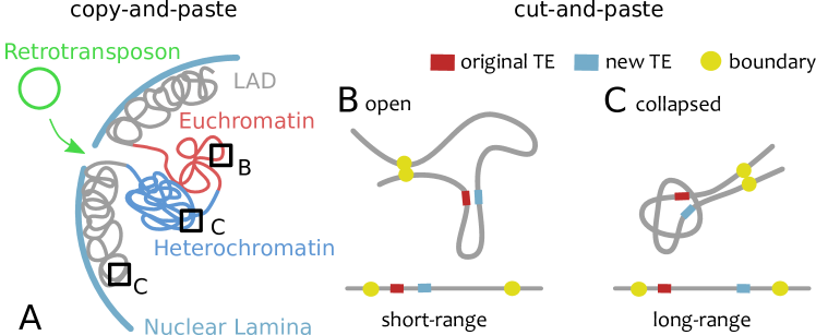
In light of the success of this polymer model, we now propose to extend it to understand the distribution of integration sites across the TE phylogeny. Importantly, different TEs have different integration strategies, which suggests that they are likely to interact differently with the 3D genome organization within the nucleus. TEs can be primarily distinguished based on the mechanism through which they proliferate, i.e. via “copy and paste” (class I or retrotransposons) or “cut and paste” (class II, DNA transposons). The former require an RNA intermediate to proliferate and thus exit the nuclear environment, whereas the latter simply relocate their DNA via endonuclease excision Wicker et al. (2007); Sultana et al. (2017).
Retroviruses, like HIV, are very similar to retrotransposons with the addition that they can exit to the extracellular space and invade other cells. Thus, a new copy of a retrotransposon (class I or copy-and-paste) or a retrovirus must travel from the periphery to the nuclear interior while the genome is “scanned” from the outside-in for integration sites (Fig. 2A). This implies that the global, nuclear-scale genome architecture is expected to be important for this re-integration process. For instance, Lamin Aassociated Domains (LAD) Kind et al. (2013) positioning with respect to nuclear pores, inverted versus conventional organisation Solovei et al. (2009), compartments Rao et al. (2014) and enhancer hot-spots Lucic et al. (2019) will likely play major roles for retrotransposons. On the contrary, a DNA transposon (class II or cut-and-paste) probes the genome in the immediate surrounding of its excision site and will diffuse from the inside-out (Fig. 2B-C). As a result of this, the mesoscale ( 1 Mbp) organisation of the genome may have a profound effect on the 1D genomic distribution of DNA transposons. For example, heterochromatin-rich chromatin is thought to be collapsed Boettiger et al. (2016); Michieletto et al. (2016) with a typical overall size that depends on the genomic length as ; on the other hand, euchromatin-rich compartments Rao et al. (2017) are more open Gilbert et al. (2004) and their size may be more similar to that of a random walk, i.e. scaling as . The contact probability of two genomic loci at distance can be estimated to scale as Mirny (2011) where is for collapsed polymers (such as heterochromatin), for ideal ones (such as euchromatin Boettiger et al. (2016)) and for self-avoiding walks Gennes (1979). Thus, a crude calculation would predict that a DNA transposon should re-integrate at distance with a probability that depends (through the exponent ) on the folding of the genome at these (TAD-size) length-scales. A similar effect is at play in the enhancement of long range contacts in oncogene-induced senescent cells Chiang et al. (2019).
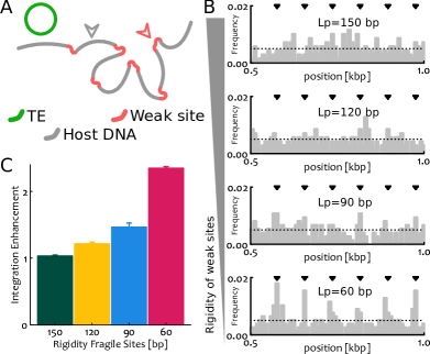
In addition to this contribution coming from large- and meso-scale folding, one may argue that there ought to be other complementary effects such as specific features of the integrase Taganov et al. (2004); Serrao et al. (2014) or tethering Gao et al. (2008) enzymes. These orthogonal elements are more local and are expected to equally affect both DNA transposons and retrotransposons. To investigate the role of local chromatin features on a generic integration event, we here performed original simulations on a heterogeneously flexible polymer that crudely mimics heterogeneous chromatin in vivo (Fig. 3A). Specifically, we consider a stretch of 1.6kbp long DNA with persistence length and regularly interspersed with weak sites that display a lower bending rigidity . This lower local DNA rigidity may be due to, for instance, to denaturation bubbles Fosado et al. (2017), R-loops Santos-Pereira and Aguilera (2015) or replication stress Chan et al. (2009). In these conditions – which may be reproduced in vitro by considering DNA with a sequence of bases that modulates its local flexibility Pruss et al. (1994a) – we ask what is the integration pattern displayed by an invading DNA element by counting the number of integration events in each segment of the polymer over many (1000) independent simulations. We observe that, by varying the value of the rigidity parameter from bp to bp, the integration patterns become less uniform, more periodic and reflecting the distribution of weak sites (Fig. 3B).
From these patterns we can compute the enhancement of integration in susceptible sites due to their different flexibility. This is simply the sum of integration frequencies in all susceptible sites divided by the one expected for a random distribution of events, i.e. where is the number of weak sites and the length of the polymer. This calculation is reported in Fig. 3C and shows that the enhancement increases with the flexibility of the susceptible sites. The output of these simulations may be readily measured in experiments in vitro on designed DNA and chromatin templates as done for HIV Pasi et al. (2016); Benleulmi et al. (2015), and may thus inform the mechanistic principles leading to DNA integration. Perhaps more importantly, however, these simulations suggest that the heterogeneity of the DNA (or chromatin) substrate in both mechanics and folding may affect TE expansion with potentially important and far-reaching consequences on the evolutionary paths and proliferative success of certain TEs in vivo.
III Conclusions
It is now becoming increasingly clear that cell function, health and fate are correlated to 3D genome folding Bonev et al. (2017); Stadhouders et al. (2019). TEs are intrinsically linked to 3D organisation as they are “living elements” within a complex multi-scale environment. In the last few years, there have been a handful of studies that started to interrogate how TEs shape genome organisation, from demarcating TAD boundaries Zhang et al. (2019); Sun et al. (2018); Kruse et al. (2019) to harboring binding sites for architectural proteins Choudhary et al. (2018). It is thus now realized that TEs have profound implications in the fate and health of a cell – not only via the traditional pathway of genomic instability and epigenetic silencing – but also through the global regulation of genome folding.
Now, while this crucial relationship will certainly receive more attention in the future, we argue that the other direction of the relationship, i.e. how 3D structure affects de novo TE insertions, is also of utmost importance. For example, biases in insertion patterns due to tissue-specific genome organisation in the germline (versus, for example, somatic cells) can create preferential pathways for genome evolution. We suggest that the dissection of this interplay could be done by employing a “perturb-and-measure” strategy of inducing TE expansion in a cell line whilst obtaining information on the 3D genome organisation and epigenetic states pre and post expansion. This approach will determine – also through the use of polymer physics models – which 3D and/or epigenetic features are associated with de novo TE insertions and thus detect insertion biases. Consequently, it will allow the generation of “topographical maps” of TE insertions in a given tissue-specific 3D genome organisation. Ultimately, understanding the preferential insertion of TEs may lead to a better understanding of genome and TE evolution or even inform better strategies to drive genomic variations in crops.
IV Acknowledgements
HWN, AB and DM acknowledge support of EPSRC and BBSRC through the Physics of Life Network under the form of Sandpit grants. HWN, DB and DM would also like to acknowledge the networking support by the “European Topology Interdisciplinary Action” (EUTOPIA) CA17139. HWN is supported by The Royal Society (award numbers UF160138). AB is supported by The Royal Society (award numbers UF160222 and RGF/R1/180006).
References
- Deniz et al. (2019) Ö. Deniz, J. M. Frost, and M. R. Branco, Nature Reviews Genetics 20 (2019).
- Schnable et al. (2009) P. S. Schnable, S. Pasternak, C. Liang, J. Zhang, L. Fulton, T. A. Graves, P. Minx, A. D. Reily, L. Courtney, S. S. Kruchowski, C. Tomlinson, C. Strong, K. Delehaunty, C. Fronick, B. Courtney, S. M. Rock, E. Belter, F. Du, K. Kim, R. M. Abbott, M. Cotton, A. Levy, P. Marchetto, K. Ochoa, S. M. Jackson, B. Gillam, W. Chen, L. Yan, J. Higginbotham, M. Cardenas, J. Waligorski, E. Applebaum, L. Phelps, J. Falcone, K. Kanchi, T. Thane, A. Scimone, N. Thane, J. Henke, T. Wang, J. Ruppert, N. Shah, K. Rotter, J. Hodges, E. Ingenthron, M. Cordes, S. Kohlberg, J. Sgro, B. Delgado, K. Mead, A. Chinwalla, S. Leonard, K. Crouse, K. Collura, D. Kudrna, J. Currie, R. He, A. Angelova, S. Rajasekar, T. Mueller, R. Lomeli, G. Scara, A. Ko, K. Delaney, M. Wissotski, G. Lopez, D. Campos, M. Braidotti, E. Ashley, W. Golser, H. Kim, S. Lee, J. Lin, Z. Dujmic, W. Kim, J. Talag, A. Zuccolo, C. Fan, A. Sebastian, M. Kramer, L. Spiegel, L. Nascimento, T. Zutavern, B. Miller, C. Ambroise, S. Muller, W. Spooner, A. Narechania, L. Ren, S. Wei, and S. Kumari, Science 326, 1112 (2009).
- Lee et al. (2016) H. E. Lee, S. Ayarpadikannan, and H. S. Kim, Genes and Genetic Systems 90, 245 (2016).
- Chuong et al. (2017) E. B. Chuong, N. C. Elde, and C. Feschotte, Nature Reviews Genetics 18, 71 (2017).
- Bousios and Gaut (2016) A. Bousios and B. S. Gaut, Current Opinion in Plant Biology 30, 123 (2016).
- Schatz and Ji (2011) D. G. Schatz and Y. Ji, Nature Reviews Immunology 11, 251 (2011).
- Payer and Burns (2019) L. M. Payer and K. H. Burns, Nature Reviews Genetics (2019).
- Wicker et al. (2007) T. Wicker, J. L. Bennetzen, P. Capy, B. Chalhoub, A. Flavell, P. Leroy, M. Morgante, O. Panaud, E. Paux, P. Sanmiguel, and A. H. Schulman, Nature Reviews Genetics 8, 973 (2007).
- Lu et al. (2017) L. Lu, J. Chen, S. M. Robb, Y. Okumoto, J. E. Stajich, and S. R. Wessler, Proceedings of the National Academy of Sciences of the United States of America 114, E10550 (2017).
- Sanchez et al. (2017) D. H. Sanchez, H. Gaubert, H. G. Drost, N. R. Zabet, and J. Paszkowski, Nature Communications 8, 1 (2017).
- Eickbush and Eickbush (2014) T. H. Eickbush and D. G. Eickbush, Microbiol Spectrum 3, 1 (2014).
- Schroder et al. (2002) A. R. W. Schroder, P. Shinn, H. Chen, C. Berry, J. R. Ecker, and F. Bushman, Cell 110, 521 (2002).
- Craigie and Bushman (2012) R. Craigie and F. D. Bushman, Cold Spring Harbor Perspectives in Medicine 2, 1 (2012).
- Kvaratskhelia et al. (2014) M. Kvaratskhelia, A. Sharma, R. C. Larue, E. Serrao, and A. Engelman, Nucleic Acids Res. 42, 10209 (2014).
- Cheung et al. (2016) S. Cheung, L. Ma, P. H. Chan, H. L. Hu, T. Mayor, H. T. Chen, and V. Measday, Journal of Biological Chemistry 291, 6396 (2016).
- Gao et al. (2008) X. Gao, Y. Hou, H. Ebina, H. L. Levin, and D. F. Voytas, Genome Research 18, 359 (2008).
- Lieberman-Aiden et al. (2009) E. Lieberman-Aiden, N. L. van Berkum, L. Williams, M. Imakaev, T. Ragoczy, A. Telling, I. Amit, B. R. Lajoie, P. J. Sabo, M. O. Dorschner, R. Sandstrom, B. Bernstein, M. a. Bender, M. Groudine, A. Gnirke, J. Stamatoyannopoulos, L. a. Mirny, E. S. Lander, and J. Dekker, Science 326, 289 (2009).
- Dekker et al. (2017) J. Dekker, A. S. Belmont, M. Guttman, V. O. Leshyk, J. T. Lis, S. Lomvardas, L. A. Mirny, C. C. O’Shea, P. J. Park, B. Ren, J. C. Ritland Politz, J. Shendure, and S. Zhong, Nature 549, 219 (2017).
- Nagano et al. (2013) T. Nagano, Y. Lubling, T. J. Stevens, S. Schoenfelder, E. Yaffe, W. Dean, E. D. Laue, A. Tanay, and P. Fraser, Nature 502, 59 (2013).
- Kind et al. (2013) J. Kind, L. Pagie, H. Ortabozkoyun, S. Boyle, S. S. De Vries, H. Janssen, M. Amendola, L. D. Nolen, W. A. Bickmore, and B. Van Steensel, Cell 153, 178 (2013).
- Flyamer et al. (2017) I. M. Flyamer, J. Gassler, M. Imakaev, S. V. Ulyanov, N. Abdennur, S. V. Razin, L. Mirny, and K. Tachibana-Konwalski, Nature 544, 1 (2017).
- Nozawa et al. (2017) R.-s. Nozawa, L. Boteva, D. C. Soares, C. Naughton, A. R. Dun, B. Ramsahoye, P. C. Bruton, R. S. Saleeb, M. Arnedo, B. Hill, R. Duncan, S. K. Maciver, and N. Gilbert, Cell 169, 1214 (2017).
- Dong et al. (2017) P. Dong, X. Tu, P. Y. Chu, P. Lü, N. Zhu, D. Grierson, B. Du, P. Li, and S. Zhong, Molecular Plant 10, 1497 (2017).
- Stadhouders et al. (2019) R. Stadhouders, G. J. Filion, and T. Graf, Nature 569, 345 (2019).
- Dixon et al. (2012) J. R. Dixon, S. Selvaraj, F. Yue, A. Kim, Y. Li, Y. Shen, M. Hu, J. S. Liu, and B. Ren, Nature 485, 376 (2012).
- Wang et al. (2015) J. Wang, C. Vicente-García, D. Seruggia, E. Moltó, A. Fernandez-Miñán, A. Neto, E. Lee, J. L. Gómez-Skarmeta, L. Montoliu, V. V. Lunyak, I. K. Jordan, and N. L. Craig, Proceedings of the National Academy of Sciences of the United States of America 112, E4428 (2015).
- Raviram et al. (2018) R. Raviram, P. P. Rocha, V. M. Luo, E. Swanzey, E. R. Miraldi, E. B. Chuong, C. Feschotte, R. Bonneau, and J. A. Skok, Genome Biology 28, 1 (2018).
- Zhang et al. (2019) Y. Zhang, T. Li, S. Preissl, M. L. Amaral, J. D. Grinstein, E. N. Farah, E. Destici, Y. Qiu, R. Hu, A. Y. Lee, S. Chee, K. Ma, Z. Ye, Q. Zhu, H. Huang, R. Fang, L. Yu, J. C. Izpisua Belmonte, J. Wu, S. M. Evans, N. C. Chi, and B. Ren, Nature Genetics 51, 1380 (2019).
- Kruse et al. (2019) K. Kruse, N. Diaz, R. Enriquez-Gasca, X. Gaume, M.-E. Torres-Padilla, and J. M. Vaquerizas, bioRxiv , 523712 (2019).
- Sun et al. (2018) J. H. Sun, L. Zhou, D. J. Emerson, S. A. Phyo, K. R. Titus, W. Gong, T. G. Gilgenast, J. A. Beagan, B. L. Davidson, F. Tassone, and J. E. Phillips-Cremins, Cell 175, 224 (2018).
- Kunarso et al. (2010) G. Kunarso, N.-Y. Y. Chia, J. Jeyakani, C. Hwang, X. Lu, Y.-S. S. Chan, H.-H. H. Ng, and G. Bourque, Nat. Genet. 42, 631 (2010).
- Schmidt et al. (2012) D. Schmidt, P. C. Schwalie, M. D. Wilson, B. Ballester, Â. Gonalves, C. Kutter, G. D. Brown, A. Marshall, P. Flicek, and D. T. Odom, Cell 148, 335 (2012).
- Choudhary et al. (2018) M. N. Choudhary, R. Z. Friedman, J. T. Wang, H. S. Jang, X. Zhuo, and T. Wang, bioRxiv , 485342 (2018).
- Nora et al. (2017) E. P. Nora, A. Goloborodko, A. L. Valton, J. H. Gibcus, A. Uebersohn, N. Abdennur, J. Dekker, L. A. Mirny, and B. G. Bruneau, Cell 169, 930 (2017).
- Cournac et al. (2016) A. Cournac, R. Koszul, and J. Mozziconacci, Nucleic Acids Research 44, 245 (2016).
- Winter et al. (2018) D. J. Winter, A. R. Ganley, C. A. Young, I. Liachko, C. L. Schardl, P. Y. Dupont, D. Berry, A. Ram, B. Scott, and M. P. Cox, PLoS Genetics 14, 1 (2018).
- Grob and Grossniklaus (2019) S. Grob and U. Grossniklaus, Genome Biology 20, 1 (2019).
- Sultana et al. (2017) T. Sultana, A. Zamborlini, G. Cristofari, and P. Lesage, Nature Reviews Genetics 18, 292 (2017).
- Charlesworth and Charlesworth (1983) B. Charlesworth and D. Charlesworth, Genet. Res. Camb. 42, 1 (1983).
- Xue and Goldenfeld (2016) C. Xue and N. Goldenfeld, Physical Review Letters 117, 1 (2016).
- Le Rouzic et al. (2007) A. Le Rouzic, T. S. Boutin, and P. Capy, Proceedings of the National Academy of Sciences of the United States of America 104, 19375 (2007).
- Roessler et al. (2018) K. Roessler, A. Bousios, E. Meca, and B. S. Gaut, Genome Biology and Evolution 10, 803 (2018).
- Naughtin et al. (2015) M. Naughtin, Z. Haftek-Terreau, J. Xavier, S. Meyer, M. Silvain, Y. Jaszczyszyn, N. Levy, V. Miele, M. S. Benleulmi, M. Ruff, V. Parissi, C. Vaillant, and M. Lavigne, PLoS ONE 10, 1 (2015).
- Bonev et al. (2017) B. Bonev, N. Mendelson Cohen, Q. Szabo, L. Fritsch, G. L. Papadopoulos, Y. Lubling, X. Xu, X. Lv, J. P. Hugnot, A. Tanay, and G. Cavalli, Cell 171, 557 (2017).
- Cremer and Cremer (2001) T. Cremer and C. Cremer, Nat. Rev. Genet. 2, 292 (2001).
- Mirny (2011) L. A. Mirny, Chromosome Res. 19, 37 (2011).
- Brackley et al. (2012) C. A. Brackley, M. E. Cates, and D. Marenduzzo, Phys. Rev. Lett. 109, 168103 (2012).
- Michieletto et al. (2016) D. Michieletto, E. Orlandini, and D. Marenduzzo, Physical Review X 6, 041047 (2016).
- Michieletto et al. (2018) D. Michieletto, M. Chiang, D. Colì, A. Papantonis, E. Orlandini, P. R. Cook, and D. Marenduzzo, Nucleic Acids Research 46, 83 (2018).
- Jost et al. (2014) D. Jost, P. Carrivain, G. Cavalli, and C. Vaillant, Nucleic Acids Res. 42, 1 (2014).
- Bianco et al. (2018) S. Bianco, D. G. Lupiáñez, A. M. Chiariello, C. Annunziatella, K. Kraft, R. Schöpflin, L. Wittler, G. Andrey, M. Vingron, A. Pombo, S. Mundlos, and M. Nicodemi, Nature Genetics 50, 662 (2018).
- Sanborn et al. (2015) A. L. Sanborn, S. S. P. Rao, S.-C. Huang, N. C. Durand, M. H. Huntley, A. I. Jewett, I. D. Bochkov, D. Chinnappan, A. Cutkosky, J. Li, K. P. Geeting, A. Gnirke, A. Melnikov, D. McKenna, E. K. Stamenova, E. S. Lander, and E. L. Aiden, Proceedings of the National Academy of Sciences 112, 201518552 (2015).
- Gennes (1979) P. G. D. Gennes, Scaling concepts in polymer physics (1979).
- Flory (1953) P. J. Flory, Principles of polymer chemistry (Cornell University Press (Ithaca), 1953).
- Michieletto et al. (2019) D. Michieletto, M. Lusic, D. Marenduzzo, and E. Orlandini, Nature Communications 10, 575 (2019).
- Brackley et al. (2016a) C. A. Brackley, J. Johnson, S. Kelly, P. R. Cook, and D. Marenduzzo, Nucleic Acids Res. 44, 3503 (2016a).
- Di Pierro et al. (2016) M. Di Pierro, B. Zhang, E. L. Aiden, P. G. Wolynes, and J. N. Onuchic, Proceedings of the National Academy of Sciences 113, 12168 (2016).
- Nuebler et al. (2018) J. Nuebler, G. Fudenberg, M. Imakaev, N. Abdennur, and L. A. Mirny, Proceedings of the National Academy of Sciences 115, E6697 (2018).
- Buckle et al. (2018) A. Buckle, C. A. Brackley, S. Boyle, D. Marenduzzo, and N. Gilbert, Molecular cell 72, 786 (2018).
- Brackley et al. (2017) C. Brackley, J. Johnson, D. Michieletto, A. Morozov, M. Nicodemi, P. Cook, and D. Marenduzzo, Phys. Rev. Lett. 119, 138101 (2017).
- Fudenberg et al. (2016) G. Fudenberg, M. Imakaev, C. Lu, A. Goloborodko, N. Abdennur, and L. A. Mirny, Cell Reports 15, 2038 (2016).
- Orlandini et al. (2019) E. Orlandini, D. Marenduzzo, and D. Michieletto, Proceedings of the National Academy of Sciences 116, 8149 (2019).
- Pereira et al. (2018) M. C. F. Pereira, C. A. Brackley, D. Michieletto, C. Annunziatella, S. Bianco, Chiariello, Andrea M, M. Nicodemi, and D. Marenduzzo, bioRxiv , 32 (2018).
- Brackley et al. (2016b) C. A. Brackley, J. M. Brown, D. Waithe, C. Babbs, J. Davies, J. R. Hughes, V. J. Buckle, and D. Marenduzzo, Genome Biol. 17, 31 (2016b).
- Chiang et al. (2018) M. Chiang, D. Michieletto, C. A. Brackley, N. Rattanavirotkul, H. Mohammed, D. Marenduzzo, and T. Chandra, bioRxiv , 468561 (2018).
- Stadhouders et al. (2018) R. Stadhouders, E. Vidal, F. Serra, B. Di Stefano, F. Le Dily, J. Quilez, A. Gomez, S. Collombet, C. Berenguer, Y. Cuartero, J. Hecht, G. J. Filion, M. Beato, M. A. Marti-Renom, and T. Graf, Nature Genetics 50, 238 (2018).
- Benedetti et al. (2014) F. Benedetti, J. Dorier, Y. Burnier, and A. Stasiak, Nucleic Acids Res. 42, 2848 (2014).
- Rao et al. (2014) S. S. P. Rao, M. H. Huntley, N. C. Durand, E. K. Stamenova, I. D. Bochkov, J. T. Robinson, A. L. Sanborn, I. Machol, A. D. Omer, E. S. Lander, and E. L. Aiden, Cell 159, 1665 (2014).
- Brangwynne et al. (2009) C. P. Brangwynne, G. H. Koenderink, F. C. MacKintosh, and D. A. Weitz, Trends Cell Biol. 19, 423 (2009).
- Caragine et al. (2018) C. M. Caragine, S. C. Haley, and A. Zidovska, Physical Review Letters 121, 148101 (2018).
- Cho et al. (2018) W.-K. Cho, J.-H. Spille, M. Hecht, C. Lee, C. Li, V. Grube, and I. I. Cisse, Science 361, 412 (2018).
- Brangwynne et al. (2015) C. P. Brangwynne, P. Tompa, and R. V. Pappu, Nature Phys. 11, 899 (2015).
- Erdel and Rippe (2018) F. Erdel and K. Rippe, Biophys J. 114, 2262 (2018).
- Michieletto and Gilbert (2019) D. Michieletto and N. Gilbert, Current Opinion in Cell Biology 58, 120 (2019).
- Khanna et al. (2019) N. Khanna, Y. Zhang, J. S. Lucas, O. K. Dudko, and C. Murre, Nature Communications 10, 1 (2019).
- Hilbert et al. (2018) L. Hilbert, Y. Sato, H. Kimura, F. Jülicher, A. Honigmann, V. Zaburdaev, and N. Vastenhouw, bioRxiv , 234112 (2018).
- Lucas et al. (2014) J. S. Lucas, Y. Zhang, O. K. Dudko, and C. Murre, Cell 158, 339 (2014).
- Shin et al. (2018) Y. Shin, Y. C. Chang, D. S. Lee, J. Berry, D. W. Sanders, P. Ronceray, N. S. Wingreen, M. Haataja, and C. P. Brangwynne, Cell 175, 1481 (2018).
- Wang et al. (2007) G. P. Wang, A. Ciuffi, J. Leipzig, C. C. Berry, and F. D. Bushman, Genome Res. 17, 1186 (2007).
- Marini et al. (2015) B. Marini, A. Kertesz-Farkas, H. Ali, B. Lucic, K. Lisek, L. Manganaro, S. Pongor, R. Luzzati, A. Recchia, F. Mavilio, M. Giacca, and M. Lusic, Nature 521, 227 (2015).
- Pruss et al. (1994a) D. Pruss, F. Bushman, and A. Wolffe, Proc. Natl. Acad. Sci. USA 91, 5913 (1994a).
- Pruss et al. (1994b) D. Pruss, R. Reeves, F. Bushman, and A. Wolffe, J. Biol. Chem. 269, 25031 (1994b).
- Lucic et al. (2019) B. Lucic, H.-C. Chen, M. Kuzman, E. Zorita, J. Wegner, V. Minneker, W. Wang, R. Fronza, S. Laufs, M. Schmidt, R. Stadhouders, V. Roukos, K. Vlahovicek, G. J. Filion, and M. Lusic, Nature Communications 10 (2019).
- Ciuffi et al. (2005) A. Ciuffi, M. Llano, E. Poeschla, C. Hoffmann, J. Leipzig, P. Shinn, J. R. Ecker, and F. Bushman, Nature Medicine 11, 1287 (2005).
- Schrijvers et al. (2012) R. Schrijvers, S. Vets, J. De Rijck, N. Malani, F. D. Bushman, Z. Debyser, and R. Gijsbers, Retrovirology 9, 1 (2012).
- Benleulmi et al. (2015) M. Benleulmi, J. Matysiak, D. Henriquez, C. Vaillant, P. Lesbats, C. Calmels, M. Naughtin, O. Leon, A. Skalka, M. Ruff, M. Lavigne, M.-L. Andreola, and V. Parissi, Retrovirology 12, 13 (2015).
- Frenkel and Smit (2001) D. Frenkel and B. Smit, Understanding molecular simulation: from algorithms to applications (Academic Press, 2001).
- Solovei et al. (2009) I. Solovei, M. Kreysing, C. Lanctôt, S. Kösem, L. Peichl, T. Cremer, J. Guck, and B. Joffe, Cell 137, 356 (2009).
- Serrao et al. (2014) E. Serrao, L. Krishnan, M. C. Shun, X. Li, P. Cherepanov, A. Engelman, and G. N. Maertens, Nucleic Acids Res. 42, 5164 (2014).
- Pasi et al. (2016) M. Pasi, D. Mornico, S. Volant, A. Juchet, J. Batisse, C. Bouchier, V. Parissi, M. Ruff, R. Lavery, and M. Lavigne, Nucleic Acids Research 44, 7830 (2016).
- Boettiger et al. (2016) A. N. Boettiger, B. Bintu, J. R. Moffitt, S. Wang, B. J. Beliveau, G. Fudenberg, M. Imakaev, L. A. Mirny, C.-t. Wu, and X. Zhuang, Nature 529, 418 (2016).
- Rao et al. (2017) S. S. Rao, S.-C. Huang, B. G. S. Hilaire, J. M. Engreitz, E. M. Perez, K.-R. Kieffer-Kwon, A. L. Sanborn, S. E. Johnstone, G. D. Bascom, I. D. Bochkov, X. Huang, M. S. Shamim, J. Shin, D. Turner, Z. Ye, A. D. Omer, J. T. Robinson, T. Schlick, B. E. Bernstein, R. Casellas, E. S. Lander, and E. L. Aiden, Cell 171, 305 (2017).
- Gilbert et al. (2004) N. Gilbert, S. Boyle, H. Fiegler, K. Woodfine, N. P. Carter, and W. A. Bickmore, Cell 118, 555 (2004).
- Chiang et al. (2019) M. Chiang, D. Michieletto, C. A. Brackley, N. Rattanavirotkul, H. Mohammed, D. Marenduzzo, and T. Chandra, Cell Reports 28, 3212 (2019).
- Taganov et al. (2004) K. D. Taganov, I. Cuesta, L. A. Cirillo, R. A. Katz, K. S. Zaret, and A. M. Skalka, J. Virol. 78, 5848 (2004).
- Fosado et al. (2017) Y. A. G. Fosado, D. Michieletto, and D. Marenduzzo, Phys. Rev. Lett. 119, 1 (2017).
- Santos-Pereira and Aguilera (2015) J. M. Santos-Pereira and A. Aguilera, Nat. Rev. Genet. 16, 583 (2015).
- Chan et al. (2009) K. L. Chan, T. Palmai-Pallag, S. Ying, and I. D. Hickson, Nature Cell Biology 11, 753 (2009).