Improvement in corrosion resistance and biocompatibility of AZ31 magnesium alloy by NH ions
Abstract
Magnesium alloys have been considered to be favorable biodegradable metallic materials used in orthopedic and cardiovascular applications. We introduce NH to the AZ31 Mg alloy surface by ion implantation at the energy of 50 KeV with doses ranging from ions/cm2 to ions/cm2 to improve its corrosion resistance and biocompatibility. Surface morphology, mechanical properties, corrosion behavior and biocompatibility are studied in the experiments. The analysis confirms that the modified surface with smoothness and hydrophobicity significantly improves the corrosion resistance and biocompatibility while maintaining the mechanical property of the alloy.
I Introduction
Magnesium and its alloys as biodegradable metals have received much attention due to their promising applications in orthopedic, cardiovascular and other medical fields Erbel et al. (2007); Waksman et al. (2006); Witte et al. (2006, 2005); Zartner et al. (2005). For example, biodegradable implants can provide mechanical support during the mending of injured tissue and can also be decomposed in the body fluid after cure rather than been removed by a second surgery Jin et al. (2017); Witte et al. (2008). The magnesium ions released from the implanted material also offer beneficial effects on bone growth Gu et al. (2009); Zhao et al. (2014); Feyerabend et al. (2010). Moreover, magnesium and its alloys have remarkable advantages over the Ti-based alloys and stainless steels in density and elastic modulus. The density of magnesium alloys range from 1.74 to 2.0 g/cm3 and the elastic modulus is from 41 to 45 GPa which are closer to those of human bones compared to the Ti-based alloys (4.4-4.5 g/cm3 and 110-117 GPa) and stainless steels (7.9-8.1 g/cm3 and 189-205 GPa) Staiger et al. (2006); Ali et al. (2019).
There are still a number of challenges in the development of magnesium alloys for medical applications. Since the intrinsic standard corrosion potential of Mg (-2.37 ) is smaller than that of other biodegradable metals (Zn: -0.76 , Fe: -0.44 ) Li et al. (2019); Zheng et al. (2017), pure magnesium and most magnesium alloys have poor performance in corrosion resistance and produce hydrogen gas during the degradation process Chen et al. (2014). Additionally, unlike applications in pyrotechnics, metallurgical industry, and automotive products, biomedical field requires magnesium and its alloys to possess not only controlled degradation rate but also excellent biocompatibility Li et al. (2012); Kainer and Buch (2004). Therefore, a lot of methods such as alloying, coating and surface treatment have been used to enhance both the corrosion resistance and the biocompatibility of magnesium and its alloys.
Ion implantation is one of the effective surface treatment methods to improve the anticorrosion and biocompatibility of magnesium alloys through the surface modification. Ion implantation can form a functional surface by inserting a beam of varied ionized particles; it also has the following outstanding advantages: 1) It does not change the size and shape of the samples, so it can be used in the last step in the product manufacturing process, especially for those small, complex and irregular parts (such as screws, nuts, etc.); 2) The implanted ions have high purity and controllable concentration; 3) The treatment process is clean and environmental friendly. Many types of ions, such as gaseous ions O2 Wu et al. (2012); Wan et al. (2007), N2 Wu et al. (2007); Fukumoto et al. (2001); Wu et al. (2009), metallic ions Gd Tao et al. (2014), Ce Wang et al. (2008), and dual ions Zr-NCheng et al. (2016), Cr-O Xu et al. (2011), have been utilized in the ion implantation technology to effectively improve the corrosion resistance of magnesium and its alloys. Additionally, a class of organic functional ion implantation has been applied in other fields such as chemical modified electrode, graphene, and bioceramic Zhang et al. (2013); Gao et al. (2008); Li and Niu (2002); Guo et al. (2013). Zhao et al. Zhao et al. (1999) reported that Al2O3 ceramics bombarded with NH ions demonstrated excellent performance in biocompatibility with living bones in the animal tests. As Li et al. Li and Niu (2002) reported that the 3T3 mouse fibroblasts and human endothelial cells achieved better attachment and proliferation cultured on the surface of the polypropylene implanted by COOH+ ions. However, to the best of our knowledge, there is limited information about the organic functional ions in Mg alloy surface modification. In the consideration of the potential of ion implantation for anticorrosion and the less toxicity of organic molecules comparing to the metal ions, this paper reports a study of improving the corrosion resistance and the biocompatibility of AZ31 Mg alloy by NH implantation. The mechanical performance of the alloy is measured by Nano indenter. The corrosion property is analyzed by both electrochemical and immersion tests, and the biocompatibility of the modified alloy is investigated by the cell viability assay in vitro.
II Experimental details
II.1 Ion implantation and surface characterization
The AZ31 Mg alloy (Mg with Al 3.12 wt.%, Zn 0.93 wt.%, and Mn 0.30 wt.%) was cut into blocks with dimensions of . The samples were ground with different grit sizes of SiC sandpapers (up to 2000 grit), ultrasonically cleaned in acetone, absolute ethanol for 10 min respectively, and dried in the air. NH ion implantation was performed on an ion implanter equipped with gaseous NH3 as the ion source. The pressure in the target chamber was maintained around Pa, and the current density of ion beam was less than 0.01 A/cm2. The NH ions identified by mass spectrometry were accelerated and implanted into the target samples with doses of , , and ions/cm2 at an energy of 50 KeV.
The elemental depth profiles and chemical compositions of the treated samples were determined by X-ray photoelectron spectroscopy with Al K X-rays as the radiation source (XPS, PHI Quantera II, Ulvac-Phi Inc.). The approximated sputtering rate for depth profiling analysis was 8.8 nm/min based on the reference standard. Atomic force microscopy (AFM, SPM-960, Shimadzu, Japan) was used to examine the surface morphology before and after the ion implantation. The water contact angle was measured by a contact angle goniometer (FTA 200, Dataphysics Inc., USA). The mechanical properties of the sample surface were determined using Nano Indenter (XP, MTS Systems Corporation, USA).
II.2 Electrochemical tests
The electrochemical tests were operated on the electrochemical workstation of a three-electrode cell system with a platinum foil as the counter electrode, a saturated calomel electrode (SCE) as the reference electrode, and the sample as the working electrode. The exposed surface area of the sample to the Hank′s solution (NaCl 8.00 g/L, KCl 0.40 g/L, CaCl2 0.14 g/L, MgCl26H2O 0.10 g/L, MgSO47H2O 0.10 g/L, KH2PO4 0.06 g/L, Na2HPO2H2O 0.06 g/L, NaHCO3 0.35 g/L, glucose 1.00 g/L) was 1 cm2 at the temperature of C. The potentiodynamic polarization curves were obtained at a scanning rate of 1 mV/s. The corrosion potential () and the corrosion current density () were measured from the polarization curve according to Tafel analysis. The electrochemical impedance spectroscopy (EIS) was performed in a range from 100 kHz to 100 mHz with a 5 mV amplitude perturbation. For each type of samples, at least three samples were tested.
II.3 Immersion tests
Immersion tests on the NH2-implanted and the untreated AZ31 Mg alloys were carried out in Hank′s solution according to ASTM-G31-72 AST (2004). The samples were submerged in the solution with the volume to area ratio of 20 mL/cm2 at a temperature of C using water bath. After immersion for 3, 7, 15, 31 days, the samples were taken out from the solution. The corrosion products on the samples were removed with chromic acid. The samples were gently rinsed with distilled water, and then dried in open air. The changes on the surface morphology were detected using scanning electron microscopy (SEM, S-4800, Hitachi, Japan). For each immersion time node, the average corrosion rate for 3 parallel specimens was calculated as follows:
| (1) |
where is the corrosion rate expressed in units of grams per square meter per hour (). represents the mass loss. is the surface area of the sample before immersion. is the immersion time.
II.4 Cytotoxicity tests
Mouse MC3T3-E1 preosteoblasts (acquired from American Type Culture Collection (ATCC)) were utilized to evaluate the cytotoxicity of untreated and NH implanted AZ31 Mg alloy by indirect cell assay. Cells were cultured in Dulbecco’s Modified Eagle’s Medium (DMEM, Gibco) with 10% fetal bovine serum (FBS) at 37∘C in a humidified atmosphere of 5% CO2. Prior to the cell viability experiment, samples were sterilized under ultraviolet radiation for 2 hrs. Extracts were produced by immersing the samples into serum-free DMEM in a humidified atmosphere of 5% CO2 for 72 hrs. The surface area of extraction medium ratios were set at 0.5 and 1.25 cm2/ml, respectively. Then the supernatant fluid was withdrawn, filtered, and refrigerated at 4 for the following assay. MC3T3-E1 cells were seeded on 96-well culture plates with a density of cells per well and incubated for 24 hrs to allow attachment. The medium was then replaced by the corresponding alloy extracts with 10% FBS. The DMEM medium with 10% FBS was set as control groups. After 1 and 3 days of incubation, 10 L MTT was added into each well and then cells were incubated for 4 hrs. 100 L dimethyl sulfoxide was added to dissolve the formed formazan crystals. The absorbance of each well was measured with a microplate reader at the wavelength 490 nm (Cytation3, Bio-Teh, USA). In the end, the cell viability was computed as follows:
| (2) |
III Results
III.1 Surface characterization
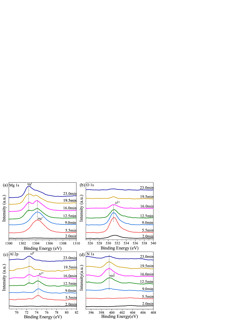
The XPS depth profile of the sample implanted with the dose of NH/cm2 is depicted in Fig.1 and the high-resolution XPS Mg 1s, O 1s, Al 2p, and N 1s spectra are shown in Fig. 2. As shown in Fig.1, the concentration of N atom increases first with the sputtering time and then slowly decreases to 0%, indicating the formation of a thin NH2-containing layer with a maximum concentration of approximately 16% at a depth of nearly 141 nm. The existence of oxygen in the top layer results from the formation of surface oxides due to the non-high vacuum condition. The oxygen and nitrogen concentrations fall down to very low levels at the depth exceeding 202 nm. The high-resolution Mg 1s spectrum in the Fig 2(a) shows that Mg is observed in the form of oxidized state (MgO/Mg(OH)2) in the top surface layer. With the longer sputtering time, the Mg 1s peak shifts toward smaller binding energy, implying that Mg gradually turns to the metallic state (Mg0). The binding energy of Al 2p at 74.4 eV suggests that Al2O3 is observed on the surface. The intensity of the Al peak shifts from the oxidized state to the metallic state with increasing in depth. The binding energy of N 1s in the spectrum is around at 398.4 eV indicating the presence of amino. This result confirms the successful grafting of organic functional groups on the surface of AZ31 magnesium alloys. As shown in Fig.2(b), the oxygen peak represents the formation of MgO, Mg(OH)2, and Al2O3 in the top layer. The oxygen peak intensity decreases with sputtering time, which is in accordance with the gradually reduced oxygen concentration as shown in Fig. 1. The formation of the amino layer and the oxide layer (MgO, Mg(OH)2, and Al2O3) provides a barrier to resist the corrosion of magnesium alloy Jin et al. (2015); Jamesh et al. (2014); Wang et al. (2017); Wong et al. (2013).

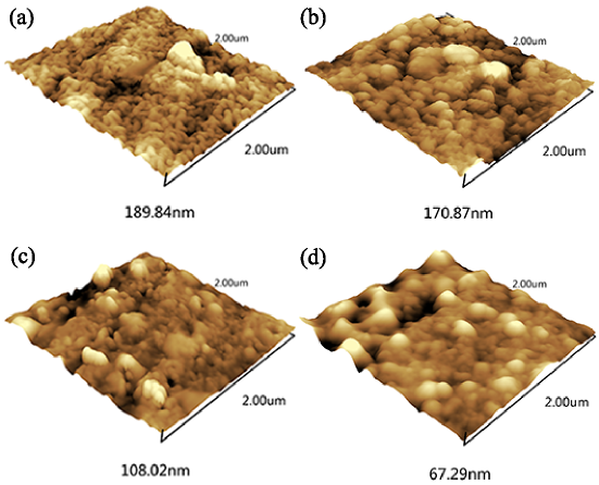
The atomic force microscopy (AFM) images of the untreated and treated samples are shown in Fig. 3, which reveals the changes in the surface topography after implanted with different doses of NH ions. The surface of the untreated sample is relatively rough with many particles. After the implantation with various doses of NH ions, more compact and finer particles are observed on the surface. The number of pits is significantly reduced with the higher implantation dose. The root-mean-square (RMS) roughness values reduce with increasing doses as shown in Table 1. This indicates that the surface becomes smoother after NH ion implantation which may be attributed to the surface restructure at raised temperature during surface modification process Xu et al. (2014). Moreover, as reported in literatures Li and Li (2006); Hong and Nagumo (1997); Sasaki and Burstein (1996), the smoother surface corresponds to the lower corrosion rate because the electrode potential has small difference between the concave and convex. Since a relatively smooth surface of sample is obtained by surface modification through NH ions, the sample is expected to be better resistant to corrosion.
| Doses (ions/cm2) | RMS roughness (nm) | (V) | (A/cm2) |
|---|---|---|---|
| 0 | -1.03 | 869.72 | |
| -1.03 | 441.34 | ||
| -1.05 | 206.00 | ||
| -1.01 | 125.23 |
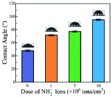
Fig. 4 shows the variation in water contact angles of AZ31 Mg alloys before and after being treated by NH ions. AZ31 Mg alloy is a type of hydrophilic material with a water contact angle of 47.95∘. With the higher implantation doses, the water contact angle of the treated sample increases. The higher contact angle corresponds to the lower wettability, which indicates that more hydrophobic surface is obtained after the NH ion implantation Salman and Okido (2012); Wang et al. (2010).
III.2 Mechanical property
The results of the hardness and the elastic tests are shown in Fig. 5. The indentation depth is much larger than the implantation depth as revealed in the XPS depth profile results, indicating that the data of hardness and the data of elastic modulus are not only based on the implanted layer but also the substrate layer. Both the hardness and the elastic modulus of the untreated sample maintain a plateau with increasing depth. While the hardness and elastic modulus of the treated samples exhibit diverse trend with the displacement into the surface. As to the implanted sample with dose of NH/cm2, the maximum hardness reaches 5.4 GPa at 35.7 nm under the surface and then decreases to a plateau value of 1.2 GPa at approximately 1017.7 nm. The maximum value of elastic modulus is 67.3 GPa at the topmost layer and then decreases to a constant value of 44.5 GPa. Similar trend can be observed for the samples with doses of and NH/cm2, respectively. Both the hardness and the elastic modulus of the treated samples decrease gradually to constant values which are the values of the substrate with increased indentation depth. Thus, the mechanical performance of the implanted layer is better than the substrate, or at least comparable to the latter Poon et al. (2005); Zhao et al. (2012).

III.3 Corrosion performance
The potentiodynamic polarization curves of different samples soaked in Hank′s solution at C are exhibited in Fig. 6. The electrochemical parameters are analyzed to characterize the corrosion nature as listed in Table 1. In contrast to untreated AZ31 magnesium alloy, the samples implanted with NH all achieve lower corrosion current density (Icorr). The Icorr values for the treated samples are shifted towards negative direction as increasing implantation dosage. This indicates that the surface layer containing more NH ions may be more beneficial to restrain the transfer of Cl- during the polarization process Nakatsugawa et al. (1996); Akavipat et al. (1985); Leitao et al. (1989). Therefore the degradation of magnesium alloy could be suppressed by NH ions implantation.

The EIS experiment is conducted to analyze the corrosion property of electrode system. In general, the impedance spectra are described either by a Nyquist (complex plane) plot in which the opposite of imaginary part of impedance (Zim) versus the real part or by a Bode plot where the modulus of the impedance and the phase angle versus frequency, respectively Girault et al. (2001); Macdonald (1987); Chang and Wang (2005); Zhu et al. (2018). Fig. 7 shows the EIS data of unimplanted and NH2-implanted samples in Hank′s solution at C. As shown in Fig. 7(a), two capacitive loops are emerged at high frequency due to the formation of magnesium hydroxide/oxide and the dissolution of Mg through cracks Correa et al. (2011). The capacitive loops are distinguishable for the treated samples with dose of and ions/cm2, whereas almost overlapping for the treated sample of ions/cm2 and untreated sample Nabizadeh et al. (2019). The capacitive loops of the implanted samples are significantly larger than that of the untreated sample. And the diameter of the capacitive semicircle is the largest for implanted sample with a dose of NH/cm2. Since the larger is the capacitive loop, the better is the corrosion resistance. The Nyquist plot implies that the anti-corrosion performance of AZ31 Mg alloy improves after NH ion implantation.
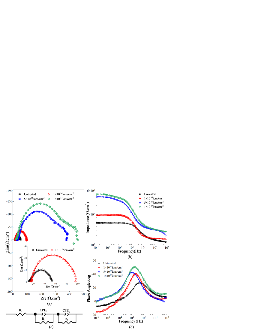
The Bode impedance plots of the untreated and the implanted sample are displayed in Fig. 7 (b). The impedances of the implanted samples are higher at all frequency range compared to that of the untreated samples. As to the sample with high implantation dose ( NH/cm2), the impedance is 8 times larger than that of the untreated sample at the low frequency of 100 mHz, and the impedance is increased evidently by 2 times compared with that of the untreated sample at the high frequency of 100 kHz. As reported in literatures Xu et al. (2014); Chen et al. (2007); Amirudin and Thierry (1995); Murray (1997); Shi et al. (2005); Park et al. (2002), the higher impedances at low and high frequencies reflect the slower process of charge transfer and the poor ability of the electrolyte to penetrate. So the Bode impedance result implies that surface protection is enhanced after NH ion implantation. The Bode phase angle evolution of the untreated and the implanted sample is shown in Fig. 7 (d). The maximum phase angles of treated samples increase with increasing implantation dose, and are all larger than that of the untreated sample. More capacitive behavior is demonstrated by larger phase angle, which suggests the occurrence of a more stable and denser layer to retard the electrolyte penetration to the substrate Wang et al. (2014); de Assis et al. (2006). Hence, the difficulties in charge transfer and electrolyte penetration confirm that ion implantation effectively improves the corrosion resistance.
To further investigate the enhanced corrosion resistance, the electrical equivalent circuit is utilized to fit the EIS spectra of the untreated sample and the implanted sample, as shown in Fig. 7 (c). The equivalent circuit displays two layers, a porous outer layer and a barrier-like inner layer. CPE1 is the constant phase element of the outer porous layer and R1 is the corresponding resistance. The CPE2 stands for the constant phase element in the inner layer in parallel connection with the resistance R2. Rs represents the solution resistance between the working electrode and the reference electrode. The fitted parameters of the electrical equivalent circuit are listed in Table 2. As disclosed in Table 2, R1 and R2 increase while CPE1 and CPE2 decrease after samples implanted by NH ion, indicating that the corrosion rates are retarded in Hank′s solution. As to the sample of high implantation dose ( ions/cm2), it achieves the highest resistance, approximately 10 times in R1 and R2 larger than that of the untreated sample. The resistance R2 of the inner layer is dramatically increased which is attributed to the formation of MgO, Al2O3, and amino based on the results of the XPS depth profile. Hence, the stable inner layer of the implanted sample can further reinforce the corrosion resistance with the corrosion evolution. These EIS results suggest that the samples modified by NH ion improve the anti-corrosion property of AZ31 Mg alloys and are in accordance with the polarization curve.
| Doses (ions/cm2) | Rs() | CPE1() | ) | CPE) | |||
|---|---|---|---|---|---|---|---|
| 0 | 11.83 | 0.98 | 19.74 | 0.54 | 24.59 | ||
| 15.67 | 0.98 | 50.78 | 0.67 | 34.11 | |||
| 35.52 | 0.96 | 159.00 | 0.59 | 207.40 | |||
| 21.93 | 0.99 | 197.10 | 0.54 | 262.20 |
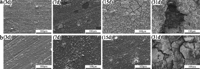
The immersion test is conducted to further study the corrosion resistance behavior. The corrosion rate is calculated by the mass loss method with sample soaked in the Hank′s solution for 3, 7, 15, 31 days at 37∘, respectively. As shown in Fig. 8, the corrosion rate decreases with increasing immersion time. The corrosion rates of NH2-implanted samples have smaller values than that of the untreated sample. Fig. 9 exhibits the corrosion morphologies of the NH2-implanted and the untreated samples after immersed into the Hank′s solution for 3, 7, 15, 31 days at C, respectively. Group (a) is the surface morphology of the untreated sample while Group (b) is the surface morphology of the NH2-implanted sample with dose of ions/cm2. As shown in Group (a) of Fig. 9, cracks are obviously observed after immersion for 3 days. The general trend of crack spacing goes bigger along with longer immersion time. The untreated sample even suffers from severe corrosion pits after immersion for 31 days. On the contrary, some tiny cracks are observed on the NH2-implanted sample after immersion for 3 and 7 days. Although the corrosion cracks grow larger, the obvious corrosion pit does not appear on the treated surface after immersion for 31 days in Group (b). Therefore, the corrosion rate of the AZ31 Mg alloy is effectively decreased after the NH ion implantation.

III.4 In vitro cytotoxicity studies
In order to evaluate the vitro cytotoxicity of the samples, the viability of MC3T3-E1 cells with different extraction medium ratios (0.50 and 1.25 cm2/ml) after incubation for 3 day is measured, as shown in Fig. 10. The cell viability of the control group is considered as 100%. As known in ISO 10993-5, cell viability is larger than 80% corresponding to Grade 0 or Grade 1 of cytotoxicity of materials, which indicates that this material can be used as biomaterials Bian et al. (2017); ISO (2009). For the extraction medium ratio of 0.5 cm2/ml, cells of the untreated samples exhibit viabilities below 80%, indicating obvious cytotoxic effects according to the ISO 10993-5. However, cell viabilities in NH-AZ31 extracts are all above 80% and higher than that in the untreated AZ31 extract during the whole incubation period. As to the extraction medium ratio of 1.25 cm2/ml (Fig. 10(b)), the cells display roughly the same viability (about 80%) for different samples after incubation for 1 day. However, after 3 days of incubation, cells grown in the extract from NH-implanted samples present significantly increased activity compared to the untreated sample, indicating that the cytotoxic effect of the implanted sample on MC3T3-E1 preosteoblasts is not developed, though the reduced cell viability is observed in the first day of incubation. This indicates that the implanted samples with the smoother surface and the reduced corrosion rate show the relatively stable and biofriendly effect on cell growth Sun et al. (2016). Overall, the cell viability of samples with NH ion implantation is significantly higher than that of the untreated samples after the incubation for 3 days, indicating the better biocompatibility of the NH-implanted AZ31 Mg alloy Bagherifard et al. (2018).

IV Discussion
IV.1 Corrosion behavior
According to the XPS spectra (Fig. 1-2), NH ions have been successfully implanted into the sample surface with the formation of the ammonia and the oxide layers. A relatively smooth and compact surface of treated sample is observed by AFM (Fig. 3). As shown in Fig. 4, a larger water contact angle of the treated sample suggests that a hydrophobic surface is obtained after the NH ions implantation.
The modulated smooth surface with hydrophobicity significantly improves the corrosion resistance of AZ31 Mg alloy which can be proved by the electrochemical tests (Fig. 6-7) and the immersion test (Fig. 8-9) in Hank′s solution at the temperature of C. The degradation process is generally accompanied by electrochemical reactions. Magnesium is one of the active metals that can quickly dissolve (Eq. (3)) at the anode electrode during immersion Song and Atrens (2003); Song et al. (1999). Mg2+ reacts with H2O to generate hydrogen gas at the cathode electrode (Eq. (4)). The Mg(OH)2 layer forms with consuming the H+ and producing the OH- (Eq. (5)), creating an alkaline environment.
| (3) |
| (4) |
| (5) |
The Mg(OH)2 can be corroded by Hank′s solution which contains abundant Cl-. According to the penetration/dissolution mechanism as reported in Duan et al. (2007), Cl- disperses and interacts in the hydroxide, then penetrates through the hydroxides to reach the substrate, which results in the dissolution of the substrate. The thinner Mg(OH)2 layer of the untreated sample is easily attacked by Cl- leading to the poor corrosion resistance of the material. However, for samples implanted by NH, the stable products including the ammonia layer and oxide layer (MgO/Al2O3) are formed as the strong barriers against the corrosion to the substrate. These layers can delay the electrolytes to Mg substrate. Therefore, the corrosion rate is decreased after the NH ion implantation.
IV.2 Cytotoxicity evaluation
Although the AZ31 magnesium alloy is a candidate of biomaterial because of its mechanical property and biodegradability, improvements in chemical stability in living tissues, biological properties including wettability, adhesion and biocompatibility are required so that it could have strong bondage with organic bone tissue. The -NH2 and -NH amidogen radicals are helpful in simulating the activity of living tissues. In order to evaluate the biocompatibility of NH implanted AZ31, we have conducted the toxicity assay which is a vital index for rapidly screening the biocompatibility of biometal materials. The toxicity of implants is mainly affected by the amount of released ions during degradation. In practice, cytotoxic effect occurs if the released ion concentration is beyond the tolerance limit. Moreover the corrosion process contributes to the local alkalization and hydrogen evolution which are harmful to cell viability.
As shown in Fig. 10, samples implanted by NH ions do not induce toxicity to MC3T3-E1 cells after cultured for 3 days. This indicates that the decreased corrosion rate leads to the limited alkalization and hydrogen evolution and delays the release of ions. Additionally, the -NH2 amidogen radical as a functional group, which is generally found in organic molecules, is less toxic in comparison with the metal ions. In contrary, the untreated sample presents a lower cell viability compared to the implanted ones, mainly resulting from the higher concentration of the released metal ions. Therefore, the biocompatibility is enhanced through grafting the -NH2 amidogen radical onto the inorganic biomaterial (AZ31).
V Conclusions
In this study, AZ31 magnesium alloy is implanted by different doses of NH ions at energy of 50 KeV. The degradation behavior and the cytotoxicity of the ion implanted samples have been investigated. The results from potentiodynamic polarization, electrochemical impedance spectroscopy, and immersion tests demonstrate that the implanted samples show the improved performance in corrosion resistance. This is attributed to the formation of a stable and protective layer composed of oxide and amino with stable chemical bonds as a barrier to prevent the material from being corroded by biological liquid. Furthermore, the modification by NH ions improves the bioactivity of the material surface. In the toxicity experiment, higher viability of implanted samples is observed from the extraction medium ratio of 1.25 cm2/ml after incubating for 3 days, which confirms the desired biocompatibility of the treated samples in vitro. Our data indicates that NH ion implantation is a favorable method of improving corrosion resistance and cytocompatibility of the AZ31 magnesium alloy. The method of implanting simple organic functional groups on Mg alloy surface may provide a new perspective for the surface treatment for biomaterials.
Acknowledgments
This work is supported by National Science Foundation (NSF) of China with the Grant No.. Additional support is provided by Ministry of Science and Technology of China .
References
- Erbel et al. (2007) R. Erbel, C. Di Mario, J. Bartunek, J. Bonnier, B. de Bruyne, F. R. Eberli, P. Erne, M. Haude, B. Heublein, M. Horrigan, C. Ilsley, D. Bose, J. Koolen, T. F. Luscher, N. Weissman, R. Waksman, Temporary scaffolding of coronary arteries with bioabsorbable magnesium stents: a prospective, non-randomised multicentre trial, Lancet 369 (2007) 1869–1875.
- Waksman et al. (2006) R. Waksman, R. Pakala, P. K. Kuchulakanti, R. Baffour, D. Hellinga, R. Seabron, F. O. Tio, E. Wittchow, S. Hartwig, C. Harder, R. Rohde, B. Heublein, A. Andreae, K. H. Waldmann, A. Haverich, Safety and efficacy of bioabsorbable magnesium alloy stents in porcine coronary arteries, Catheter Cardio Inte 68 (2006) 607–617.
- Witte et al. (2006) F. Witte, J. Fischer, J. Nellesen, H. A. Crostack, V. Kaese, A. Pisch, F. Beckmann, H. Windhagen, In vitro and in vivo corrosion measurements of magnesium alloys, Biomaterials 27 (2006) 1013–1018.
- Witte et al. (2005) F. Witte, V. Kaese, H. Haferkamp, E. Switzer, A. Meyer-Lindenberg, C. J. Wirth, H. Windhagen, In vivo corrosion of four magnesium alloys and the associated bone response, Biomaterials 26 (2005) 3557–3563.
- Zartner et al. (2005) P. Zartner, R. Cesnjevar, H. Singer, M. Weyand, First successful implantation of a biodegradable metal stent into the left pulmonary artery of a preterm baby, Catheter Cardio Inte 66 (2005) 590–594.
- Jin et al. (2017) W. H. Jin, G. M. Wang, Z. J. Lin, H. Q. Feng, W. Li, X. Peng, A. M. Qasim, P. K. Chu, Corrosion resistance and cytocompatibility of tantalum-surface-functionalized biomedical zk60 mg alloy, Corros Sci 114 (2017) 45–56.
- Witte et al. (2008) F. Witte, N. Hort, C. Vogt, S. Cohen, K. U. Kainer, R. Willumeit, F. Feyerabend, Degradable biomaterials based on magnesium corrosion, Curr Opin Solid St M 12 (2008) 63–72.
- Gu et al. (2009) X. N. Gu, Y. F. Zheng, Y. Cheng, S. P. Zhong, T. F. Xi, In vitro corrosion and biocompatibility of binary magnesium alloys, Biomaterials 30 (2009) 484–498.
- Zhao et al. (2014) Y. Zhao, M. I. Jamesh, W. K. Li, G. S. Wu, C. X. Wang, Y. F. Zheng, K. W. K. Yeung, P. K. Chu, Enhanced antimicrobial properties, cytocompatibility, and corrosion resistance of plasma-modified biodegradable magnesium alloys, Acta Biomater 10 (2014) 544–556.
- Feyerabend et al. (2010) F. Feyerabend, J. Fischer, J. Holtz, F. Witte, R. Willumeit, H. Drucker, C. Vogt, N. Hort, Evaluation of short-term effects of rare earth and other elements used in magnesium alloys on primary cells and cell lines, Acta Biomater 6 (2010) 1834–1842.
- Staiger et al. (2006) M. P. Staiger, A. M. Pietak, J. Huadmai, G. Dias, Magnesium and its alloys as orthopedic biomaterials: A review, Biomaterials 27 (2006) 1728–1734.
- Ali et al. (2019) M. Ali, M. Hussein, N. Al-Aqeeli, Magnesium-based composites and alloys for medical applications: A review of mechanical and corrosion properties, Journal of Alloys and Compounds (2019).
- Li et al. (2019) P. Li, C. Schille, E. Schweizer, E. Kimmerle-Muller, F. Rupp, A. Heiss, C. Legner, U. E. Klotz, J. Geis-Gerstorfer, L. Scheideler, Selection of extraction medium influences cytotoxicity of zinc and its alloys, Acta Biomater (2019).
- Zheng et al. (2017) Y. Zheng, X. Xu, Z. Xu, J. Wang, H. Cai, Development of Zn-Based Degradable Metallic Biomaterials, Metallic Biomaterials in: Directions and Technologies, John Wiley Sons, New, 2017.
- Chen et al. (2014) Y. J. Chen, Z. G. Xu, C. Smith, J. Sankar, Recent advances on the development of magnesium alloys for biodegradable implants, Acta Biomater 10 (2014) 4561–4573.
- Li et al. (2012) Y. Li, C. Wen, D. Mushahary, R. Sravanthi, N. Harishankar, G. Pande, P. Hodgson, Mg-zr-sr alloys as biodegradable implant materials, Acta Biomater 8 (2012) 3177–3188.
- Kainer and Buch (2004) K. Kainer, F. V. Buch, The current state of technology and potential for further development of magnesium applications, Magnesium-Alloys and Technology, Weinheim: Wiley-VCH Verlag, 2004.
- Wu et al. (2012) G. S. Wu, K. Feng, A. Shanaghi, Y. Zhao, R. Z. Xu, G. Y. Yuan, P. K. Chu, Effects of surface alloying on electrochemical corrosion behavior of oxygen-plasma-modified biomedical magnesium alloy, Surf Coat Tech 206 (2012) 3186–3195.
- Wan et al. (2007) G. J. Wan, M. F. Maltz, H. Sun, P. P. Li, N. Huang, Corrosion properties of oxygen plasma immersion ion implantation treated magnesium, Surf Coat Tech 201 (2007) 8267–8272.
- Wu et al. (2007) G. Wu, X. Zeng, S. Yao, H. Han, Ion implanted az31 magnesium alloy, Materials Science Forum 546-549 (2007) 551–554.
- Fukumoto et al. (2001) S. Fukumoto, A. Yamamoto, M. Terasawa, T. Mitamura, H. Tsubakino, Microstructures and corrosion resistance of magnesium implanted with nitrogen ions, Materials Transactions 42 (2001) 1232–1236.
- Wu et al. (2009) G. S. Wu, K. J. Ding, X. Q. Zeng, X. M. Wang, S. S. Yao, Improving corrosion resistance of titanium-coated magnesium alloy by modifying surface characteristics of magnesium alloy prior to titanium coating deposition, Scripta Mater 61 (2009) 269–272.
- Tao et al. (2014) X. W. Tao, Z. Z. Wang, X. B. Zhang, Z. X. Ba, Y. M. Wang, Nanomechanical and corrosion properties of zk60 magnesium alloy improved by gd ion implantation, Surf Rev Lett 21 (2014).
- Wang et al. (2008) X. M. Wang, X. Q. Zeng, S. S. Yao, G. S. Wu, Y. J. Lai, The corrosion behavior of ce-implanted magnesium alloys, Mater Charact 59 (2008) 618–623.
- Cheng et al. (2016) M. Q. Cheng, Y. Q. Qiao, Q. Wang, H. Qin, X. L. Zhang, X. Y. Liu, Dual ions implantation of zirconium and nitrogen into magnesium alloys for enhanced corrosion resistance, antimicrobial activity and biocompatibility, Colloid Surface B 148 (2016) 200–210.
- Xu et al. (2011) R. Z. Xu, G. S. Wu, X. B. Yang, T. Hu, Q. Y. Lu, P. K. Chu, Controllable degradation of biomedical magnesium by chromium and oxygen dual ion implantation, Mater Lett 65 (2011) 2171–2173.
- Zhang et al. (2013) M. X. Zhang, C. M. Guo, J. B. Hu, Indium tin oxide electrode modified by a nh ion implantation technique for determination of daunorubicin, J Electrochem Soc 160 (2013) H1–H5.
- Gao et al. (2008) D. M. Gao, Y. Sun, Q. Y. Zhao, J. B. Hu, Q. L. Li, Determination of hemoglobin at a novel nh2/ito ion implantation modified electrode, Microchim Acta 160 (2008) 241–246.
- Li and Niu (2002) D. J. Li, L. F. Niu, Effects of cooh+ ion implantation on hemocompatibility of polypropylene, Sci China Ser E 45 (2002) 666–670.
- Guo et al. (2013) M. X. Guo, M. S. Li, X. Q. Liu, M. L. Zhao, D. J. Li, D. S. Geng, X. L. Sun, H. Q. Gu, N-containing functional groups induced superior cytocompatible and hemocompatible graphene by nh2 ion implantation, J Mater Sci-Mater M 24 (2013) 2741–2748.
- Zhao et al. (1999) Q. Zhao, G. J. Zhai, D. H. L. Ng, X. Z. Zhang, Z. Q. Chen, Surface modification of al2o3 bioceramic by nh2+ ion implantation, Biomaterials 20 (1999) 595–599.
- Li and Niu (2002) D. J. Li, L. F. Niu, Cell attachment of polypropylene surface-modified by cooh+ ion implantation, Nucl Instrum Meth B 192 (2002) 393–401.
- AST (2004) ASTM-G31-72. Standard practice for laboratory immersion corrosion testing of metals, Annual book of ASTM standards., American Society for Testing and Materials, Philadephia, Pennsylvania, U S A, 2004.
- Jin et al. (2015) W. H. Jin, G. S. Wu, H. Q. Feng, W. H. Wang, X. M. Zhang, P. K. Chu, Improvement of corrosion resistance and biocompatibility of rare-earth we43 magnesium alloy by neodymium self-ion implantation, Corros Sci 94 (2015) 142–155.
- Jamesh et al. (2014) M. I. Jamesh, G. S. Wu, Y. Zhao, W. H. Jin, D. R. McKenzie, M. M. M. Bilek, P. K. Chu, Effects of zirconium and nitrogen plasma immersion ion implantation on the electrochemical corrosion behavior of mg-y-re alloy in simulated body fluid and cell culture medium, Corros Sci 86 (2014) 239–251.
- Wang et al. (2017) C. M. Wang, J. Shen, X. K. Zhang, B. Duan, J. X. Sang, In vitro degradation and cytocompatibility of a silane/mg(oh)2 composite coating on az31 alloy by spin coating, J Alloy Compd 714 (2017) 186–193.
- Wong et al. (2013) H. M. Wong, Y. Zhao, V. Tam, S. L. Wu, P. K. Chu, Y. F. Zheng, M. K. T. To, F. K. L. Leung, K. D. K. Luk, K. M. C. Cheung, K. W. K. Yeung, In vivo stimulation of bone formation by aluminum and oxygen plasma surface-modified magnesium implants, Biomaterials 34 (2013) 9863–9876.
- Xu et al. (2014) R. Z. Xu, X. B. Yang, P. H. Li, K. W. Suen, S. Wu, P. K. Chu, Eelectrochemical properties and corrosion resistance of carbon-ion-implanted magnesium, Corros Sci 82 (2014) 173–179.
- Li and Li (2006) W. Li, D. Y. Li, Influence of surface morphology on corrosion and electronic behavior, Acta Mater 54 (2006) 445–452.
- Hong and Nagumo (1997) T. Hong, M. Nagumo, Effect of surface roughness on early stages of pitting corrosion of type 301 stainless steel, Corros Sci 39 (1997) 1665–1672.
- Sasaki and Burstein (1996) K. Sasaki, G. T. Burstein, The generation of surface roughness during slurry erosion-corrosion and its effect on the pitting potential, Corros Sci 38 (1996) 2111–2120.
- Salman and Okido (2012) S. A. Salman, M. Okido, Self-assembled monolayers formed on az31 mg alloy, J Phys Chem Solids 73 (2012) 863–866.
- Wang et al. (2010) Y. H. Wang, W. Wang, L. Zhong, J. Wang, Q. L. Jiang, X. Y. Guo, Super-hydrophobic surface on pure magnesium substrate by wet chemical method, Appl Surf Sci 256 (2010) 3837–3840.
- Poon et al. (2005) R. W. Y. Poon, K. W. K. Yeung, X. Y. Liu, P. K. Chu, C. Y. Chung, W. W. Lu, K. M. C. Cheung, D. Chan, Carbon plasma immersion ion implantation of nickel-titanium shape memory alloys, Biomaterials 26 (2005) 2265–2272.
- Zhao et al. (2012) T. T. Zhao, Y. Li, Y. Liu, X. Q. Zhao, Nano-hardness, wear resistance and pseudoelasticity of hafnium implanted niti shape memory alloy, J Mech Behav Biomed 13 (2012) 174–184.
- Nakatsugawa et al. (1996) I. Nakatsugawa, R. Martin, E. J. Knystautas, Improving corrosion resistance of az91d magnesium alloy by nitrogen ion implantation, Corrosion 52 (1996) 921–926.
- Akavipat et al. (1985) S. Akavipat, E. B. Hale, C. E. Habermann, P. L. Hagans, Effects of iron implantation on the aqueous corrosion of magnesium, Mater Sci Eng 69 (1985) 311–316.
- Leitao et al. (1989) E. Leitao, M. Barbosa, M. F. Dasilva, J. C. Soares, J. P. Muller, Electrochemical studies of magnesium implanted with high-doses of light-ions, Nucl Instrum Meth B 39 (1989) 559–562.
- Girault et al. (2001) P. Girault, J. L. Grosseau-Poussard, J. F. Dinhut, L. Marechal, Influence of a chromium ion implantation on the passive behaviour of nickel in artificial sea-water: An eis and xps study, Nucl Instrum Meth B 174 (2001) 439–452.
- Macdonald (1987) J. Macdonald, Chapters 1-4: Impedance spectroscopy-Emphasising solid Materials and systems, John Wiley Sons, New York, 1987.
- Chang and Wang (2005) Y. Y. Chang, D. Y. Wang, Corrosion behavior of electroless nickel-coated aisi 304 stainless steel enhanced by titanium ion implantation, Surf Coat Tech 200 (2005) 2187–2191.
- Zhu et al. (2018) H. Zhu, T. Zhao, Q. Wei, N. Liu, L. Ma, Z. Hu, Y. Wang, Z. Yu, Corrosion resistance improvement of mg alloy az31 by combining bilayer amorphous dlc: H/sinx film with n+ ions implantation, Journal of Alloys and Compounds 762 (2018) 171–183.
- Correa et al. (2011) P. S. Correa, C. F. Malfatti, D. S. Azambuja, Corrosion behavior study of az91 magnesium alloy coated with methyltriethoxysilane doped with cerium ions, Prog Org Coat 72 (2011) 739–747.
- Nabizadeh et al. (2019) M. Nabizadeh, A. A. Sarabi, H. E. Mohammadloo, Comparative investigation of cu ion and adipic acid addition on electrochemical and microstructure characteristics of vanadium conversion coating on az31 mg alloy, Surf Coat Tech 357 (2019) 1–11.
- Chen et al. (2007) Y. Chen, X. H. Wang, J. Li, J. L. Lu, F. S. Wang, Long-term anticorrosion behaviour of polyaniline on mild steel, Corros Sci 49 (2007) 3052–3063.
- Amirudin and Thierry (1995) A. Amirudin, D. Thierry, Application of electrochemical impedance spectroscopy to study the degradation of polymer-coated metals, Prog Org Coat 26 (1995) 1–28.
- Murray (1997) J. N. Murray, Electrochemical test methods for evaluating organic coatings on metals: an update. part iii: Multiple test parameter measurements, Prog Org Coat 31 (1997) 375–391.
- Shi et al. (2005) A. Shi, S. Koka, J. Ullett, Performance evaluation on the weathering resistance of two usaf coating systems (standard 85285 topcoat versus fluorinated apc topcoat) via electrochemical impedance spectroscopy, Prog Org Coat 52 (2005) 196–209.
- Park et al. (2002) J. H. Park, G. D. Lee, A. Nishikata, T. Tsuru, Anticorrosive behavior of hydroxyapatite as an environmentally friendly pigment, Corros Sci 44 (2002) 1087–1095.
- Wang et al. (2014) Z. B. Wang, H. X. Hu, C. B. Liu, Y. G. Zheng, The effect of fluoride ions on the corrosion behavior of pure titanium in 0.05 m sulfuric acid, Electrochim Acta 135 (2014) 526–535.
- de Assis et al. (2006) S. L. de Assis, S. Wolynec, I. Costa, Corrosion characterization of titanium alloys by electrochemical techniques, Electrochim Acta 51 (2006) 1815–1819.
- Bian et al. (2017) D. Bian, W. R. Zhou, J. X. Deng, Y. Liu, W. T. Li, X. Chu, P. Xiu, H. Cai, Y. H. Kou, B. G. Jiang, Y. F. Zheng, Development of magnesium-based biodegradable metals with dietary trace element germanium as orthopaedic implant applications, Acta Biomater 64 (2017) 421–436.
- ISO (2009) ISO 10993-5:2009(E). Biological evaluation of medical devices, Part 5: Tests for in vitro cytotoxicity, International Organization for Standardization, 2009.
- Sun et al. (2016) J. D. Sun, Y. Zhu, L. Meng, P. Chen, T. T. Shi, X. Y. Liu, Y. F. Zheng, Electrophoretic deposition of colloidal particles on mg with cytocompatibility, antibacterial performance, and corrosion resistance, Acta Biomater 45 (2016) 387–398.
- Bagherifard et al. (2018) S. Bagherifard, D. J. Hickey, S. Fintova, F. Pastorek, I. Fernandez-Pariente, M. Bandini, T. J. Webster, M. Guagliano, Effects of nanofeatures induced by severe shot peening (ssp) on mechanical, corrosion and cytocompatibility properties of magnesium alloy az31, Acta Biomater 66 (2018) 93–108.
- Song and Atrens (2003) G. L. Song, A. Atrens, Understanding magnesium corrosion - a framework for improved alloy performance, Adv Eng Mater 5 (2003) 837–858.
- Song et al. (1999) G. L. Song, A. Atrens, M. Dargusch, Influence of microstructure on the corrosion of diecast az91d, Corros Sci 41 (1999) 249–273.
- Duan et al. (2007) H. P. Duan, C. W. Yan, F. H. Wang, Effect of electrolyte additives on performance of plasma electrolytic oxidation films formed on magnesium alloy az91d, Electrochim Acta 52 (2007) 3785–3793.