Blood Vessel Detection using Modified Multiscale MF-FDOG Filters for Diabetic Retinopathy
Abstract
Blindness in diabetic patients caused by retinopathy (characterized by an increase in the diameter and new branches of the blood vessels inside the retina) is a grave concern. Many efforts have been made for the early detection of the disease using various image processing techniques on retinal images. However, most of the methods are plagued with the false detection of the blood vessel pixels. Given that, here, we propose a modified matched filter with the first derivative of Gaussian. The method uses the top-hat transform and contrast limited histogram equalization. Further, we segment the modified multiscale matched filter response by using a binary threshold obtained from the first derivative of Gaussian. The method was assessed on a publicly available database (DRIVE database). As anticipated, the proposed method provides a higher accuracy compared to the literature. Moreover, a lesser false detection from the existing matched filters and its variants have been observed.
Index Terms:
retinopathy, top-hat transform, retinal vessel extraction, Blood vasculature, matched filter, first derivative of Gaussian.I Introduction
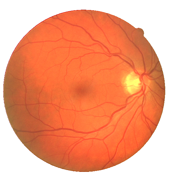
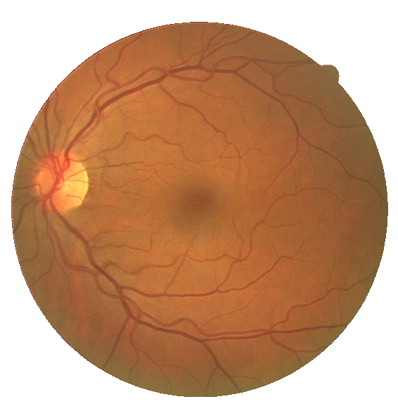
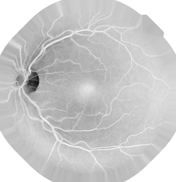
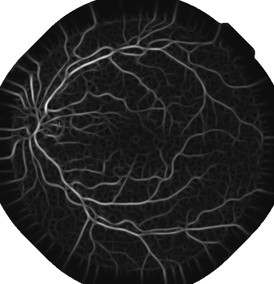
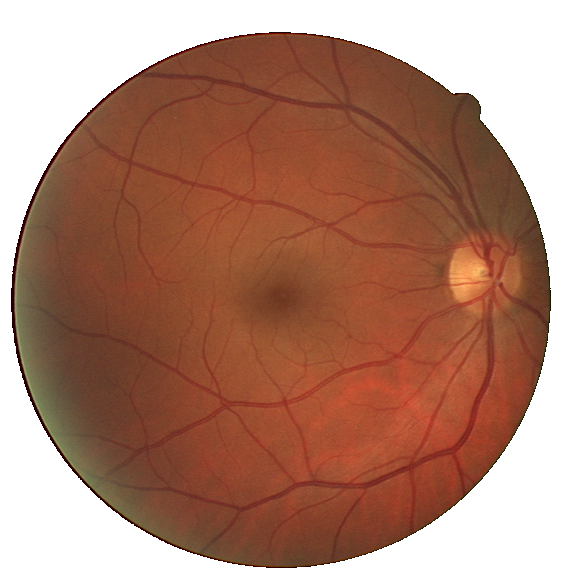
Lately, digital image processing has been a very popular toolbox in the field of medicine, navigation system, agriculture, object tracking, etc. [1]. Researchers have successfully detected diseases like cancer, diabetic retinopathy, brain tumor, breast calcification, etc. using various image processing techniques [2]. Blood vessel detection (BVD) is an emerging area of research in the field to identify the illnesses like hypertension, arteriosclerosis, diabetes, etc. [3]. Here, the estimates are made by observing a change in the diameter and branching of the blood vessels (a tree-like structure) inside the retina (refer to Fig. 1).
The reported BVD approaches can broadly be classified in 4 categories viz., (i) tracking based approach, (ii) morphological based approach, (iii) filtering based approach, and (iv) machine learning approach. Out of these, the first three approaches are based on unsupervised learning; however, the last method is based on supervised learning [4, 5]. Nevertheless, filtering based approaches are still very popular [6, 7, 8]. Literature indicates that various proposed algorithms in the domain are based on line detection techniques [9], mainly due to similarities in BVD problem and line detection problem. Further, the use of matched filter has been demonstrated [10]. In addition, various thresholding techniques, such as adaptive thresholding, entropy based thresholding, etc., have been applied on matched filter response to obtain the binary map (blood vessel and background) [11]. However, matched filter response fails to distinguish between non-vessels and vessels edges in retinal images [3]. To mitigate the shortcoming, matched filter with first-order derivative of Gaussian (MF-FDOG) [3] and modified matched filter with double-sided thresholding [12] have been proposed. Further reduction in the false positive detection was observed by combining the zero crossing property of the Laplacian of Gaussian with matched filter [6]. However, all of these methods assume that the intensity profile across the blood vessel follow the Gaussian distribution, which may not be true always. On the other hand, matched filter based approach is free from the deceptive constraints.
Given that, here, we propose an automated unsupervised approach based on matched filter for automatic segmentation of blood vessel map from retinal image. In this paper, we a) introduce a modified multiscale MF-FDOG filter which process the retinal blood vessels by considering neighborhood pixels of blood vessel having Gaussian profile, b) use top-hat transform followed by contrast limited adaptive histogram equalization (CLAHE) for retinal image enhancement in preprocessing stage, and c) perform the quantitative analysis of the proposed framework on publicly available Digital Retinal Image for Vessels Extraction (DRIVE) database [13]. We find that the proposed method significantly improves the true positive detection and reduces the false detection.
II Background
Matched filter with first-order derivative of Gaussian (MF-FDOG) has been found an effective method to extract the retinal vessels [12]. The MF-FDOG as defined by Zhang et.al. [12] is given below.
| (1) |
where and are the scale and the mean of the matched filter kernel . It is defined as
| (2) |
In equation (1), is subtracted from the kernel to obtain the zero mean filter to remove the smooth background. Here, is the spatial coordinates of the filter which depends on the parameter and respectively. is used to maintain the confidence interval of the Gaussian distribution along the -axis, however, the parameter is the length of kernel along the -axis. In usual practice, the value of is set as 3 so that 99% of the area under the Gaussian curve lies within the range . Further, the first derivative of Equation (1) is
| (3) |
Now, we need to estimate the threshold value for the matched filter to implement it effectively. Here, the threshold value for the matched filter is obtained by using response of the image to FDOG. Filter bank for matched filter and FDOG are obtained by rotating the kernels and . Filter bank response and of retinal image are obtained by apply filter kernel and respectively. The filter response is averaged using mean filter of size as
| (4) |
The normalized response is obtained by normalizing each pixel value of the averaged response . Then, threshold is computed as
| (5) |
where is the reference threshold computed as , where is the mean value of matched filter response , and is a constant. To obtain the binary vessel map , the threshold is applied to as
| (6) |
It can be noticed here that for vessel pixels in retinal image, the threshold value is lower, however, for non-vessel pixels the threshold value is higher. Therefore, blood vessel pixel sustain whereas non-vessel pixels vanished in the binary vessel map due to higher threshold.
III Proposed Modified MF-FDOG
Despite multiple advantages of the MF-FDOG [12], the filtering process suffers with inherent limitations. The method considers the parameter dependent on due to which filter kernel do not consider neighborhood pixels of vessels into filtering process. To overcome the same, here, we consider that is independent of resulting in the consideration of the neighborhood pixels of vessels in the filtering process (see Fig. 2 and Fig. 3). To make independent of , the limiting value of in Equations (1) and (3) are modified to
| (7) |
while keeping all the other parameters same. The designed filters are rotated at different angles to detect the vessels of different orientations. The filters bank of MF and FDOG for and are shown in Fig. 2 and Fig. 3 respectively.


IV Segmentation
To extract the blood vessels from retinal fundus images, the proposed framework is shown in Fig. 4. The main parts of our approach are discussed in following sections.
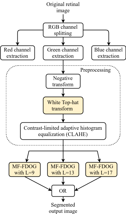
IV-A Preprocessing
Initially, an original RGB color retinal image is separated into red, green, and blue channels where green channel image have higher contrast compare to other two channel images. In green channel image, the blood vessel pixels are visible and easily distinguishable from the background pixels. Because in our eyes, lens pigments absorb light colors differently [14]. Therefore, red vessels in color retinal image are more visible in the green channel image. The green channel image is inverted followed by superimposed with fundus mask to make the blood vessel brighter and keep the focus on the region of interest . In image filtering, boundary pixels have a grate importance which may deteriorate the segmentation performance if they are not taken care before filtering process. Therefore, to deal with boundary pixels, inverted green channel image are fake padded radially using boundary pixels (shown in Fig. 6). Thereafter, white top-hat transformation is used to enhance only the blood vessels. Usually, vessels have small thickness compared to the other structures in retinal images such as exudates, fovea, optic disc, etc. Therefore, a disc shaped structuring element having diameter at least equal to the diameter of the thickest blood vessel in retinal image is preferred for top-hat transformation. The top-hat transformation highlight the object smaller than structuring element and suppress the other structures. The white top-hat transformed image can be obtained as
| (8) |
where, is inverted green channel image, is the structuring element, and denotes the morphological opening operator.
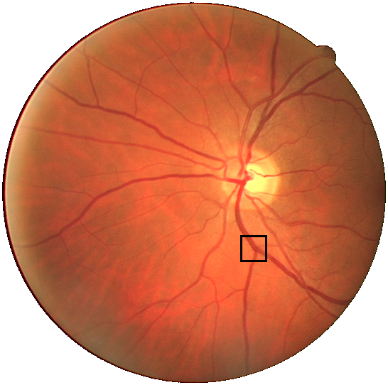


In this paper, the diameter of the structuring element is chosen 11 pixels wide as the maximum width of the blood vessel is less than 11 pixels. For the DRIVE database, the width of the blood vessel varies in the range of 1-10 pixels [15]. Fig. 5 shows the width of a wide blood vessel in the retinal image as 7 pixels wide. The diameter of the structuring element may vary for the different database having different image resolution. After applying top-hat transformation, blood vessel have poor contrast compare to background. Therefore, contrast limited adaptive histogram equalization (CLAHE) is applied for further image enhancement.
IV-B Blood vessel extraction
After preprocessing, enhanced images is further processed through bank of filters obtained using the proposed modified multiscale MF-FDOG. In image processing, filtering operation on edge pixels always degrade the performance of many algorithm. To handle the boundary pixels near field of view (FOV) edges, the retinal image is radially padded using boundary pixels of retina inside FOV in radial direction. The modified MF-FDOG method discussed in section III is applied on radially padded retinal image (Fig. 6). Matched filters of different scales are used to detect thick along with thin vessels. All intermediate steps of the proposed framework (with the help of an example image from DRIVE database) are shown in Fig. 6.
V Results and Discussion
V-A Database
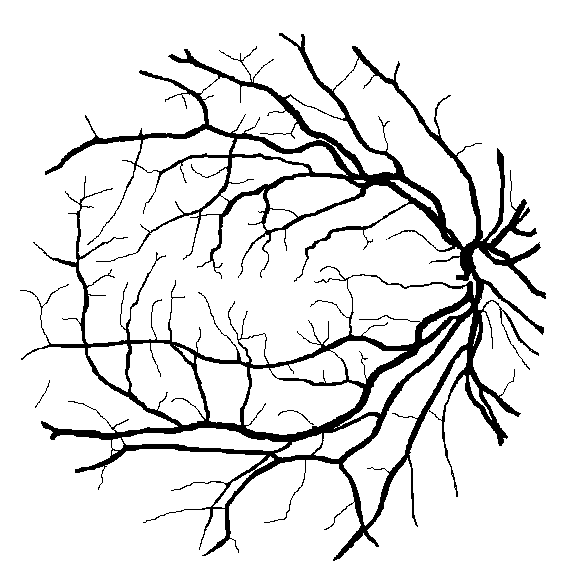
In this study, the retinal fundus images are obtained from DRIVE database [13]. The database is constructed under diabetic retinopathy screening program conducted in Netherlands. For the database, 40 images have been randomly selected from the total of 400 diabetic patients that have been screened. Each image was captured using a Canon CR5 non-mydriatic 3CCD camera with a 45 degree FOV having 8 bit per color plane of resolution pixels. The results of the manual segmentation (also called gold standard ground truth) are available for the database.
V-B Results
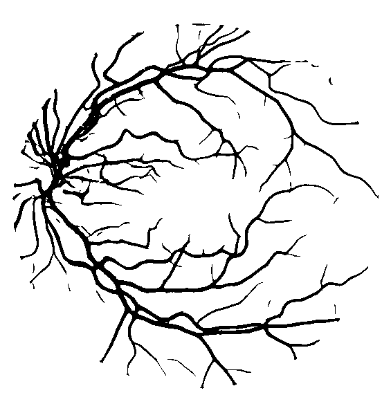
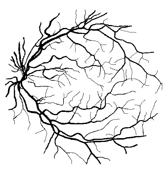
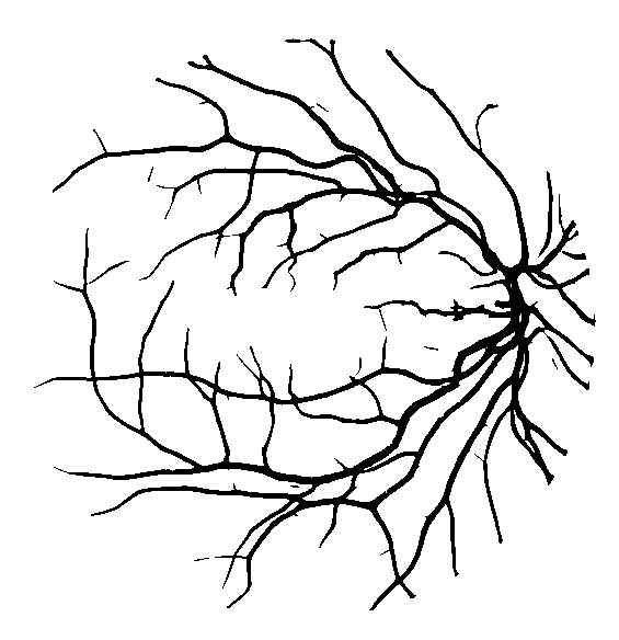
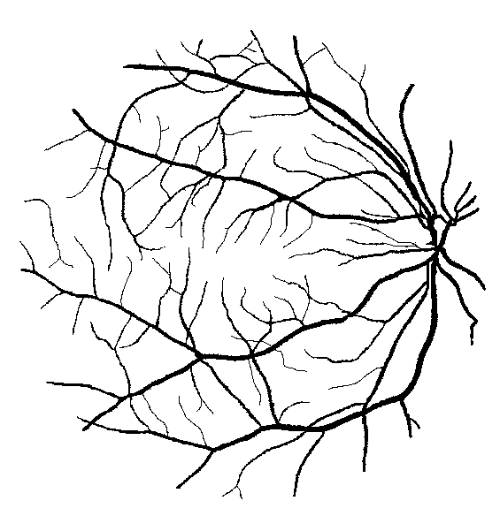
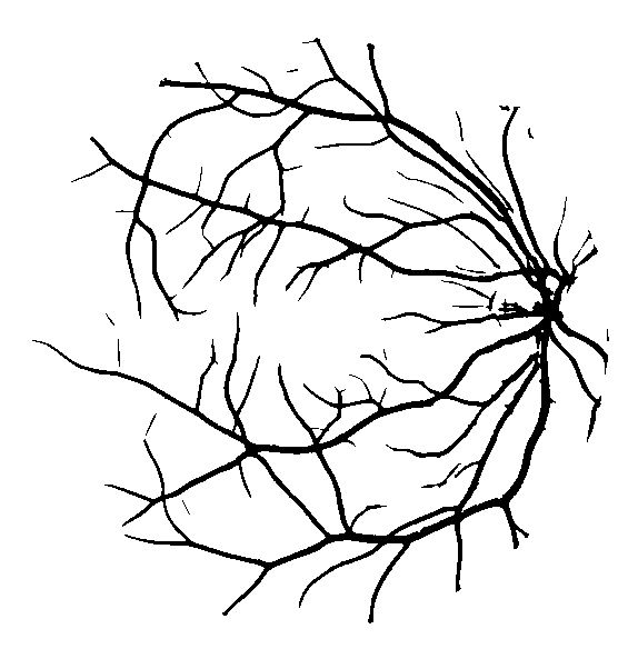
Out of the selected 40 images, 20 images are kept for training and 20 for testing. The proposed framework is evaluated on only test images because the framework is fully unsupervised. Three different scales, 9, 13, and 17 along with 1, 1.5, and 2 are used respectively for multiscale MF-FDOG filter design. A bank of MF-FDOG filter kernels are obtained by rotating the kernels at increment to obtain 12 different kernels at different orientations. The retinal images from the test set are filtered using those kernels to identify the vessel at different orientation. The segmented blood vessels obtained using the proposed framework are provided along with its gold standard ground truth in Figure 7. The corresponding results are evaluated and compared with competitive algorithms using three different metrics: accuracy (), sensitivity (), specificity () (see table I). Accuracy, sensitivity, and specificity are calculated using false positive (), false negative (), true positive (), and true negative () values. denotes the number of pixels correctly identified as vessel pixels, and denotes the number of pixels belongs to the background but wrongly identified as vessel pixel. represents the number of pixels correctly identified as background pixels, and represents the number of pixels belongs to the vessels but incorrectly assigned to background pixels. The evaluation metrics are measured using the following mathematical expressions
The performance metrics for 20 test images of DRIVE database are tabulated in Table I. The performance metrics illustrate that average segmentation accuracy, sensitivity, and specificity of the presented technique are 94.34%, 71.93%, and 97.64% respectively. Our method resulted in a highly precise identification (deviation in accuracy, ). Further, Fig. 7 shows that our proposed algorithm is competent in identifying thick as well as thin vessels in the presence of exudates, fovea, and optic disc. In addition, we provide a comparison table (Table II) of the presented algorithm with different competing methods. Indeed, the results show that our proposed algorithm outperforms many unsupervised methods on DRIVE database concerning accuracy and specificity.
| Image | Se | Sp | Acc |
| 1 | 0.8015 | 0.9656 | 0.9441 |
| 2 | 0.7598 | 0.9801 | 0.9471 |
| 3 | 0.7281 | 0.9702 | 0.935 |
| 4 | 0.6206 | 0.9909 | 0.9415 |
| 5 | 0.6611 | 0.9873 | 0.9431 |
| 6 | 0.6729 | 0.9814 | 0.9379 |
| 7 | 0.6779 | 0.9784 | 0.9386 |
| 8 | 0.6837 | 0.9749 | 0.9383 |
| 9 | 0.6959 | 0.9807 | 0.9472 |
| 10 | 0.6759 | 0.9844 | 0.9475 |
| 11 | 0.6985 | 0.9748 | 0.939 |
| 12 | 0.7612 | 0.9681 | 0.9422 |
| 13 | 0.6654 | 0.9832 | 0.9382 |
| 14 | 0.7784 | 0.9675 | 0.9452 |
| 15 | 0.7439 | 0.9748 | 0.9508 |
| 16 | 0.6908 | 0.9815 | 0.9435 |
| 17 | 0.711 | 0.9736 | 0.9412 |
| 18 | 0.7591 | 0.9683 | 0.9443 |
| 19 | 0.8306 | 0.9739 | 0.9567 |
| 20 | 0.7688 | 0.9689 | 0.9475 |
| Average | 0.7193 | 0.9764 | 0.9434 |
| Std. deviation | 0.0515 | 0.0068 | 0.0049 |
| Se - sensitivity, Sp - specificity, Acc - accuracy | |||
| Unsupervised Methods | |||
| Methods | Se | Sp | Acc |
| 2nd observer | 0.7760 | 0.9725 | 0.9473 |
| Chaudhuri et al.[10]∗ | 0.6168 | 0.9741 | 0.8773 |
| Zhang et al. [12] | 0.7120 | 0.9724 | 0.9382 |
| Gou et al. [7] | 0.7526 | 0.9669 | 0.9393 |
| Presented method | 0.7193 | 0.9764 | 0.9434 |
| ∗results are taken from [16] | |||
VI Conclusion
To summarize, here, we have performed the blood vessel extraction on a set of 20 test images of DRIVE database. We found that the top-hat transform followed by CLAHE is a comparatively effective approach to eliminate the structure other than blood vessels in the preprocessing stage. Further, the modified match filter parameters improved the MF-FDOG filters response. Indeed, the proposed framework provides a better average accuracy (94.34%) and specificity (97.64%). Moreover, the method can be extended to precisely estimate the object having Gaussian distribution.
References
- [1] T. Acharya and A. K. Ray, Image processing: principles and applications. John Wiley & Sons, 2005.
- [2] F. Ritter, T. Boskamp, A. Homeyer, H. Laue, M. Schwier, F. Link, and H. . Peitgen, “Medical image analysis,” IEEE Pulse, vol. 2, pp. 60–70, Nov 2011.
- [3] L. Zhang, Q. Li, J. You, and D. Zhang, “A modified matched filter with double-sided thresholding for screening proliferative diabetic retinopathy,” IEEE Transactions on Information Technology in Biomedicine, vol. 13, no. 4, pp. 528–534, 2009.
- [4] M. M. Fraz, P. Remagnino, A. Hoppe, B. Uyyanonvara, A. R. Rudnicka, C. G. Owen, and S. A. Barman, “Blood vessel segmentation methodologies in retinal images–a survey,” Computer methods and programs in biomedicine, vol. 108, no. 1, pp. 407–433, 2012.
- [5] J. Almotiri, K. Elleithy, and A. Elleithy, “Retinal vessels segmentation techniques and algorithms: a survey,” Applied Sciences, vol. 8, no. 2, p. 155, 2018.
- [6] D. Kumar, A. Pramanik, S. S. Kar, and S. P. Maity, “Retinal blood vessel segmentation using matched filter and laplacian of gaussian,” in Signal Processing and Communications (SPCOM), 2016 International Conference on, pp. 1–5, IEEE, 2016.
- [7] D. Gou, Y. Wei, H. Fu, and N. Yan, “Retinal vessel extraction using dynamic multi-scale matched filtering and dynamic threshold processing based on histogram fitting,” Machine Vision and Applications, vol. 29, no. 4, pp. 655–666, 2018.
- [8] M. Raman, R. Korah, and K. Tamilselvan, “An automatic localization of optic disc in low resolution retinal images by modified directional matched filter,” INTERNATIONAL ARAB JOURNAL OF INFORMATION TECHNOLOGY, vol. 16, no. 1, pp. 1–7, 2019.
- [9] U. T. Nguyen, A. Bhuiyan, L. A. Park, and K. Ramamohanarao, “An effective retinal blood vessel segmentation method using multi-scale line detection,” Pattern recognition, vol. 46, no. 3, pp. 703–715, 2013.
- [10] S. Chaudhuri, S. Chatterjee, N. Katz, M. Nelson, and M. Goldbaum, “Detection of blood vessels in retinal images using two-dimensional matched filters,” IEEE Transactions on medical imaging, vol. 8, no. 3, pp. 263–269, 1989.
- [11] A. Hoover, V. Kouznetsova, and M. Goldbaum, “Locating blood vessels in retinal images by piecewise threshold probing of a matched filter response,” IEEE Transactions on Medical imaging, vol. 19, no. 3, pp. 203–210, 2000.
- [12] B. Zhang, L. Zhang, L. Zhang, and F. Karray, “Retinal vessel extraction by matched filter with first-order derivative of Gaussian,” Computers in biology and medicine, vol. 40, no. 4, pp. 438–445, 2010.
- [13] J. Staal, M. D. Abràmoff, M. Niemeijer, M. A. Viergever, and B. Van Ginneken, “Ridge-based vessel segmentation in color images of the retina,” IEEE transactions on medical imaging, vol. 23, no. 4, pp. 501–509, 2004.
- [14] Y. Q. Zhao, X. H. Wang, X. F. Wang, and F. Y. Shih, “Retinal vessels segmentation based on level set and region growing,” Pattern Recognition, vol. 47, no. 7, pp. 2437–2446, 2014.
- [15] A. Fathi and A. R. Naghsh-Nilchi, “Automatic wavelet-based retinal blood vessels segmentation and vessel diameter estimation,” Biomedical Signal Processing and Control, vol. 8, no. 1, pp. 71–80, 2013.
- [16] M. Niemeijer, J. Staal, B. van Ginneken, M. Loog, and M. D. Abramoff, “Comparative study of retinal vessel segmentation methods on a new publicly available database,” in Medical Imaging 2004: Image Processing, vol. 5370, pp. 648–657, International Society for Optics and Photonics, 2004.