Investigations on physical and biological range uncertainties in Krakow proton beam therapy centre ††thanks: Presented at 3rd Jagiellonian Symposium on Fundamental and Applied Subatomic Physics, June 23 – 28, 2019 in Collegium Maius, Kraków, Poland
Abstract
Physical and biological range uncertainties limit the clinical potential of Proton Beam Therapy (PBT). In this proceedings, we report on two research projects, which we are conducting in parallel and which both tackle the problem of range uncertainties. One aims at developing software tools and the other at developing detector instrumentation. Regarding the first, we report on our development and pre-clinical application of a GPU-accelerated Monte Carlo (MC) simulation toolkit Fred. Concerning the letter, we report on our investigations of plastic scintillator based PET detectors for particle therapy delivery monitoring. We study the feasibility of Jagiellonian-PET detector technology for proton beam therapy range monitoring by means of MC simulations of the activity induced in a phantom by proton beams and present preliminary results of PET image reconstruction. Using a GPU-accelerated Monte Carlo simulation toolkit Fred and plastic scintillator based PET detectors we aim to improve patient treatment quality with protons.
1 Introduction
The increasing numbers of proton facilities and successful proton treatments [1] indicate that the relevance of Proton Beam Therapy (PBT) as technique for tumor radiation therapy is a rapidly growing. Kraków proton facility is in clinical operation since Oct. 2016 and more than 10 patients a day are currently treated.
Range uncertainties, i.e. uncertainty of the distance that protons travel inside patient body, currently represent one of the biggest caveats for the exploitation of the full potential of proton therapy treatments [2]. Proton beams range is particularly affected by biological and physical uncertainties in a heterogeneous patient body. Therefore, to assure target coverage, medical physicists currently apply up to about 1 cm safety margins around the tumor volume, which lead to the unwanted irradiation of the healthy tissues surrounding the tumor [2].
The biological dose, expressed in , delivered to the patient is the actual quality of clinical interest. It is calculated as , where is the physical dose expressed in and is the Relative Biological Effectiveness. By definition, in conventional therapy with photons therefore, physical and biological doses are equal and correlated with clinical response. Protons have an increased biological effectiveness compared to photons, i.e. RBE is larger than one. Currently in clinical routine, the RBE of protons is assumed to be constant and equal to 1.1 [3]. This convention neglects complex, often nonlinear dependency of the RBE on such parameters as penetration depth, Linear Energy Transfer (LET), dose, fractionation scheme, tissue type and endpoint, cell cycle phase or oxygenation level. These dependencies might affect the effective proton range, i.e., introduce biological range uncertainty and thus affect the dose to the surrounding tissue and Organs at Risk (OAR). Modification of proton physical dose by RBE, which is an uncertain weighting factor, makes the correlation of proton biological dose and clinical effect of tumor irradiation uncertain, and unification of clinical studies comparing the effectiveness of different radiation modalities challenging.
An improvement resulting from correctly applying radiobiological assumptions in PBT could be achieved only under the condition that the physical dose is accurately delivered to the patient. In fact, physical range uncertainties, occurring due to patient mispositioning or Computed Tomography (CT) scanner calibration introducing Hounsfield Unit (HU) to stopping power conversion, could cause differences between treatment plan and treatment delivery. Monte Carlo simulations and range monitoring methods are essential in PBT to guarantee that the physical dose is accurately delivered and therefore to reduce the biological and physical range uncertainties in a patient body.
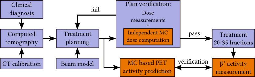
Fig. 1 illustrates which role could play each of the two topics reported in this manuscript in the clinical treatment workflow.
2 Monte Carlo simulations to address physical and biological range uncertainties of proton beams
It is recognized that Monte Carlo (MC) methods can offer improved dose calculation accuracy in heterogeneous media and therefore predict more accurately therapeutic dose distributions in patients compared to the analytical algorithms that are typically employed in the Treatment Planning Systems (TPS) used in clinical routine [2]. Nowadays, the use of a variable RBE is being discussed among the PBT scientific community. We perform biological dose calculations with variable RBE and investigate biological range uncertainties by means of MC simulations of patient CT images exploiting the MC dose calculation tool Fred [4, 5]. Fred offers a unique combination of features: accuracy of a MC code including biological dose computation, flexibility of a research tool, and high dose calculation speed due to GPU-acceleration. These characteristics are impossible to achieve with the currently available commercial TPS and general purpose MC codes like Geant4/FLUKA [6, 7].
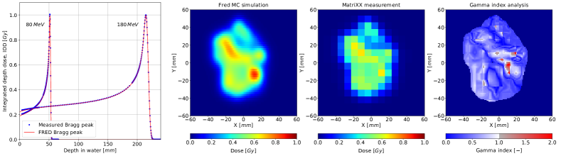
We have successfully implemented in Fred the proton beam model used clinically for patient treatment in the Kraków facility. The Integrated Depth Dose profiles (IDD) of single proton pencil beams in water simulated with Fred MC code for different energies are in excellent agreement with the IDDs obtained during the commissioning measurements (see fig. 2 left), showing differences of less than 2%.
The emittance model, describing lateral beam propagation in air, was implemented in Fred, in accordance with the clinical system. The longitudinal and lateral pencil beam shapes are modelled in Fred MC with a submillimeter precision.
We validated the beam model experimentally using the transversal patient treatment plan verification measurements. Such measurements are routinely performed by medical physicists for treatment plan quality assurance with an array of 1020 ionization chambers (MatriXX IBA) placed in a water phantom. A transversal dose plane extracted from Fred MC and the dose distribution measured with the MatriXX detector at the same depth in water, as well as the Gamma Index (GI) map obtained from GI test are presented in fig. 2. The GI passing rate ( mm criteria) greater than 98% was obtained for 182 dose plane measurements for 10 patients. Based on these results, we can assure that dose distributions of clinical treatment plans can be recalculated accurately.
We are currently performing treatment planning studies to quantify the biological range uncertainties exploring various biological models with variable RBE and clinical data of patients treated in Kraków.
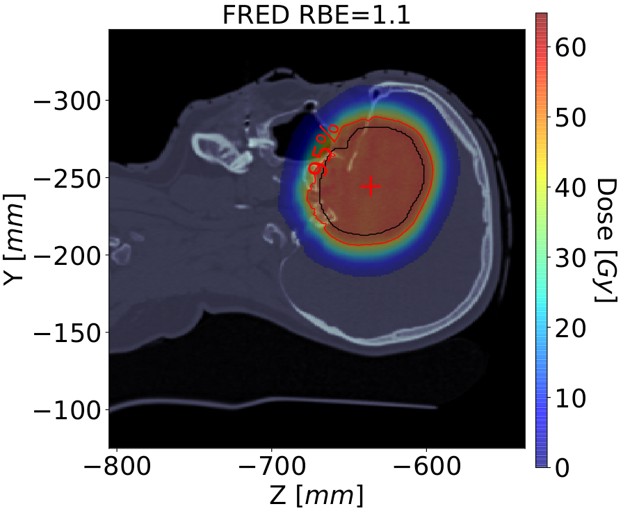
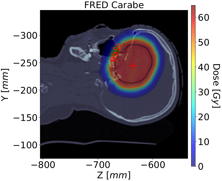
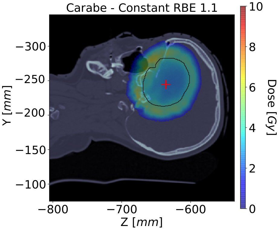
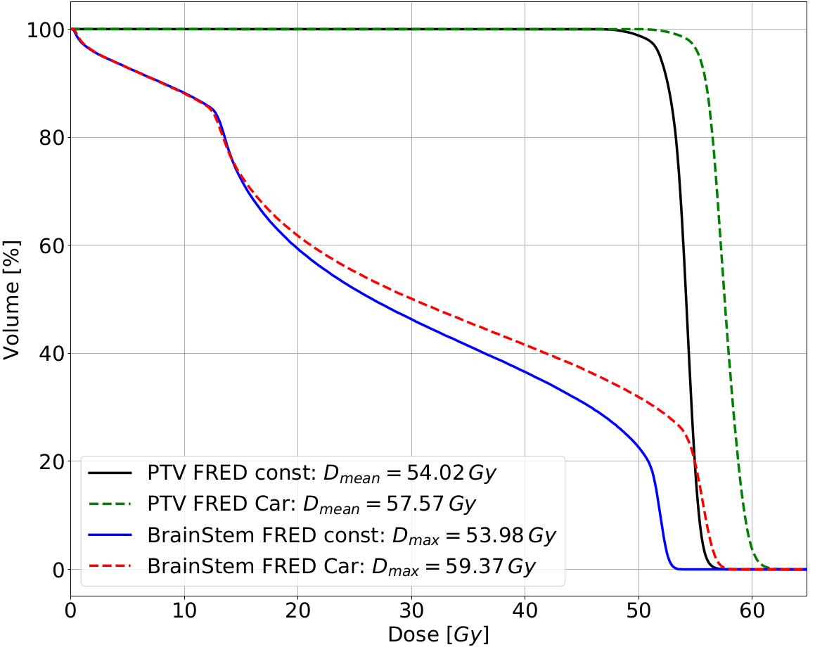
An example of a selected Head and Neck (H&N) patient is presented in fig. 3, where the radiobiological dose distributions computed with constant and variable RBE using Fred as well as corresponding Dose Volume Histograms (DVHs) are shown. The MC calculation time for this case was about 10 min (9108 primary protons, 2106 tracking rate). The mean dose to Planning Target Volume (PTV), calculated with Fred using variable RBE model proposed by Carabe [8], is increased with respect to the clinically applied constant RBE=1.1 assumption of about 3 Gy(RBE), whereas the maximum dose to the brain stem OAR increases by about 4 Gy(RBE). Our results show that incorporation of the variable RBE model in patient dose calculation increases dose in PTV and OAR with respect to constant RBE assumption. This is especially important when OARs are very close to or overlap with the PTV as it occurs frequently in H&N patients. Considering variable RBE hypothesis in the proton therapy clinic could be essential as increased biological dose deposited to OARs that exceeds clinical dose constraints can be potentially associated with increased normal tissue complication probability an therefore an increased risk of necrosis or secondary cancer.
The use of the fast MC dose computation tool Fred for physical and biological dose recalculation of patient treatment plans (retrospectively and prospectively) can provide additional clinical information for medical physicists and medical doctors and can potentially prevent inaccuracies in patient treatment.
3 J-PET detector to address physical range uncertainties of proton beams
The proton interactions with patient tissues allow range monitoring during or just after the treatment detecting emitted secondary radiation. Tracking of prompt-gamma, PET-gamma and secondary protons and neutrons are examples [9, 10, 11]. Prototype systems for prompt-gamma and PET-gamma range monitoring were tested clinically and obtained satisfying precision of Bragg-peak position monitoring on-line in the PBT treatment room [12, 13, 14]. At the Jagiellonian University in Kraków, a novel solution for diagnostic PET imaging, Jagiellonian-PET (J-PET) is being developed.
A single detection unit of the J-PET scanner [15] consists of a 50 cm long and 624 mm2 intersection size scintillator strip. The light pulses produced in the strip by 511 keV back-to-back photons propagate to its edges where they are converted into electrical signals by photomultipliers (PMT). The interaction position of the photon with the detector is estimated from the time difference between the PMT signals located at the ends of the strip. A J-PET module consists of 13 scintillator strips read-out through a single front-end electronics and a FPGA-based DAQ system. A modular, lightweight and portable design of J-PET enables flexibility in detector configuration and easy installation. Increasing the number of J-PET detector layers increases the detection efficiency of the system.
We performed comprehensive MC simulations using the GATE toolkit [16] and reconstructions of 3D activity distributions using the CASTOR software [17]. The aim was to characterize the sensitivity of the J-PET for proton beam range detection. We investigated single and multi-layer cylindrical and dual-head configurations of the J-PET modules that can be possibly applied for in-room range monitoring. The list-mode TOF-MLEM reconstruction (5 iterations with 500 ps TOF resolution without regularization), takes into account random events, scatter, attenuation and normalization corrections. Eventually, the reconstructed PET-activity profiles can be correlated with the position of dose distal fall-off (Fig. 5, right) and used for proton beam range monitoring.
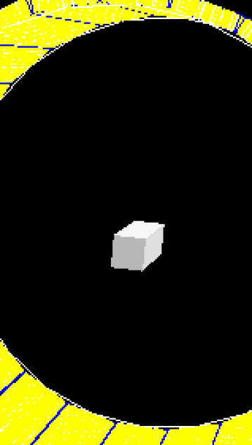
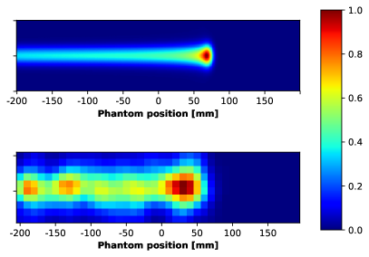
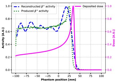
In this manuscript, we present preliminary results for one of the investigated setup configurations, i.e. a single layer J-PET barrel (fig. 4). Fig. 5 illustrates a cross-section through the centre of the beam of 3D dose distribution deposited in a 5520 cm3 PMMA phantom by 108 protons of nominal energy 150 MeV (top left) and the same cross-section of reconstructed 3D distribution of activity produced by the beam in the phantom (bottom left). The activity map was reconstructed in 555 mm3 voxel grid. The scintillator strips’ detection efficiency is taken into account in MC simulations. The expected spatial and time resolutions of J-PET with wavelength-shifting strips (WLS) [18] is taken into account through plastic length discretization used in the simulations and image reconstruction.
The fig. 5 (right) presents the MC simulated profiles of: (i) proton dose deposition in the PMMA phantom, (ii) activity produced in the phantom and (iii) actual signal detected by J-PET barrel from activity. The results show that the J-PET detector is feasible to acquire the activity produced during proton therapy treatment and that the offline 3D reconstruction of PET activity images is possible using CASTOR toolkit. The characterization of J-PET sensitivity for proton beam range detection is currently an ongoing research activity.
4 Summary
Within the research projects conducted in the Institute of Nuclear Physics PAN in Kraków we investigate physical and biological range uncertainties of proton beams through positron emission tomography (PET) based solutions and Monte Carlo (MC) simulations. Taking advantage of the Fred accuracy and time performance possible due to GPU acceleration we aim to improve quality assurance and treatment planing in Kraków PBT facility. A Monte Carlo study of J-PET detector feasibility performed in the frame of the project suggests that this technique might be considered as a novel proton beam therapy range monitoring approach.
5 Acknowledgments
The authors acknowledge Dosimetry and Quality Control Laboratory of CCB for supporting project activities. The FRED Monte Carlo project is carried out within the Reintegration programme of the Foundation for Polish Science co-financed by the European Union under the European Regional Development Fund grant no. POIR.04.04.00-00-2475/16-00. The project on range monitoring with J-PET detector is funded by the National Centre for Research and Development (NCBiR), grant no. LIDER/26/0157/L-8/16/NCBR/2017. MG and JB acknowledge the support within InterDokMed programme, project no. POWR.03.02.00-00-I013/16. PM acknowledges Foundation for Polish Science for support within TEAM programme, project no. TEAM/2017-4/39. We acknowledge the support of NVIDIA Corporation with the donation of the GPU used for this research. AR acknowledges Prof. Reinhard Schulte from Loma Linda University, CA, USA for mentoring and support.
References
- [1] Durante M., et al. Charged-particle therapy in cancer: clinical uses and future perspectives Nat. Rev. Clin. Oncol.. 2017;14:483.
- [2] Paganetti H.. Range uncertainties in proton therapy and the role of Monte Carlo simulations Phys. Med. Biol.. 2012;57:99–117.
- [3] Paganetti H., et al. Relative biological effectiveness (RBE) values for proton beam therapy Int. J. Radiat. Oncol. Biol. Phys.. 2002;53:407–421.
- [4] Schiavi A., et al. Fred: a GPU-accelerated fast-Monte Carlo code for rapid treatment plan recalculation in ion beam therapy Phys. Med. Biol.. 2017;62:7482–7504.
- [5] Garbacz M., et al. Proton therapy treatment plan verification in CCB Krakow using fred Monte Carlo TPS tool in IFMBE Proceedings;68 2019.
- [6] Agostinelli S., et al. Geant4 - a simulation toolkit Nucl. Inst. and Meth. in Phys. Res. A. 2003.
- [7] Battistoni G., et al. The FLUKA code: description and benchmarking AIP Conference Proceedings. 2007;896:31–49.
- [8] Carabe-Fernandez A., et al. Range Uncertainty in Proton Therapy Due to Variable Biological Effectiveness Phys. Med. Biol.. 2012;57:1159–1172.
- [9] Bauer J., et al. Implementation and initial clinical experience of offline PET/CT-based verification of scanned carbon ion treatment Radiath. Oncol.. 2013;107:218–226.
- [10] Krimmer J., et al. Prompt-gamma monitoring in hadrontherapy: A review Nucl. Inst. and Meth. in Phys. Res. A. 2018;878:58–73.
- [11] Moskal P., et al. Positronium in medicine and biology Nat. Rev. Phys.. 2019;1:527–529.
- [12] Ferrero V., et al. Online proton therapy monitoring: clinical test of a Silicon-photodetector-based in-beam PET Scientific Reports. 2018;8:4100.
- [13] Richter C., et al. First clinical application of a prompt gamma based in vivo proton range verification system Radiath. Oncol.. 2016;118:232–237.
- [14] Hueso-González F., et al. A full-scale clinical prototype for proton range verification using prompt gamma-ray spectroscopy Phys. Med. Biol.. 2018;63:185019.
- [15] Kowalski P., et al. Estimating the NEMA characteristics of the J-PET tomograph using the GATE package Phys. Med. Biol.. 2018;63.
- [16] Sarrut D., et al. A review of the use and potential of the GATE Monte Carlo simulation code for radiation therapy and dosimetry applications Med. Phys.. 2014;41:64301.
- [17] Merlin T., et al. CASToR: A generic data organization and processing code framework for multi-modal and multi-dimensional tomographic reconstruction Phys. Med. Biol.. 2018;63.
- [18] Smyrski J., et al. Measurement of gamma quantum interaction point in plastic scintillator with WLS strips Nucl. Inst. and Meth. in Phys. Res. A. 2017;851:39–42.