Perceptual-assisted Adversarial Adaptation for Choroid Segmentation in optical coherence tomography
Abstract
Accurate choroid segmentation in optical coherence tomography (OCT) image is vital because the choroid thickness is a major quantitative biomarker of many ocular diseases. Deep learning has shown its superiority in the segmentation of the choroid region but subjects to the performance degeneration caused by the domain discrepancies (e.g., noise level and distribution) among datasets obtained from the OCT devices of different manufacturers. In this paper, we present an unsupervised perceptual-assisted adversarial adaptation (PAAA) framework for efficiently segmenting the choroid area by narrowing the domain discrepancies between different domains. The adversarial adaptation module in the proposed framework encourages the prediction structure information of the target domain to be similar to that of the source domain. Besides, a perceptual loss is employed for matching their shape information (the curvatures of Bruch’s membrane and choroid-sclera interface) which can result in a fine boundary prediction. The results of quantitative experiments show that the proposed PAAA segmentation framework outperforms other state-of-the-art methods.
Index Terms— Choroid segmentation, deep learning, adversarial adaptation, perceptual loss
1 Introduction
The choroid in OCT contains abundant blood vessels which are extremely important for the supply of oxygen and nutrients to the outer retina [1]. Accurate choroid segmentation is a prerequisite for the diagnosis of many ocular diseases. However, manually choroid segmentation by professional ophthalmologists is a heavy and tedious task. An efficient automatic choroid segmentation algorithm is urgently needed.
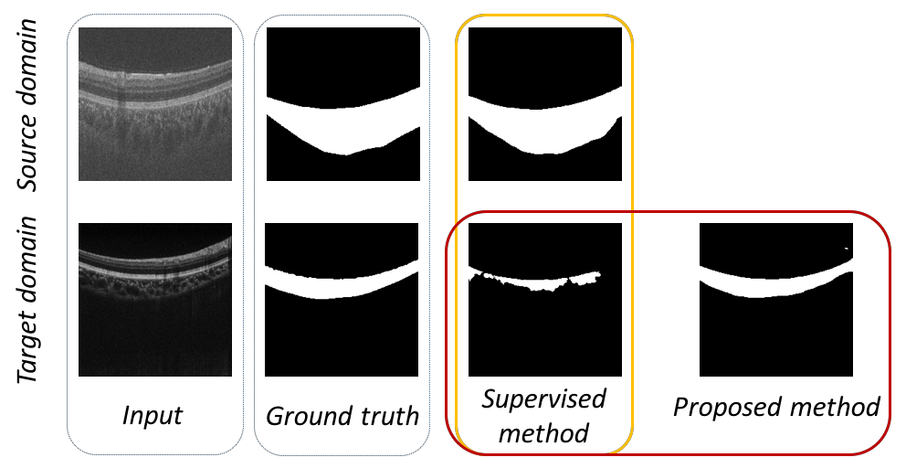
In recent years, deep learning has shown its advantage for medical image analysis [3], especially the convolutional neural network (CNN) which is trained in an end to end manner has been widely used for medical image segmentation. Massod et al. [4] and Caneiro et al. [5] have shown the advantage of deep learning methods compared to non-deep learning methods in choroid segmentation task. In [4], the authors proposed a two-stage segmentation network with a combination of CNNs and morphological operations. In [5], patch-based supervised machine learning methods were used to segment retinal and choroid boundary. The supervised methods based on deep learning have brought significant improvement for choroid segmentation. Although the supervised segmentation methods (e.g., FCN, U-Net) have a outstanding performance for choroid segmentation, there are two challenges for supervised methods. 1) supervised deep learning fails to obtain satisfactory segmentation results on new datasets due to the domain discrepancy between different datasets. 2) pixel-level annotations are time-consuming and labor-intensive in existing supervised methods for choroid segmentation. The orange rounded rectangle in Fig. 1 show that segmentation degrades when the supervised models are adapted from the source domain to the target domain.
To overcome the shortcomings of supervised deep learning, an unsupervised domain adaptation method aimed at minimizing the domain shift (domain discrepancy) has been proposed over the past few years [6]. Tsai et al. [7] considered semantic segmentation as structured outputs and conduct adversarial learning in output space for semantic segmentation. Wang et al. [8] introduced an unsupervised domain adaptation method using a patch discriminator for joint optic disc and cup segmentation. Although the unsupervised domain adaptation methods mentioned above can obtain better performance when facing the domain discrepancy, adversarial adaptation can only narrow the prediction distribution between the source domain and the target domain. It is difficult for adversarial learning to capture details. In this paper, we present a perceptual-assisted adversarial adaptation for choroid segmentation. Our PAAA framework incorporates adversarial adaptation to address the domain discrepancy by encouraging the structure information of the target domain close to that of the source domain. Furthermore, we utilize the perceptual loss module to match shape information of the source and target domains which can result in a fine boundary prediction.
Contributions: 1) We propose a perceptual-assisted adversarial adaptation method to minimize the performance degradation caused by domain discrepancy. 2) To the best of our knowledge, our work is the first of its kind to leverage the combined advantage of adversarial adaptation and perceptual loss for choroid segmentation in OCT in an unsupervised manner. 3) We apply our method to segment choroid area in OCT data. Results show that our proposed method outperforms the state-of-the-art methods.
2 METHOLODY
2.1 Problem Formulation
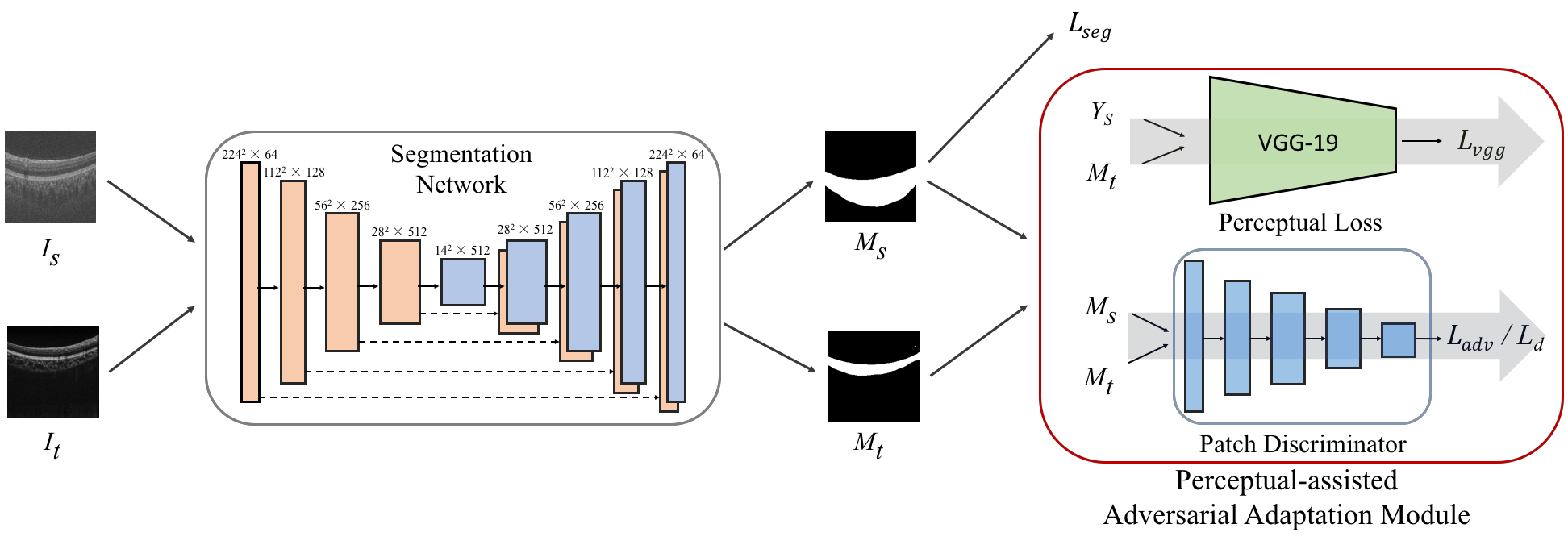
As shown in Fig. 2, our proposed method includes two parts: the segmentation network and the perceptual-assisted adversarial adaptation part which combines patch discriminator module with perceptual loss module to implement domain adaptation in output space. Formally, the source domain and target domain are denoted as and , respectively. The input images from source domain are denoted as while the ones from target domain are denoted as . and represent the predictions of the source image and the target image while denotes the label of source image.
2.2 Learning at Source Domain
We first adopt the cross-entropy loss as the segmentation loss for images from the source domain to train the segmentation network:
| (1) |
where is the ground truth annotaions for source images and is the output of segmentation network. represents the segmentation network.
2.3 Adversarial Adaptation for Target Domain
The segmentation loss is only applied on the source domain. To optimized the network for images in target domain, we forward them into to obtain the segmentation prediction . Then we first leverage the adversarial training to minimize the discrepancy between the prediction of the target domain and the one of the source domain in output space. Adversarial training is achieved by utilizing a PatchGAN loss [9]. Two predictions and generated from the network are fed into the discriminator . To make the distribution of closer to , we use an adversarial loss as :
| (2) |
This loss is designed to train the segmentation network and fool the discriminator by maximizing the probability of the target prediction being considered as the source prediction. Through adversarial optimization, the output space of the target domain learns to mimic the distribution of the source output space.
2.4 Perceptual-assisted Adaptation for Target Domain
Besides, a perceptual loss [10] is employed for matching their shape information (the curvatures of Bruch’s membrane and choroid-sclera interface) which can result in a fine boundary prediction. As the name suggests, penalizes results that are not perceptually similar to labels by defining a distance measure between feature maps of target prediction mask and source label after passing a pretrained VGG network. The constriction matches the shape information between the source domain and target domain which drives the prediction of the choroid area close to the true physiological structure and makes a better boundary prediction. Perceptual loss is defined as
| (3) |
where is the feature map of the i’-th layer of the pre-trained network. For this work, denotes the feature maps from layers relu1_1, relu2_1, relu3_1, relu4_1 and relu5_1 of the VGG-19 [11] network pretrained on the ImageNet [12] dataset.
The overall training objective for segmentation network is:
| (4) |
By incorporating adversarial adaptation with perceptual loss based on VGG network, the proposed method can minimize the discrepancy between the source domain and the target domain in output space.
3 Experiments
a
b
c
d
e
f
3.1 Datasets
Two different datasets obtained from topcon OCT device and nidek OCT device are utilized to implement experiments in this paper. The two datasets are called TOPCON and NIDEK, respectively. Both of TOPCON and NIDEK datasets have pixel-level manual annotations. We set the train part of TOPCON dataset as the source domain and the train part of NIDEK dataset as the target domain. The train part of TOPCON dataset consists of 640 OCT images with 512 512 resolution and the train part of NIDEK dataset includes 120 OCT images with 512 1024 resolution. The test part of NIDEK dataset has 30 OCT images and are used for evaluation.
3.2 Implementation details
The architecture of the proposed method can be referred to Fig 2. The proposed method leverage U-Net [2] as the backbone. The method was implemented in Python based on PyTorch. In the experiments, both of source image and target image were resized to 224 224 resolution. We trained the entire network end-to-end using Adam optimizer [13]. The beta1 and beta2 of Adam optimizer was set to 0.9 and 0.99, respectively. The weight decay factor was 0.0001. The learning rate of the segmentation network was set to and the one of the discriminator was set to 0.0001. , , in training objective were set to 100, 0.01, 0.06. Only the segmentation module was utilized in the evaluation phase.
| Methods | Source domain | Target domain | ||||
| Oracle | NIDEK | NIDEK | 2.65 | 89.30 | - - - | - - - |
| FCN [14] | TOPCON | NIDEK | 53.08 | 42.97 | 50.43 | 46.33 |
| U-Net [2] | TOPCON | NIDEK | 51.26 | 51.11 | 48.61 | 38.19 |
| FCN + AA [7] | TOPCON | NIDEK | 24.50 | 51.19 | 16.98 | 28.57 |
| U-Net + AA | TOPCON | NIDEK | 19.63 | 60.73 | 16.98 | 28.57 |
| Proposed method | TOPCON | NIDEK | 3.21 | 85.77 | 0.56 | 3.53 |
-
1
AUSDE (pixels)
-
2
IOU ()
3.3 Metrics
In order to properly evaluate the performance of our proposed method, the following metrics will be utilized in experiments.
Intersection over uniou (IOU): Intersection over uniou is a common evaluation metric to evaluate the segmentation quality. The IOU is defined as: / , where and represent area of overlap and area of union between the prediction and the reference standard.
Average unsigned surface detection error (AUSDE) [15]: Average unsigned surface detection error was computed for each lower boundary of choroid by measuring absolute Euclidean distance in the z-axis between the results of the algorithm and the reference standard. AUSDE is efficient metric to evaluate performance of choroid segmentation between different algorithms.
Moreover, ( and ) is another criterion to evaluate the adaptation performance of models. is the gap of between the model and the fully-supervised model, where denotes ausde or iou.
3.4 Results and Discusions
We evaluate the performance of our proposed method using the metrics mentioned in 3.3 and compare our methods with other state-of-the-art methods when models are adapted from the source domain to the target domain.
Quantitative evaluation: In Table. 1, we compare the proposed method with oracle (upper bound), supervised methods and unsupervised domain adaptation methods. Except for oracle, all experiments take TOPCON as source domain and NIDEK as target domain. The oracle represents the results of U-Net trained and evaluated on the same target domain. The comparison results between the supervised method (U-Net) and oracle quantitatively show the performance degradation due to the domain discrepancy. Our method achieves over 48 pixels decreasing for AUSDE and over 34 IOU improvement compared with the supervised methods (FCN, U-Net), demonstrating the effectiveness of perceptual-assisted adversarial adaptation method used to narrow domain discrepancy. We also compare our method with the state-of-the-art unsupervised domain adaptation method AdaptSegNet (FCN + AA) and our method outperforms the AdaptSegNet by more than 21 pixels decreasing for AUSDE and more than 34 IOU improvement, which show that the proposed PAAA method can obtain a better segmentation performance than AdaptSegNet. The ablation experiments were implemented, the proposed method can decrease the AUSDE by more than 16 pixels and improve the IOU by more than 25 compared with the baseline method (U-Net + AA) without perceptual loss module. The ablation experiments demonstrate that the perceptual loss help to result in a fine boundary prediction and obtain a better segmentation result. The and indicate that the segmentation results of our proposed method are closer to that of oracle than other methods. what’s more, the proposed method don not require pixel-level annotations in the target domain while the oracle needs the labor-intensive and time-consuming annotations to be trained.
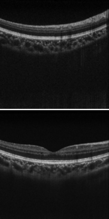
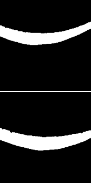
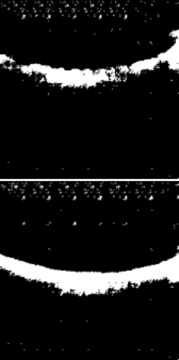
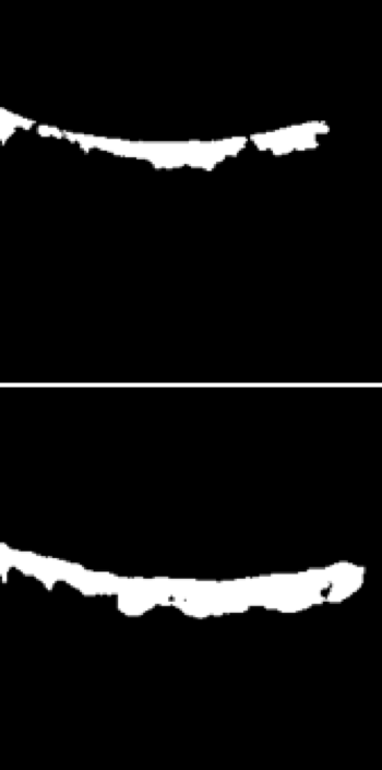
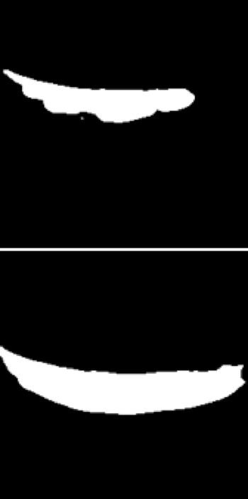
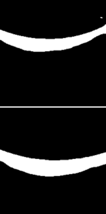
Qualitative evaluation: Fig. 3 shows the visualization of choroid segmentation results when models are adapted from source domain image to target domain image. The predictions of our methods are visually better than that of other methods and more closer to the label (ground truth). The comparison shows that the proposed method has a better segmentation performance than other methods.
4 Conclusion
In this paper, we have proposed a novel framework Perceptual-assisted Adversarial Adaptation to narrow the discrepancy between the source domain and the target domain and to obtain a better segmentation performance. The experiment results have demonstrated that our proposed method has an outstanding performance for choroid segmentation compared with other methods when models are adapted from the source domain to the target domain.
References
- [1] Javier Mazzaferri, Luke Beaton, Gisele Hounye, Diane N. Sayah, and Santiago Costantino, “Open-source algorithm for automatic choroid segmentation of oct volume reconstructions,” Scientific Reports, vol. 7, pp. 1 – 10, 2017.
- [2] Olaf Ronneberger, Philipp Fischer, and Thomas Brox, “U-net: Convolutional networks for biomedical image segmentation,” in MICCAI. 2015, vol. 9351, pp. 234–241, Springer International Publishing.
- [3] Geert Litjens, Thijs Kooi, Babak Ehteshami Bejnordi, Arnaud Arindra Adiyoso Setio, Francesco Ciompi, Mohsen Ghafoorian, Jeroen A.W.M. van der Laak, Bram van Ginneken, and Clara I. Sánchez, “A survey on deep learning in medical image analysis,” Medical Image Analysis, vol. 42, pp. 60 – 88, 2017.
- [4] Saleha Massood, Ruogu Fang, Huating Li, Bin Sheng, Akash Mathavan, Xiangning Wang, Po Yang, Qiang Wu, Jing Qin, and Weiping Jia, “Automatic choroid layer segmentation from optical coherence tomography images using deep learning,” Scientific Reports, vol. 9, pp. 1–18, 2019.
- [5] David Alonso-Caneiro, Jason Kugelman, Jared Hamwood, Scott A. Read, Stephen J. Vincent, Fred K. Chen, and Michael J. Collins, “Automatic retinal and choroidal boundary segmentation in oct images using patch-based supervised machine learning methods,” in Computer Vision – ACCV 2018 Workshops. 2019, pp. 215–228, Springer International Publishing.
- [6] Mei Wang and Weihong Deng, “Deep visual domain adaptation: A survey,” Neurocomputing, vol. 312, pp. 135–153, 2018.
- [7] Yi-Hsuan Tsai, Wei-Chih Hung, Samuel Schulter, Kihyuk Sohn, Ming-Hsuan Yang, and Manmohan Chandraker, “Learning to adapt structured output space for semantic segmentation,” in CVPR, 2018.
- [8] Shujun Wang, Lequan Yu, Xin Yang, Chi-Wing Fu, and Pheng-Ann Heng, “Patch-based output space adversarial learning for joint optic disc and cup segmentation,” IEEE Transactions on Medical Imaging, 2019.
- [9] Phillip Isola, Jun-Yan Zhu, Tinghui Zhou, and Alexei A. Efros, “Image-to-image translation with conditional adversarial networks,” in CVPR, 2017.
- [10] Leon A. Gatys, Alexander S. Ecker, and Matthias Bethge, “Image style transfer using convolutional neural networks,” in CVPR, 2016.
- [11] Karen Simonyan and Andrew Zisserman, “Very deep convolutional networks for large-scale image recognition,” in ICLR, 2015.
- [12] Jia Deng, Wei Dong, Richard Socher, Lijia Li, Kai Li, and Li Feifei, “ImageNet: A Large-Scale Hierarchical Image Database,” in CVPR, 2009.
- [13] Diederik P Kingma and Jimmy Lei Ba, “Adam: A method for stochastic optimization,” in ICLR, 2015.
- [14] Jonathan Long, Evan Shelhamer, and Trevor Darrell, “Fully convolutional networks for semantic segmentation,” in CVPR, 2015.
- [15] Dehui Xiang, Haihong Tian, Xiaoling Yang, Fei Shi, Weifang Zhu, Haoyu Chen, and Xinjian Chen, “Automatic segmentation of retinal layer in oct images with choroidal neovascularization,” IEEE Transactions on Image Processing, vol. 27, no. 12, pp. 5880–5891, Dec 2018.