Topological constraints in eukaryotic genomes and how they can be exploited to improve spatial models of chromosomes
Abstract
Several orders of magnitude typically separate the contour length of eukaryotic chromosomes and the size of the nucleus where they are confined. The ensuing topological constraints can slow down the relaxation dynamics of genomic filaments to the point that mammalian chromosomes are never in equilibrium over a cell’s lifetime. In this opinion article, we revisit these out-of-equilibrium effects and discuss how their inclusion in physical models can enhance the spatial reconstructions of interphase eukaryotic genomes from phenomenological constraints collected during interphase.
I Introduction
From viruses to eukaryotes, genomic DNA filaments are confined in spaces of linear dimension much smaller than their contour lengths. In bacteriophages, the m-long genome is stored in nm-wide viral capsids and the corresponding packing density is so high that viral DNA filaments that have little chance to be entangled when in solution (knotting probability ) become almost certainly knotted ( probability) once confined inside capsids (Rybenkov et al., 1993; Arsuaga et al., 2002; Marenduzzo et al., 2009, 2010). In humans, instead, the various cm-long chromosomes that make up the genome are kept inside m-wide nuclei (Alberts et al., 2014). Despite the major change of scale with respect to viruses, the volume fraction occupied by this eukaryotic genome is still large, about (Rosa and Everaers, 2008).
These considerations pose several conundrums: How can chromosomal DNA be at the same time packed and yet accessible to the regulatory and transcriptional machineries? What is its typical degree of genomic entanglement and how much does it interfere with DNA transactions? To what extent are these aspects shaped by general passive physical mechanisms versus active ones, e.g. involving topoisomerase enzymes?
II Intra- and inter-chromosome architecture
II.1 Phenomenology
Addressing these questions has proved challenging because of the wide range of length and time scales involved in genome architecture. Classical experimental tools provide details of chromosome architecture at two opposite scales (Marti-Renom and Mirny, 2011). At the smallest one (nm) X-ray crystallography revealed that DNA achieves local packing by wrapping around histones, while at the largest one (m) fluorescence in situ hybridization (FISH) showed that each chromosome occupies a compact region of the nucleus, termed territory (Cremer and Cremer, 2001, 2010).
More recently, experimental breakthroughs such as super-resolution imaging, electron microscopy tomography plus selective labelling and chromosome conformational capture (Hi-C) techniques have significantly extended our “multiscale” knowledge of genome architecture (Boettiger et al., 2016; Bintu et al., 2018; Nir et al., 2018; Ou et al., 2017; Dekker et al., 2002; Lieberman-Aiden et al., 2009).
These and other advancements helped establishing various results that foster the present discussion of genomic entanglement.
Regarding inter-chromosome organization we recall that:
-
(i)
the positioning of chromosome territories correlates significantly with sequence-dependent properties of the underlying DNA (most notably, gene density (Bolzer et al., 2005));
- (ii)
For intra-chromosome aspects we instead know that:
-
(iii)
on the scale of a few kilo-basepairs up to about 1 mega-basepair, chromosomes are organized into self-interacting regions, called topologically-associating domains or TADs (Dixon et al., 2012; Nora et al., 2012). On the tens of mega-basepairs scale, chromatin is organized into compartments of varying compactness depending on their functional and epigenomic state (Lieberman-Aiden et al., 2009; Wang et al., 2016).
-
(iv)
despite this variability, when averaged over chromosomes and experimental realizations, the mean contact probability of two chromosomal loci at genomic distance scales approximately as (Lieberman-Aiden et al., 2009), and the mean square separation scales as (Sachs et al., 1995; Münkel et al., 1999).
II.2 Relating genomic architecture and relaxation dynamics with polymer physics
The interpretation of these experimental results has been aided by an intense theoretical and computational activity that demonstrated how salient genomic architecture properties can be reproduced by a broad range of polymer models, and hence are likely governed by general physical mechanisms (Mirny, 2011; Rosa and Zimmer, 2014; Bianco et al., 2017; Jost et al., 2017; Haddad et al., 2017; Tiana and Giorgetti, 2018). This applies in particular to the aforementioned properties (i-iv) which can be rationalised as manifestations of the topological constraints that rule the behavior of semi-dilute or dense polymer systems by having a dramatic impact on their relaxation time scales (Doi and Edwards, 1986).
In fact, a solution of initially disentangled chains of contour length can reach the fully-mixed, homogeneous equilibrium state only via reptation, a slow and stochastic slithering-like motion with characteristic time scale equal to , where is a microscopic collision time and is the typical contour length between entanglement points (De Gennes, 1971; Doi and Edwards, 1986).
Thus, based on this fundamental polymer physics result, it was estimated that the characteristic relaxation, or equilibration, time of mammalian chromosomes exceeds 100 years (Rosa and Everaers, 2008). The orders-of-magnitude difference between this time scale and the typical duration of the cell cycle ( day) has several implications for genome organization, as it was realised even before Hi-C probing methods became available (Rosa and Everaers, 2008). It is clear, in fact, that mammalian chromosomes are never fully relaxed as they undergo the cyclic structural rearrangements from the separate compact rod-like mitotic architecture to the decondensed interphase one (Grosberg et al., 1993; Rosa and Everaers, 2008).
II.3 Implications for (minimal) intra- and inter-chromosome entanglement
From this standpoint, the emergence of chromosome territories is quantitatively explained as due to the kinetically trapped decondensation of the compact mitotic chromatin (Rosa and Everaers, 2008): interphase chromosomes retain the memory and limited mutual overlap of the earlier mitotic state, consistent with experimental results (Cremer and Cremer, 2001; Branco and Pombo, 2006; Cremer and Cremer, 2010). In addition, the ordered linear organization of the mitotic rods should also inform the intra-chromosomal architecture, making it more local than equilibrated polymers. This is consistent with the experimental fact that the effective scaling behaviour of the contact probability with the genomic separation in interphase chromosomes has a more local character () than the one expected () for equilibrated polymers (Lieberman-Aiden et al., 2009). Intuitively, the same “memory” mechanism ought to facilitate the subsequent separation of interphase chromosomes and their recondensation upon re-entering the mitotic phase in the cell cycle (Rosa and Everaers, 2008).
For the present discussion, we stress that these out-of-equilibrium effects should impact not only the architecture but also the physical entanglement of eukaryotic genomes. In fact, mammalian chromosomes should be more unlinked (for the limited inter-chromosomal intermingling) and unknotted (for the enhanced intra-chromosomal local contacts) than at equilibrium. These heuristic conclusions are supported by various studies showing that the aforementioned scaling relationships obtained by FISH and Hi-C experiments can be ascribed to the topological constraints at play in solutions of unknotted and unlinked polymers (Khokhlov and Nechaev, 1985; Vettorel et al., 2009; Halverson et al., 2014; Rosa and Everaers, 2014).
II.4 Implications for genomic structural modelling and its improvement
These considerations appear particularly relevant for the structural modelling of eukaryotic genomes based on phenomenological data, such as spatial proximity constraints, which are typically too sparse to pin down even coarse-grained models of interphase chromosomes (Lieberman-Aiden et al., 2009).
A key question is whether such structural models should additionally be informed by the notion that interphase chromosomes must originate and eventually return to the separate and condensed mitotic state.
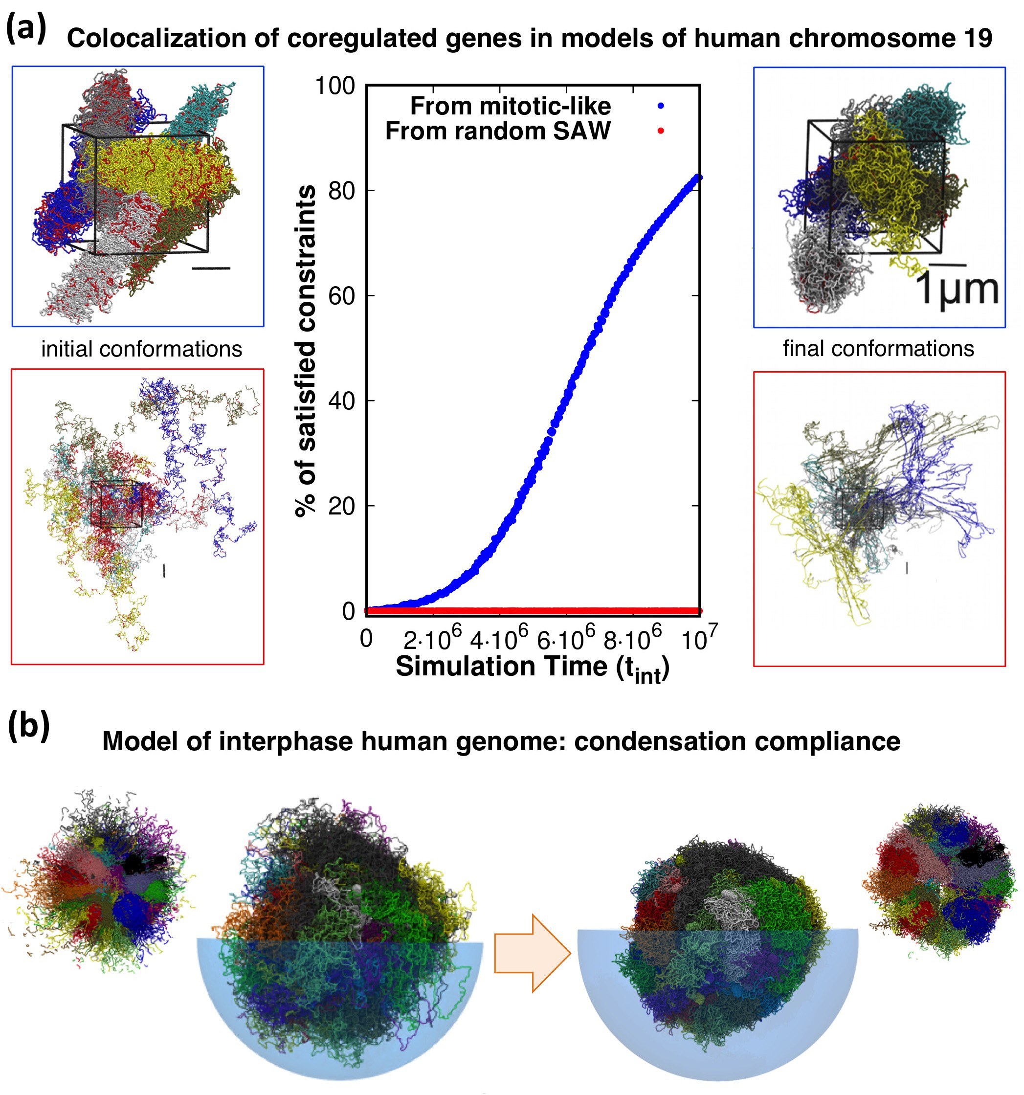
Evidence presented in our earlier work help shed some light on the matter. With our co-workers, we considered a model system of 6 copies of human chromosome 19 in a cubic simulation box with periodic boundary conditions to explore the connection between coregulation and colocalization of genes (Di Stefano et al., 2013). Each copy was initially prepared as a mitotic-like conformation (Rosa and Everaers, 2008), consisting of a polymer filament forming a solenoidal pattern with rosette-like cross-section featuring chromatin loops of about kilo-basepairs, see Fig. 1a. We then used a molecular-dynamics steering protocol to bring in proximity pairs of intra-chromosomal loci that were known to be significantly co-regulated. Importantly, topological constraints were accounted for by avoiding unphysical chain crossings during the steering process.
Remarkably, and consistently with the gene kissing hypothesis (Cavalli, 2007), we found that most ( 80%) pairs of significantly coregulated genes could indeed be colocalised in space within the contact range of 120nm and further showed that this colocalization compliance followed from the presence of gene cliques in the coregulatory network (Di Stefano et al., 2013).
Conversely, the same protocol applied to the same set of chains but initially prepared as generic self-avoiding random walks failed to give colocalization (Di Stefano et al., 2013): physically, this happens because the intra- and inter-chain entanglements present in the original system, by mimicking an “artificially” equilibrated set of chromosomes, were too numerous and conflicting to be successfully negotiated on a viable simulation time scale, see Fig. 1a.
Further elements come from the genome-wide structural modelling of human chromosomes of Di Stefano et al. (2016). In this study too, the model chromosomes were initially prepared in mitotic-like states and were then steered to bring in proximity those pairs of loci that corresponded to significantly enhanced entries of two independent Hi-C datasets (Dixon et al., 2012; Rao et al., 2014). The architecture of the final conformations were, as expected, significantly changed by the steering protocol. Yet, as illustrated in Fig. 1b, we verified that each model chromosome could be brought to a condensed compact shape as needed for the interphase-mitotic transition without significant hindrance from intra- or inter-chromosomal topological constraints (Di Stefano et al., 2016).
We note that the limitedly-entangled architecture of models of long eukaryotic chromosomes has emerged lately (Di Pierro et al., 2016) as the consequence of microphase separation of regions of different chromatin types (Jost et al., 2014) in a block co-polymer model with pair interactions tuned to reproduce the contact propensities of point (iv). The point is reinforced by studies on the yeast genome showing that knots and links have a generally low incidence especially in comparison to equivalent systems of equilibrated chains (Duan et al., 2010; Segal et al., 2014; Pouokam et al., 2019). Finally, besides the indication from structural models, other mechanisms such as loop extrusion have been advocated to be instrumental for maintaining a low degree of chromosomal entanglement (Racko et al., 2018; Orlandini et al., 2019).
To some inevitable extent though, physical entanglements are still expected to arise in eukaryotic chromosomes.
The recent work of Roca’s lab showed that knots do occur in eukaryotic minichromosomes in vivo, for instance during transcription, due to transient accumulation of entanglement (Valdés et al., 2017, 2019). On broader scales, various knots (Siebert et al., 2017), and even links (Niewieczerzal et al., 2019), were found in model mouse chromosomes obtained from single cell Hi-C (Stevens et al., 2017). The genuineness of the entangled states was suggested by the systematic recurrence of certain knot types in independent instances of the reconstructed chromosomal structures (Siebert et al., 2017). These were obtained by imposing phenomenological constraints on an initially disconnected set of effective monomers, so we expect that a more defined knot spectrum could be obtained by using disentangled self-avoiding chains as the reference model.
III Conclusions
To conclude, we have discussed experimental evidence and general physical mechanisms based on polymer theory that consistently point to an unusually low degree of entanglement expected in long eukaryotic chromosomes. Such property, which is arguably essential for the capability of chromosomes to reconfigure as needed at various stages of the cell cycle, appears important for genomic modelling too.
We argued, in fact, that the structural modelling of long chromosomes can benefit, both for realism and computational efficiency, by starting off with disentangled self-avoiding chains, e.g. mitotic-like ones, because their plasticity makes it possible to accommodate a large number of phenomenological constraints in a physically-viable manner, i.e. without deformations involving intra- or inter-chain crossings.
The latter are, of course, possible in in vivo systems thanks to the action of topoisomerase enzymes. An important open question regards, in fact, the extent to which these active mechanisms are involved in the shaping the overall intra- and inter-chromosome architecture. This point, we believe, can be significantly advanced in future studies with a tight synergy of experiments and models (Goloborodko et al., 2016; Jost et al., 2017; Valdés et al., 2019).
Conflict of Interest Statement
The authors declare that the research was conducted in the absence of any commercial or financial relationships that could be construed as a potential conflict of interest.
Author Contributions
All authors listed have made a substantial, direct and intellectual contribution to the work, and approved it for publication.
Funding
The authors acknowledge support from the Italian Ministry of Education, MIUR.
References
- Rybenkov et al. (1993) V. V. Rybenkov, N. R. Cozzarelli, and A. V. Vologodskii, Proc. Natl. Acad. Sci. USA 90, 5307 (1993).
- Arsuaga et al. (2002) J. Arsuaga, M. Vázquez, S. Trigueros, D. W. L. Sumners, and J. Roca, Proc. Natl. Acad. Sci. USA 99, 5373 (2002).
- Marenduzzo et al. (2009) D. Marenduzzo, E. Orlandini, A. Stasiak, D. W. Sumners, L. Tubiana, and C. Micheletti, Proc. Natl. Acad. Sci. USA 106, 22269 (2009).
- Marenduzzo et al. (2010) D. Marenduzzo, C. Micheletti, and E. Orlandini, J. Phys. Condens. Matter. 22, 283102 (2010).
- Alberts et al. (2014) B. Alberts, A. D. Johnson, J. Lewis, D. Morgan, M. Raff, K. Roberts, and P. Walter, Molecular Biology of the Cell, ed. (Garland Science, New York, 2014).
- Rosa and Everaers (2008) A. Rosa and R. Everaers, Plos Comput. Biol. 4, e1000153 (2008).
- Marti-Renom and Mirny (2011) M. A. Marti-Renom and L. A. Mirny, Plos Comput. Biol. 7, e1002125 (2011).
- Cremer and Cremer (2001) T. Cremer and C. Cremer, Nature Rev. Genet. 2, 292 (2001).
- Cremer and Cremer (2010) T. Cremer and M. Cremer, CSH Perspect. Biol. 2, a003889 (2010).
- Boettiger et al. (2016) A. N. Boettiger, B. Bintu, J. R. Moffitt, S. Wang, B. J. Beliveau, G. Fudenberg, M. Imakaev, L. A. Mirny, C.-t. Wu, and X. Zhuang, Nature 529, 418 (2016).
- Bintu et al. (2018) B. Bintu, L. J. Mateo, J.-H. Su, N. A. Sinnott-Armstrong, M. Parker, S. Kinrot, K. Yamaya, A. N. Boettiger, and X. Zhuang, Science 362, 419 (2018).
- Nir et al. (2018) G. Nir, I. Farabella, C. Pérez Estrada, C. G. Ebeling, B. J. Beliveau, H. M. Sasaki, S. D. Lee, S. C. Nguyen, R. B. McCole, S. Chattoraj, J. Erceg, J. AlHaj Abed, N. M. C. Martins, H. Q. Nguyen, M. A. Hannan, S. Russell, N. C. Durand, S. S. P. Rao, J. Y. Kishi, P. Soler-Vila, M. Di Pierro, J. N. Onuchic, S. P. Callahan, J. M. Schreiner, J. A. Stuckey, P. Yin, E. Lieberman-Aiden, M. A. Marti-Renom, and C.-t. Wu, Plos Genet. 14, 1 (2018).
- Ou et al. (2017) H. D. Ou, S. Phan, T. J. Deerinck, A. Thor, M. H. Ellisman, and C. C. O’Shea, Science 357, eaag0025 (2017).
- Dekker et al. (2002) J. Dekker, K. Rippe, M. Dekker, and N. Kleckner, Science 295, 1306 (2002).
- Lieberman-Aiden et al. (2009) E. Lieberman-Aiden, N. L. van Berkum, L. Williams, M. Imakaev, T. Ragoczy, A. Telling, I. Amit, B. R. Lajoie, P. J. Sabo, M. O. Dorschner, R. Sandstrom, B. Bernstein, M. A. Bender, M. Groudine, A. Gnirke, J. Stamatoyannopoulos, L. A. Mirny, E. S. Lander, and J. Dekker, Science 326, 289 (2009).
- Bolzer et al. (2005) A. Bolzer, G. Kreth, I. Solovei, D. Koehler, K. Saracoglu, C. Fauth, S. Muller, R. Eils, C. Cremer, M. R. Speicher, and T. Cremer, Plos Biol. 3, e157 (2005).
- Branco and Pombo (2006) M. R. Branco and A. Pombo, Plos Biol. 4, e138 (2006).
- Dixon et al. (2012) J. R. Dixon, S. Selvaraj, F. Yue, A. Kim, Y. Li, Y. Shen, M. Hu, J. S. Liu, and B. Ren, Nature 485, 376 (2012).
- Nora et al. (2012) E. P. Nora, B. R. Lajoie, E. G. Schulz, L. Giorgetti, I. Okamoto, N. Servant, T. Piolot, N. L. van Berkum, J. Meisig, J. Sedat, et al., Nature 485, 381 (2012).
- Wang et al. (2016) S. Wang, J.-H. Su, B. J. Beliveau, B. Bintu, J. R. Moffitt, C.-t. Wu, and X. Zhuang, Science 353, 598 (2016).
- Sachs et al. (1995) R. K. Sachs, G. van den Engh, B. Trask, H. Yokota, and J. E. Hearst, Proc. Natl. Acad. Sci. USA 92, 2710 (1995).
- Münkel et al. (1999) C. Münkel, R. Eils, S. Dietzel, D. Zink, C. Mehring, G. Wedemann, T. Cremer, and J. Langowski, J. Mol. Biol. 285, 1053 (1999).
- Mirny (2011) L. A. Mirny, Chromosome Res. 19, 37 (2011).
- Rosa and Zimmer (2014) A. Rosa and C. Zimmer, Int. Rev. Cell Mol. Biol. 307, 275 (2014).
- Bianco et al. (2017) S. Bianco, A. M. Chiariello, C. Annunziatella, A. Esposito, and M. Nicodemi, Chromosome Res. 25, 25 (2017).
- Jost et al. (2017) D. Jost, A. Rosa, C. Vaillant, and R. Everaers, in Nuclear Architecture and Dynamics, Vol. 2, edited by C. Lavelle and J.-M. Victor (Academic Press, 2017) p. 149.
- Haddad et al. (2017) N. Haddad, D. Jost, and C. Vaillant, Chromosome Res. 25, 35 (2017).
- Tiana and Giorgetti (2018) G. Tiana and L. Giorgetti, Curr. Opin. Struct. Biol. 49, 11 (2018).
- Doi and Edwards (1986) M. Doi and S. F. Edwards, The Theory of Polymer Dynamics (Oxford University Press, New York, 1986).
- De Gennes (1971) P.-G. De Gennes, J. Chem. Phys. 55, 572 (1971).
- Grosberg et al. (1993) A. Grosberg, Y. Rabin, S. Havlin, and A. Neer, Europhys. Lett. 23, 373 (1993).
- Khokhlov and Nechaev (1985) A. R. Khokhlov and S. K. Nechaev, Phys. Lett. 112A, 156 (1985).
- Vettorel et al. (2009) T. Vettorel, A. Y. Grosberg, and K. Kremer, Phys. Today 62, 72 (2009).
- Halverson et al. (2014) J. D. Halverson, J. Smrek, K. Kremer, and A. Y. Grosberg, Rep. Prog. Phys. 77, 022601 (2014).
- Rosa and Everaers (2014) A. Rosa and R. Everaers, Phys. Rev. Lett. 112, 118302 (2014).
- Di Stefano et al. (2013) M. Di Stefano, A. Rosa, V. Belcastro, D. di Bernardo, and C. Micheletti, Plos Comput. Biol. 9, e1003019 (2013).
- Cavalli (2007) G. Cavalli, Curr. Opin. Genet. Dev. 17, 443 (2007).
- Di Stefano et al. (2016) M. Di Stefano, J. Paulsen, T. G. Lien, E. Hovig, and C. Micheletti, Sci. Rep. 6, 35985 (2016).
- Rao et al. (2014) S. S. P. Rao, M. H. Huntley, N. C. Durand, E. K. Stamenova, I. D. Bochkov, J. T. Robinson, A. L. Sanborn, I. Machol, A. D. Omer, E. S. Lander, and E. Lieberman-Aiden, Cell 159, 1665 (2014).
- Di Pierro et al. (2016) M. Di Pierro, B. Zhang, E. Lieberman-Aiden, P. G. Wolynes, and J. Onuchic, Proc. Natl. Acad. Sci. USA 113, 12168 (2016).
- Jost et al. (2014) D. Jost, P. Carrivain, G. Cavalli, and C. Vaillant, Nucleic Acids Res. 42, 9553 (2014).
- Duan et al. (2010) Z. Duan, M. Andronescu, K. Schutz, S. McIlwain, Y. J. Kim, C. Lee, J. Shendure, S. Fields, C. A. Blau, and W. S. Noble, Nature 465, 363 (2010).
- Segal et al. (2014) M. R. Segal, H. Xiong, D. Capurso, M. Vazquez, and J. Arsuaga, Biostatistics 15, 442 (2014).
- Pouokam et al. (2019) M. Pouokam, B. Cruz, S. Burgess, M. R. Segal, M. Vazquez, and J. Arsuaga, Sci. Rep. 9, 6795 (2019).
- Racko et al. (2018) D. Racko, F. Benedetti, D. Goundaroulis, and A. Stasiak, Polymers 10, 1126 (2018).
- Orlandini et al. (2019) E. Orlandini, D. Marenduzzo, and D. Michieletto, Proc. Natl. Acad. Sci. USA 116, 8149 (2019).
- Valdés et al. (2017) A. Valdés, J. Segura, S. Dyson, B. Martínez-García, and J. Roca, Nucleic Acids Res. 46, 650 (2017).
- Valdés et al. (2019) A. Valdés, L. Coronel, B. Martínez-García, J. Segura, S. Dyson, O. Díaz-Ingelmo, C. Micheletti, and J. Roca, Nucleic Acids Res. 47, 6946 (2019).
- Siebert et al. (2017) J. T. Siebert, A. N. Kivel, L. P. Atkinson, T. J. Stevens, E. D. Laue, and P. Virnau, Polymers 9, 317 (2017).
- Niewieczerzal et al. (2019) S. Niewieczerzal, W. Niemyska, and J. I. Sulkowska, Sci. Rep. 9, 11753 (2019).
- Stevens et al. (2017) T. J. Stevens, D. Lando, S. Basu, L. P. Atkinson, Y. Cao, S. F. Lee, M. Leeb, K. J. Wohlfahrt, W. Boucher, A. O’Shaughnessy-Kirwan, J. Cramard, A. J. Faure, M. Ralser, E. Blanco, L. Morey, M. Sansó, M. G. S. Palayret, B. Lehner, L. Di Croce, A. Wutz, B. Hendrich, D. Klenerman, and E. D. Laue, Nature 544, 59 (2017).
- Goloborodko et al. (2016) A. Goloborodko, M. V. Imakaev, J. F. Marko, and L. Mirny, Elife 5, e14864 (2016).