Spin-wave directional anisotropies in antiferromagnetic Ba3NbFe3Si2O14
Abstract
Ba3NbFe3Si2O14 (langasite) is structurally and magnetically single domain chiral with the magnetic helicity induced through competing symmetric exchange interactions. Using neutron scattering, we show that the spin-waves in antiferromagnetic langasite display directional anisotropy. On applying a time reversal symmetry breaking magnetic field along the -axis, the spin wave energies differ when the sign is reversed for either the momentum transfer or applied magnetic field H. When the field is applied within the crystallographic -plane, the spin wave dispersion is directionally isotropic and symmetric in H. However, a directional anisotropy is observed in the spin wave intensity. We discuss this directional anisotropy in the dispersion in langasite in terms of a field induced precession of the dynamic unit cell staggered magnetization resulting from a broken twofold symmetry. Directional anisotropy, or often referred to as non reciprocal responses, can occur in antiferromagnetic phases in the absence of the Dzyaloshinskii-Moriya interaction or other effects resulting from spin-orbit coupling.
I Introduction
Mechanisms for controlling the flow of excitations, analogous to diodes in electric circuits, have been sought after to create anisotropic devices for magnonic Gladii et al. (2016); Schneider et al. (2008); Mruczkiewicz et al. (2017), acoustic Fleury et al. (2014); Liang et al. (2010, 2009), and optical Fan et al. (2012); Rikken et al. (2002); Roth and Rikken (2000) applications. Tokura and Nagaosa (2018) An example of anisotropic excitations in bulk materials are spin waves in low crystallographic symmetry crystals Katsura et al. (2005, 2007); Takahasi et al. (2012); Gitgeatpong et al. (2017) where magnons propagating in differing directions have dissimilar velocities. Such excitations have been defined as non reciprocal Deak and Fulop (2012); Szaller et al. (2013) given that the motion in one direction differs from that in the opposite Cheong et al. (2018) in the presence of broken time reversal symmetry. Onsager (1931) Other directional anisotropies have been reported in optical measurements where the response depends on the direction of the incident probing beam Vinegrad et al. (2018) resulting in contrasting absorption for counter propagating beams. Bordacs et al. (2012); Saito et al. (2008); Szaller et al. (2013)
Directional anisotropic excitations have been predicted and measured in the presence of relativistic spin-orbit coupling. For spin-wave excitations, this has been reported in MnSi Weber et al. (2018); Sato et al. (2016), LiFe5O8 Iguchi et al. (2015), CuV2O7 Gitgeatpong et al. (2017) where the antisymmetric Dzyaloshinskii-Moriya interaction has been implicated as the origin of the non reciprocal effects under applied magnetic fields. All of these materials also host a net ferromagnetic moment at low temperatures providing a direct means of coupling the magnetism to a net applied magnetic field. We find that directional anisotropy of the magnetic fluctuations can occur in the absence of such spin interactions through the application of neutron scattering at high magnetic fields in antiferromagnetic iron based langasite.
Ba3NbFe3Si2O14 Lee et al. (2010); Zhou et al. (2009); Marty et al. (2010); Chaix et al. (2013) (space group # 150 P321) is structurally single domain chiral with the 6 symmetry elements including the unity operation , two 3-fold rotational symmetry axes along the crystallographic axis , three 2-fold axes within the plane and also time reversal symmetry. The low temperature (TN=27 K) magnetic incommensurate order (with symmetry P3211′) removes the 2-fold axes of the underlying crystallographic structure with the exception of those Fe3+ spins aligned exactly along a crystallographic 2-fold axes. We discuss this point further below. However, the magnetism does preserve the 3-fold symmetry for all Fe3+ spins within the -plane and time reversal symmetry (denoted as 1′ in the magnetic space group). Marty et al. (2010) The magnetic structure is based on locally isolated triangles of 120∘ oriented Fe3+ (=5/2, =0) spins forming a hexagonal framework in the the -plane. Chaix et al. (2016); Scagnoli et al. (2013) The Fe3+ triangles are stacked along the -axis, linked via two O2- ions giving a helical exchange pathway breaking inversion symmetry.
Given the underlying nuclear structure, the magnetic spins form a single domain chiral spin pattern below TN. Quirion et al. (2017); Flack (2003) The original papers discussing the magnetic structure Marty et al. (2008) defined two terms to characterize the magnetic structure - helicity and chirality. The helicity was used to define the orientation of Fe3+ moments on neighboring planes and is fixed through competing symmetric exchange interactions with 1 meV. The chirality on an individual Fe3+ triangle is set by a weak antisymmetric exchange, allowed through the distortion of the local crystal field environment Yosida (1991), with =0.004 meV. Stock et al. (2011); Jensen (2011) The dominant role that symmetric exchange plays in fixing the magnetic handiness makes langasite unique over other chiral magnets such as MnSi, LiFe5O8, and CuV2O7 where antisymmetric exchange fixes the underlying chiral magnetic structures.
The spin excitations in magnetically ordered iron based langasite Jensen (2011); Loire et al. (2011); Stock et al. (2011) are defined by three modes termed the (denoted as modes hereafter) and modes following Ref. Jensen, 2011. The modes are the gapless in energy Nambu-Goldstone modes corresponding to a continuous rotation of the spins within the plane. On averaging over the three spins of the individual trimer, there is no net displacement of the magnetic moment in the plane and the neutron cross section along (0,0,L) is therefore weak following magnetic structure factors of neutron scattering. The mode is chiral reflecting the underlying magnetic structure and has been confirmed by polarized neutron spectroscopy. Loire et al. (2011); Moon et al. (1969); Blume (1963) In contrast, the two modes are gapped in energy and achiral, corresponding to transverse excitations of the Fe3+ spins with net zero out-of-plane displacement. The modes correspond to out of plane excitations and hence require a finite energy to overcome any anisotropy that fixes the moments in the crystallographic plane. The resulting energy gap value for the modes is therefore determined by a Dzyaloshinskii-Moriya (DM) anisotropy estimated to be of order 0.004 meV with a direction defined by =(0,0,1). Jensen (2011); Zorko et al. (2011)
In the absence of an applied magnetic field, time reversal symmetry is maintained and Kramers theorem implies at least a twofold degenerate spectrum, with spin waves excited with momentum transfer having the same response as -. With the application of a magnetic field on a lattice which is non centrosymmetric (and hence ), time reversal symmetry is broken and inversion and time reversal operators do not commute with the Hamiltonian and are hence not necessarily conserved. Therefore, differing spin responses excited with , and hence directional anisotropy, become possible. Shelankov and Pikus (1992) We now illustrate this effect in iron based langasite.
II Experimental Details
The experiments were carried out on a single crystal grown using the floating zone technique aligned such that reflections of the form (H,0,L) lay within the horizontal scattering plane. Vertical magnetic field experiments used the Panda (FRM2) and MACS (NIST) cold triple-axis spectrometers with the field applied perpendicular to the chiral crystallographic -axis along (-1,2,0). Experiments with the field aligned within the (H,0,L) horizontal scattering plane were done at RITA2 (PSI) and FLEXX/MultiFLEXX (HZB). For horizontal field measurements on FLEXX, the field was also rotated 90∘ and aligned along (1,0,0). We note that (-1,2,0) is parallel to a real space 2-fold axis and therefore preserves this crystallographic symmetry. The (100) field orientation is located 30∘ from the real space and 2-fold axes, but corresponds to a projection of along a 2-fold axis. All experiments were done with a fixed final energy defining the energy transfer as .
III Results
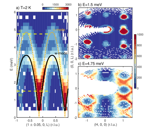
We first discuss the magnetic excitations in zero applied magnetic field. Figure 1 illustrates the neutron spectroscopic response at T taken on MACS. Fig. 1 shows a constant- slice along (1 0.05, 0, L) illustrating two distinct magnetic modes. The lower energy mode corresponds to the modes while the high-energy mode extending up to 5 meV is the gapless Goldstone mode following the notation described above. The solid and dotted curves are the linear spin-wave calculations from Ref. Jensen, 2011 for both the and branches. Constant energy slices are displayed in panels Figs. 1 at E=1.5 meV slicing through both and modes and a higher E=4.75 meV which only crosses through the mode. These scans illustrate that the -mode lacks intensity along (0,0,L) (Fig. 1 ) showing that, on averaging over the Fe3+ spins, no fluctuations perpendicular to the crystallographic axis occur consistent with this being the gapless Goldstone mode associated with a continuous rotation within the plane. This is in contrast to the lower energy -mode which does show intensity along (0,0,L) (Fig. 1 ) indicating a net component with in the plane. These results are consistent with predictions of spin wave theory. Jensen (2011)
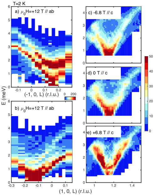
We now discuss the effects of a magnetic field on the spin dynamics. Figure 2 displays spin waves in langasite for a magnetic field aligned along the axis and also within the plane. Figures 2 show constant slices taken on Panda with a vertical field (aligned in the crystallographic along the (-1,2,0) direction) of 12 T for both Friedal pairs = (1, 0, -1/7) corresponding to opposite total momentum transfers of the incident beam. The two panels show that the higher energy -mode’s dispersion is comparatively weakly affected by the change in sign of at 12 T. The -mode excitation is gapped in comparison to zero field (Fig. 1) owing to the presence of a field induced spin anisotropy. However, the lower energy -mode shows a contrasting response with (-1, 0, 1/7) displaying a maximum in the dispersion of 3 meV while for the Friedel pair (1, 0, -1/7) the spin waves only reach a maximum of 2 meV. Figures 2 illustrates differing spin wave responses in a strong magnetic field when the sign of the total momentum transfer is reversed.
In Figs. 2 , we illustrate the dynamics when the magnetic field is aligned along the chiral -axis. Another means of searching for directionally anisotropic spin waves is to fix and reverse the sign of the time reversal symmetry breaking magnetic field. Figs. 2 show the effect of reversing the field on the dispersion near the magnetic Bragg peak of =(1, 0, 1/7) illustrating that the low-energy -mode is affected by the field displaying a differing response for = 6.8 T and 0 T. Fig. 2 shows that when a magnetic field is applied both within the crystallographic plane and along the chiral axis, an anisotropy is observed in the spin wave response. We now investigate these two field orientations in detail.
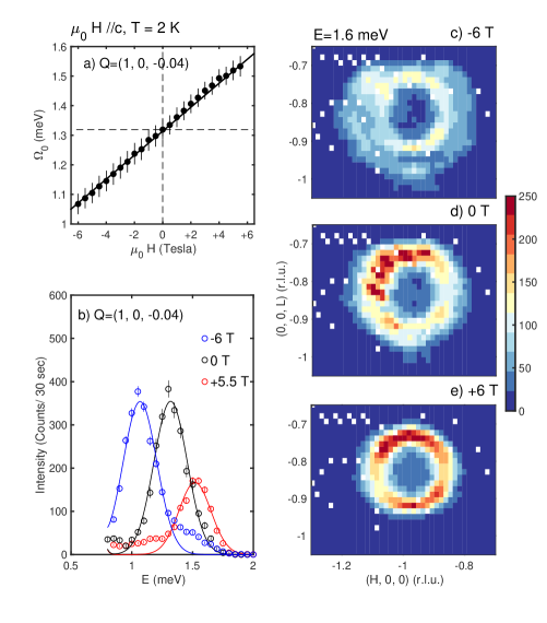
The field dependence of the mode, when the magnetic field is oriented along the chiral crystallographic -axis, is shown in Fig. 3 and by investigating constant =(1,0,-0.04) scans. The energy position is plotted as a function of applied magnetic field in panel Fig. 3 illustrating a linear scaling with field with representative constant momentum scans shown in Fig. 3 . The anisotropic in field response of the spin wave branch is further illustrated through constant E=1.6 meV scans shown in panels Fig. 3 at 6 and 0 T. With an applied field of -6 T (Fig. 3 ) a separation between the and higher velocity modes is observed. With increasing fields (Fig. 3 ) this circle of intensity fills in with the effect being linear with field.
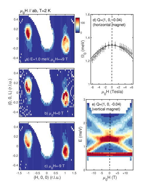
Having observed linear scaling of the -mode with the field oriented along the chiral -axis, we investigate the scaling with the field oriented within the crystallographic plane. Figure 4 shows the response of the lowest energy -mode to a vertical field applied within the crystallographic -plane. Figs. 4 illustrate constant E=1 meV scans taken using the MACS spectrometer (NIST) which simultaneously measures excitations around the points = (1, 0, -1/7) with magnetic fields of =0, 9 T. At an applied field of =0 T, symmetric spin wave cones are observable at the = (1, 0, -1/7) Friedel pairs. However, for an applied field of +9T, an apparent contraction of the cone is observed at =(-1, 0, 1/7) while the cone appears increases in diameter at =(1, 0, -1/7). The opposite response is observed for =-9T with a contraction of the spin-wave cone at =(1, 0, -1/7) and an increase in diameter at the Friedel pair (-1, 0, 1/7).
We apply higher resolution scans in Fig. 4 to plot the field dependence of the -mode near =(1,0, -0.04), the same momentum position investigated above for the horizontal field. These measurements were performed under the same conditions as in Fig. 3 , but with the horizontal field rotated by 90∘ within the scattering plane to be aligned along [1,0,0]. Fig. 4 shows a symmetric response with field with the dashed line a fit to . Further high resolution measurements using a vertical field allowing larger magnetic fields confirm this symmetric response as shown in Fig. 4 . This figure also shows that 14.5 T is enough to split the degeneracy of the low-energy -mode illustrating their symmetric energy position with . However, while the energy dispersion is symmetric with field, the intensity distribution between the two low energy modes is not giving rise to the apparent asymmetric response observed with lower resolution techniques discussed above. A similar asymmetry in intensity is seen for the -modes for in Fig. 2 .
IV Discussion and Conclusions
We have observed two different types of directional anisotropy in Ba3NbFe3Si2O14. When the field is applied within the crystallographic plane, both the and modes display different scattering structure factors for a given for . The energy position of the spin wave branches scales as and is directionally isotropic. When the field is oriented along the crystallographic -axis, the energy position of the low energy mode displays directional anisotropy with the spin wave energy scaling as .
To understand these results, we note that the mode that displays directional anisotropy in the energy dispersion is the -mode discussed above. This mode consists of transverse excitations of the Fe3+ moments such (summing over a given isolated triangle Fe3+ spins). The -mode corresponds to a tilting of the plane connecting the Fe3+ spins on a given trimer in the P321 unit cell. No such tilting occurs for the -mode where . Under a magnetic field along the crystallographic -axis, this dynamic tilting would precess around the crystallographic -axis Cheon et al. (2018) effectively giving the -mode a helical character absent at zero field. The size of the effect was predicted based on linear spin wave theory in Ref. Cheon et al., 2018 and is in agreement with Fig. 3 . Because of the globally broken 2-fold symmetry at low-temperatures, spin waves with different directions with differing , are not equivalent. We note that such a precession does not occur for the gapless -mode and this is consistent with our experiment which finds, with high resolution neutron spectroscopy, the dispersion to be isotropic under . Such a precession would also not occur if the magnetic field was perpendicular to the crystallographic -axis as this symmetry axis is preserved in the low temperature magnetic phase. The importance of the broken 2-fold axis is further highlighted by the observation of directional dichroism for electromagnons. Narita et al. (2016)
The lack of a twofold axis in langasite at low temperatures is inconsistent with an incommensurate magnetic structure where some spins would inevitably align along one of the twofold axes in a bulk crystal. However, the situation is different when the magnetic structure is truly commensurate (=1/7). Then the twofold symmetry is broken by the magnetic structure when the global phase of the helix is chosen such that there is never a magnetic moment aligned exactly parallel or perpendicular to the twofold axis at =1/2 of the sevenfold magnetic unit cell. It would be interesting to compare the results here against other magnetic systems with definitively incommensurate magnetic structures.
The results here are distinct from MnSi Weber et al. (2018); Sato et al. (2016), LiFe5O8 Iguchi et al. (2015), or Cu2V2O7 Gitgeatpong et al. (2017); Vasil’ev et al. (1999) where directional anisotropy, or non reciprocal effects, are due to an underlying antisymmetric Dzyaloshinskii-Moriya interaction. These materials also host a net ferromagnetic moment in the absence of a magnetic field at low temperatures. Such an effect originates from spin-orbit coupling and indeed has been implicated as being the origin of non reciprocal effects on spin waves in two-dimensional electron gases. Perez et al. (2016) We find that an underlying spin-orbit coupling is not required for the presence of directional anisotropy under a time reversal breaking symmetry field. In both MnSi and Cu2V2O7, the non collinear magnetic structures are stabilized by such interactions unlike the case for iron langasite where symmetric exchange stabilizes the helical magnetic structure.
In summary, we report directional anisotropy for the magnetic excitations in langasite in the magnetically ordered state. With the field aligned the -axis, directional anisotropy is observed for the spin wave energy response, with the effect scaling linearly with field. When the field is aligned within the plane, the spin wave energies are directionally isotropic, but the intensity is not. The effect originates from a precession of the dynamic unit cell staggered magnetization and does not originate from antisymmetric exchange as required in other magnetic systems.
V Acknowledgements
We are grateful for funding from the EPSRC, the Carnegie Trust for the Universities of Scotland, the STFC and through the NSF (DMR-09447720). The work at Rutgers University was supported by the DOE under Grant No. DOE: DE-FG02-07ER46382. Access to MACS was provided by the Center for High Resolution Neutron Scattering, a partnership between the National Institute of Standards and Technology and the National Science Foundation under Agreement No. DMR-1508249. We also thankfully acknowledge the financial support from HZB.
References
- Gladii et al. (2016) O. Gladii, M. Haidar, Y. Henry, M. Kostylev, and M. Bailleul, Phys. Rev. B 93, 054430 (2016).
- Schneider et al. (2008) T. Schneider, A. A. Serga, T. Neumann, B. Hillebrands, and M. P. Kostylev, Phys. Rev. B 77, 214411 (2008).
- Mruczkiewicz et al. (2017) M. Mruczkiewicz, P. Graczyk, P. Lupo, A. Adeyeye, G. Gubbiotti, and M. Krawczyk, Phys. Rev. B 96, 104411 (2017).
- Fleury et al. (2014) R. Fleury, D. L. Sounas, C. F. Sieck, M. R. Haberman, and A. Alu, Science 343, 516 (2014).
- Liang et al. (2010) B. Liang, X. S. Guo, J. Tu, D. Zhang, and J. C. Cheng, Nat. Mater. 9, 989 (2010).
- Liang et al. (2009) B. Liang, B. Yuan, and J.-C. Cheng, Phys. Rev. Lett. 103, 104301 (2009).
- Fan et al. (2012) L. Fan, J. Wang, L. T. Varghese, H. Shen, B. Niu, Y. Xuan, A. M. Weiner, and M. Qi, Science 335, 447 (2012).
- Rikken et al. (2002) G. L. J. A. Rikken, C. Strohm, and P. Wyder, Phys. Rev. Lett. 89, 133005 (2002).
- Roth and Rikken (2000) T. Roth and G. L. J. A. Rikken, Phys. Rev. Lett. 85, 4478 (2000).
- Tokura and Nagaosa (2018) Y. Tokura and N. Nagaosa, Nat. Commun. 9, 3740 (2018).
- Katsura et al. (2005) H. Katsura, N. Nagaosa, and A. V. Balatsky, Phys. Rev. Lett. 95, 057205 (2005).
- Katsura et al. (2007) H. Katsura, A. V. Balatsky, and N. Nagaosa, Phys. Rev. Lett. 98, 027203 (2007).
- Takahasi et al. (2012) Y. Takahasi, R. Shimano, Y. Kaneko, H. Murakawa, and Y. Tokura, Nat. Phys. 8, 121 (2012).
- Gitgeatpong et al. (2017) G. Gitgeatpong, Y. Zhao, P. Piyawongwatthana, Y. Qiu, L. W. Harriger, N. P. Butch, T. J. Sato, and K. Matan, Phys. Rev. Lett. 119, 047201 (2017).
- Deak and Fulop (2012) L. Deak and T. Fulop, Ann. Physics 327, 1050 (2012).
- Szaller et al. (2013) D. Szaller, S. Bordacs, and I. Kezsmarki, Phys. Rev. B 87, 014421 (2013).
- Cheong et al. (2018) S. W. Cheong, D. Talbayev, V. Kirykhin, and A. Saxena, npj Quantum Materials 3, 19 (2018).
- Onsager (1931) L. Onsager, Phys. Rev. 37, 405 (1931).
- Vinegrad et al. (2018) E. Vinegrad, D. Vestler, A. Ben-Moshe, A. R. Barnea, G. Markovich, and O. Cheshnoysky, ACS Photonics 5, 2151 (2018).
- Bordacs et al. (2012) S. Bordacs, I. Kezsmarki, D. Szaller, L. Demko, N. Kida, H. Murakawa, Y. Onose, R. Shimano, T. Room, U. Nagel, S. Miyahara, N. Furukawa, and Y. Tokura, Nat. Phys. 8, 734 (2012).
- Saito et al. (2008) M. Saito, K. Taniguchi, and T. Arima, J. Phys. Soc. Jpn. 77, 013705 (2008).
- Weber et al. (2018) T. Weber, J. Waizner, G. S. Tucker, R. Georgii, M. Kugler, A. Bauer, C. Pfleiderer, M. Garst, and P. Boni, Phys. Rev. B 97, 224403 (2018).
- Sato et al. (2016) T. J. Sato, D. Okuyama, T. Hong, A. Kikkawa, Y. Taguchi, T. H. Arima, and Y. Tokura, Phys. Rev. B 94, 144420 (2016).
- Iguchi et al. (2015) Y. Iguchi, S. Uemura, K. Ueno, and Y. Onose, Phys. Rev. B 92, 184419 (2015).
- Lee et al. (2010) C. Lee, E. Kan, H. Xiang, and M.-H. Whangbo, Chem. Mater. 22, 5290 (2010).
- Zhou et al. (2009) H. D. Zhou, L. L. Lumata, P. L. Kuhns, A. P. Reyes, E. S. Choi, D. S. Dalal, J. Lu, Y. J. Jo, L. Balicas, J. S. Brooks, and C. R. Wiebe, Chem. Mater. 21, 156 (2009).
- Marty et al. (2010) K. Marty, P. Bordet, V. Simonet, M. Loire, R. Ballou, C. Darie, J. Kljun, P. Bonville, O. Isnard, P. Lejay, B. Zawilski, and C. Simon, Phys. Rev. B 81, 054416 (2010).
- Chaix et al. (2013) L. Chaix, S. de Brion, F. Lévy-Bertrand, V. Simonet, R. Ballou, B. Canals, P. Lejay, J. B. Brubach, G. Creff, F. Willaert, P. Roy, and A. Cano, Phys. Rev. Lett. 110, 157208 (2013).
- Chaix et al. (2016) L. Chaix, R. Ballou, A. Cano, S. Petit, S. de Brion, J. Ollivier, L.-P. Regnault, E. Ressouche, E. Constable, C. V. Colin, A. Zorko, V. Scagnoli, J. Balay, P. Lejay, and V. Simonet, Phys. Rev. B 93, 214419 (2016).
- Scagnoli et al. (2013) V. Scagnoli, S. W. Huang, M. Garganourakis, R. A. de Souza, U. Staub, V. Simonet, P. Lejay, and R. Ballou, Phys. Rev. B 88, 104417 (2013).
- Quirion et al. (2017) G. Quirion, C. Bidaud, J. A. Quilliam, P. Lejay, V. Simonet, and R. Ballou, Phys. Rev. B 96, 134433 (2017).
- Flack (2003) H. D. Flack, Helv. Chim. Acta 86, 905 (2003).
- Marty et al. (2008) K. Marty, V. Simonet, E. Ressouche, R. Ballou, P. Lejay, and P. Bordet, Phys. Rev. Lett. 101, 247201 (2008).
- Yosida (1991) K. Yosida, Theory of Magnetism (Springer Verlag, 1991).
- Stock et al. (2011) C. Stock, L. C. Chapon, A. Schneidewind, Y. Su, P. G. Radaelli, D. F. McMorrow, A. Bombardi, N. Lee, and S. W. Cheong, Phys. Rev. B 83, 104426 (2011).
- Jensen (2011) J. Jensen, Phys. Rev. B 84, 104405 (2011).
- Loire et al. (2011) M. Loire, V. Simonet, S. Petit, K. Marty, P. Bordet, P. Lejay, J. Ollivier, M. Enderle, P. Steffens, E. Ressouche, A. Zorko, and R. Ballou, Phys. Rev. Lett. 106, 207201 (2011).
- Moon et al. (1969) R. M. Moon, T. Riste, and W. C. Koehler, Phys. Rev. 181, 920 (1969).
- Blume (1963) M. Blume, Phys. Rev. 130, 1670 (1963).
- Zorko et al. (2011) A. Zorko, M. Pregelj, A. Potočnik, J. van Tol, A. Ozarowski, V. Simonet, P. Lejay, S. Petit, and R. Ballou, Phys. Rev. Lett. 107, 257203 (2011).
- Shelankov and Pikus (1992) A. L. Shelankov and G. E. Pikus, Phys. Rev. B 46, 3326 (1992).
- Cheon et al. (2018) S. Cheon, H.-W. Lee, and S.-W. Cheong, Phys. Rev. B 98, 184405 (2018).
- Narita et al. (2016) H. Narita, Y. Tokunaga, A. Kikkawa, Y. Taguchi, Y. Tokura, and Y. Takahashi, Phys. Rev. B 94, 094433 (2016).
- Vasil’ev et al. (1999) A. N. Vasil’ev, L. A. Ponomarenko, A. I. Smirnov, E. V. Antipov, Y. A. Velikodny, M. Isobe, and Y. Ueda, Phys. Rev. B 60, 3021 (1999).
- Perez et al. (2016) F. Perez, F. Baboux, C. A. Ullrich, I. D’Amico, G. Vignale, G. Karczewski, and T. Wojtowicz, Phys. Rev. Lett. 117, 137204 (2016).