Ionization of biological molecules by multicharged ions using the stoichiometric model
Abstract
In the present work, we investigate the ionization of molecules of biological interest by the impact of multicharged ions in the intermediate to high energy range. We performed full non–perturbative distorted–wave calculations (CDW) for thirty–six collisional systems composed by six atomic targets: H, C, N, O, F, and S –which are the constituents of most of the DNA and biological molecules– and six charged projectiles (antiprotons, H, He, B, C, and O). On account of the radiation damage caused by secondary electrons, we inspect the energy and angular distributions of the emitted electrons from the atomic targets. We examine seventeen molecules: DNA and RNA bases, DNA backbone, pyrimidines, tetrahydrofuran (THF), and CnHn compounds. We show that the simple stoichiometric model (SSM), which approximates the molecular ionization cross sections as a linear combination of the atomic ones, gives reasonably good results for complex molecules. We also inspect the extensively used Toburen scaling of the total ionization cross sections of molecules with the number of weakly bound electrons. Based on the atomic CDW results, we propose new active electron numbers, which leads to a better universal scaling for all the targets and ions studied here in the intermediate to the high energy region. The new scaling describes well the available experimental data for proton impact, including small molecules. We perform full molecular calculations for five nucleobases and test a modified stoichiometric formula based on the Mulliken charge of the composite atoms. The difference introduced by the new stoichiometric formula is less than 3%, which indicates the reliability of the SSM to deal with this type of molecules. The results of the extensive ion–target examination included in the present study allow us to assert that the SSM and the CDW–based scaling will be useful tools in this area.
pacs:
34.50Gb, 34.80Gs, 34.80DpI Introduction
The damage caused by the impact of multicharged heavy projectiles on biological targets has become a field of interest due to its recent implementation in ion–beam cancer therapy. The effectiveness of the radiation depends on the choice of the ions. In particular, theoretical and experimental studies with different projectiles have concluded that charged carbon ions could be the most suitable ions to be used Mohamad2017 . Nonetheless, the study of such systems represents a challenge from the theoretical point of view.
The ionization of biological molecules by multicharged ions constitutes the primary damage mechanism. The most widely used method to predict such processes is the first Born approximation. At high energies, this perturbative method warrants the laws, where is the projectile charge. However, the damage is concentrated in the vicinities of the Bragg peak –at energies of hundreds of keV/amu–, precisely where the Born approximation starts to fail. Another theoretical issue arises due to the targets themselves; we are dealing with complex molecules, and the description of such targets represents a hard task for ab initio calculations.
Different approaches have been proposed to deal with the ionization of molecular targets within the independent atom model. For example, Galassi et al. galassi2000 obtain molecular cross sections by combining CDW-EIS atomic ones based on the population of the molecular orbitals. More recently, Lüdde et al. ludde2016 ; ludde2018 propose a combination of atomic cross sections with geometrical screening corrections.
The objective of this article is to face with two aspects of the ionization of biological molecules; first, we perform more appropriate calculations on the primary damage mechanism, which can replace the Born results. Second, we inspect and test a stoichiometric model to describe the ionization of molecular targets.
To overcome the limitations of first order perturbative approximations, and since the projectiles are multicharged ions, we resort to the Continuum Distorted Wave–Eikonal Initial State (CDW) galassi2000 ; fainstein1988 ; miraglia2008 ; miraglia2009 , which includes higher perturbative corrections. Details on the CDW calculation are given in Section II. We start from the premise that the ionization process is the mechanism that deposits the most significant amount of primary energy. Moreover, the residual electrons from the ionization are known to be a source of significant local biological damage Denifl2011 . The secondary electrons are included in Monte Carlo simulations, and hence their behavior requires further investigation. In Section II.1 and II.2, we calculate the mean energy and angular distributions of the ejected electrons. Surprisingly, we found a substantial dependence on the projectile charge, which is unexpected in the first Born approximation.
In Section III.1, we deal with the molecular structure complexity of the targets by implementing the simplest stoichiometric model (SSM): the molecules are assumed to be composed of isolated independent atoms, and the total cross section by a linear combination of stoichiometric weighted atomic calculations. By implementing the CDW and the SSM, we calculate ionization cross section of several molecules of biological interest, including DNA and RNA molecules, such as adenine, cytosine, guanine, thymine, uracil, tetrahydrofuran (THF), pyrimidine, and DNA backbone, by the impact of antiprotons, H+, He2+, Be4+, C6+, and O8+. In Section III.2, we test the Toburen scaling rule toburen1975 ; toburen1976 , which states that the ratio between the ionization cross section and the number of weakly bound electrons can be arranged in a narrow universal band in terms of the projectile velocity. We applied this rule to several hydrocarbons and nucleobases and noted that the width of the resulting universal band could be significantly reduced if we consider the number of active electrons in the collision based on the CDW results for the different atoms. The new scaling was then tested theoretically and by comparison with experimental data available.
The approach SSM considers the atoms in the molecule as neutral, which is not correct. In Section III.3, we used the molecular electronic structure code gamess gamess to calculate the excess or defect of electron density on the atoms composing the molecules. Then, we modified the SSM to account for the departure from the neutrality of the atoms. We find that the modified SSM for the DNA molecules does not introduce substantial changes in the cross sections.
II Theory: Ionization of Atoms
In the present study, we consider six atoms, H, C, N, O, P, and S, and six projectiles, antiprotons , H+, He2+, Be4+, C6+, and O8+. Most of the organic molecules are composed of these atoms. Some particular molecules also include halogen atoms such as fluorine and bromine; ionization cross sections of these elements have been previously published miraglia2008 .
The total ionization cross sections of these atoms were calculated using the CDW. The initial bound and final continuum radial wave functions were obtained by using the radialf code, developed by Salvat and co–workers salvat1995 , and a Hartree–Fock potential obtained from the Depurated Inversion Method mendez2016 ; mendez2018 . We used a few thousand pivot points to solve the Schrödinger equation, depending on the number of oscillations of the continuum state. The radial integration was performed using the cubic spline technique. We expand our final continuum wave function as usual,
| (1) |
The number of angular momenta considered varied from 8, at very low ejected–electron energies, up to , for the highest energies considered. The same number of azimuth angles were required to obtain the four–fold differential cross section. The calculation performed does not display prior–post discrepancies at all. Each atomic total cross section was calculated using 35 to 100 momentum transfer values, 28 fixed electron angles, and around 45 electron energies depending on the projectile impact energy. Further details of the calculation are given in Ref. montanari2017 .
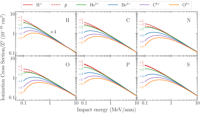
We display our total CDW ionization cross sections for the six essential elements by the impact of the six projectiles in Fig. 1. To reduce the resulting 36 magnitudes into a single consistent figure, we considered the fact that in the first Born approximation the ionization cross section scales with the square of the projectile charge, . The impact energies considered range between 0.1 to 10 MeV/amu, where the CDW is supposed to hold. In fact, for the highest projectile charges the minimum impact energy where the CDW is expected to be valid could be higher than 100 keV. We also performed similar calculations with the first Born approximation, and we corroborated that it provides quite reliable results only for energies higher than a couple of MeV/amu. We use the same line color to indicate the projectile charge throughout all the figures of this work: dashed–red, solid–red, blue, magenta, olive and orange for antiprotons, H+, He2+, Be4+, C6+, and O8+, respectively. Notably, there is no complete tabulation of ionization of atoms by the impact of multicharged ions. We hope that the ones presented in this article will be of help for future works.
Simultaneously, we will be reporting state to state ionization cross sections for the 36 ion–target systems considered in the present work miraglia2019 . A great numerical effort was paid to obtain these results, and we expect that they will be useful to estimate molecule fragmentation.
II.1 Emitted electron energies
In a given biological medium, direct ionization by ion impact accounts for just a fraction of the overall damage. Secondary electrons, as well as recoil target ions, also contribute substantially to the total damage Denifl2011 . We can consider the single differential cross section of the shell of the atom , , to be a function of the ejected electron energy as a simple distribution function surdutovic2018 . Then, we can define the mean value as in Ref. abril2015 ,
| (2) | |||||
| (3) |
where takes into account the sum of the different sub–shell contributions of the element .
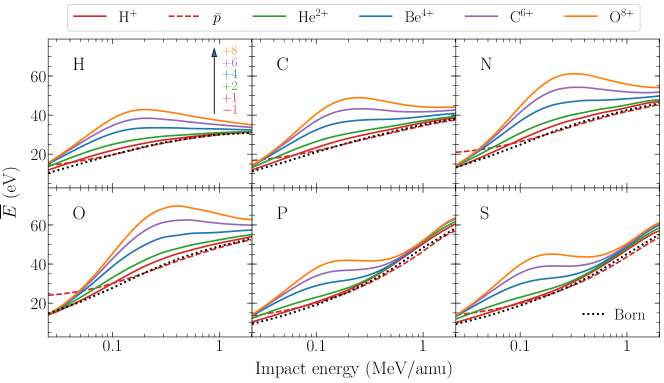
The mean emitted electron energies for H, C, N, O, P and S are shown in Fig. 2. The range of impact velocities was shortened to a.u. due to numerical limitations in the spherical harmonics expansion of Eq. (1). As the impact velocity increases, so do and , which results in the inclusion of very oscillatory functions in the integrand. Furthermore, the integrand of includes the kinetic energy (see Eq. (2)), which cancels the small energy region and reinforces the large values, making the result more sensible to large angular momenta. Regardless, for a.u., the first Born approximation holds.
In Fig. 2, we estimate of the emitted electron in the 10–70 eV energy range, for all the targets. Our results agree with the experimental findings surdutovic2018 . As can be noted in the figure, the mean energy value is surprisingly sensitive to the projectile charge , which can duplicate the proton results in the intermediate region, i.e., 100–400 keV/amu. The effect observed can be attributed to the depletion caused by the multicharged ions to the yields of low energy electrons. This behavior cannot be found in the first Born approximation, where the law cancels the dependence in Eq. (2). At high energies, tends to a universal value for all ions, as can be seen in Fig. 2.
II.2 Emitted electron angles
As mentioned before, secondary electrons contribute to the total damage. Then, not only the ejection energy is essential but also the angle of emission. Once again, we can consider the single differential cross section in terms of the ejected electron solid angle , , to be expressed as a distribution function, and the mean angle can be defined as
| (4) | |||||
| (5) |
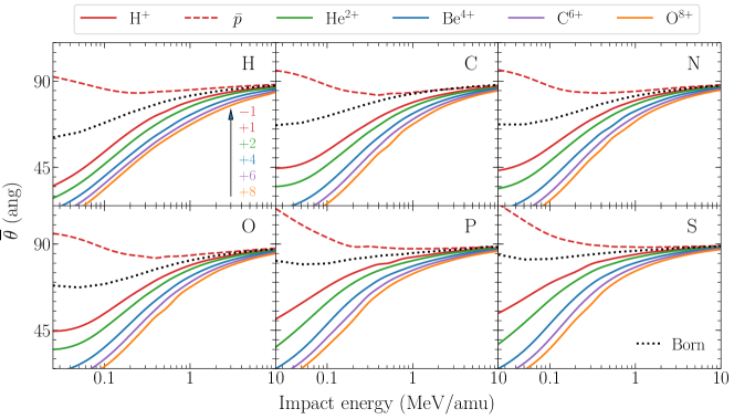
The mean emitted electron angles for the six atoms and six ions of interest are shown in Fig. 3. A significant dependence of with is noticed for all the cases. Once again, this effect could not be observed in the first Born approximation (dotted line).
For low energy electron emission, the angular dispersion is nearly isotropic Rudd1992 . A typical value for the ejection angle considered in the literature is surdutovic2018 , and it is quite correct in the range of validity of the first Born approximation for any target. However, when a distorted wave approximation is used, decreases substantially with in the intermediate energy region, as shown in Fig. 3. The higher the charge , the smaller will be. Of course, this effect only holds at intermediate energies and not at high impact energies.
To illustrate this feature, consider the impact of 500 keV C6+ on oxygen. The first Born approximation predicts emitted electrons with mean energies of 46.7 eV and mean angles of 78°, while the CDW gives 62.5 eV and 60°. These results imply deeper penetration of the secondary electrons with an orientation closer to the direction of the ion. We can attribute this forward direction correction to the capture to the continuum effect.
Furthermore, Fig. 3 provides an illustrative description of the behavior of antiprotons: the projectile repels the electrons, being . Note the opposite effect of proton and antiprotons respect to the first Born approximation; this phenomenon constitutes an angular Barkas effect.
III Ionization of Molecules
III.1 The stoichiometric model
Lets us consider a molecule composed by atoms of the element , the SSM approaches the total ionization cross section of the molecule as a sum of ionization cross sections of the isolated atoms weighted by ,
| (6) |
We classified the molecular targets of our interest in three families: CH, CHN, and DNA, as in Table 1.
| CH | CH4 (methane), C2H2 (acetylene), |
| C2H4 (ethene), C2H6 (ethane), | |
| C6H6 (benzene) | |
| CHN | C5H5N (pyridine), C4H4N2 (pyrimidine), |
| C2H7N (dimenthylamine), | |
| CH5N (monomethylamine) | |
| DNA | C5H5N5 (adenine), C4H5N3O (cytosine), |
| C5H5N5O (guanine), C5H6N2O2 (thymine), | |
| C4H4N2O2 (uracil), C4H8O (THF), | |
| C5H10O5P (DNA backbone), | |
| C20H27N7O13P2 (dry DNA) |
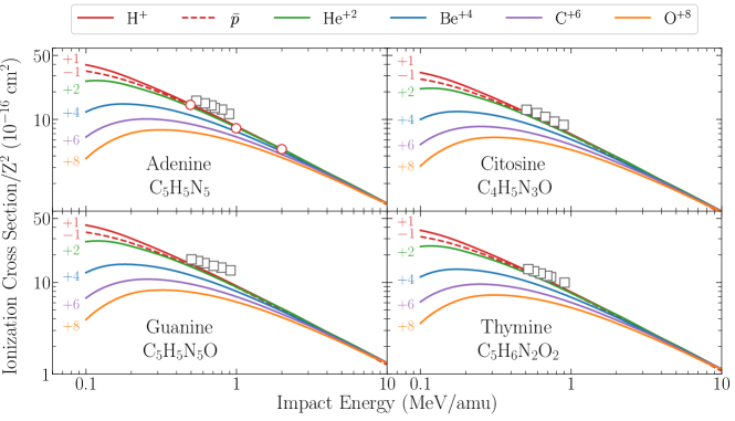
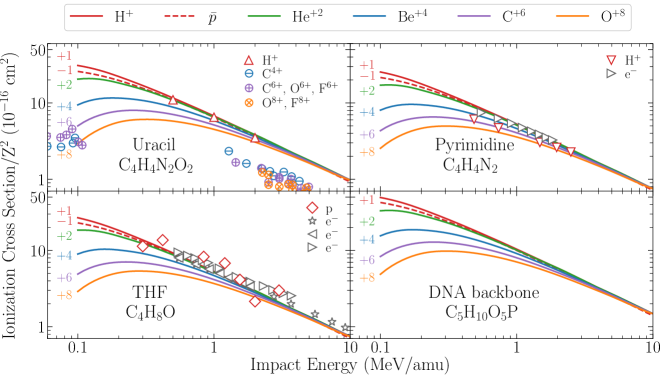
In Fig. 4, we report the reduced total ionization cross sections for adenine, cytosine, guanine, and thymine by the impact of multicharged ions obtained combining the SSM given by Eq. (6) and the CDW results. For adenine, the agreement with the experimental data available for proton impact iriki2011 is excellent. To the best of our knowledge, there are no experimental data on ion–collision ionization for the rest of the molecules. We have also included in this figure electron impact measurements rahman2016 with the corresponding equivelocity conversion for electron incident energies higher than 300 eV. In this region, the proton and electron cross section should converge. Although the electron impact measurements are above our findings for all the molecular targets, it is worth stating that our results agree very well with other electron impact theoretical predictions mozejko2003 ; tan2018 .
The reduced total ionization cross sections for uracil, DNA backbone, pyrimidine, and THF are displayed in Fig. 5. For uracil, the agreement with the experimental proton impact measurements by Itoh et al. itoh2013 is good. However, for the same target, our theory is a factor of two above the experimental ionization measurements by Tribedi and collaborators agnihotri2012 ; agnihotri2013 by the impact of multicharged ions. Nonetheless, it should be stated that our theoretical results coincide with calculations by Champion, Rivarola, and collaborators agnihotri2012 ; champion2012 , which may indicate a possible misstep of the experiments.
For pyrimidine, we show a comparison of our results with experimental data for proton impact by Wolff wolff2014 and also for electron impact ionization bug2017 at high energies. The electron impact measurements agree with our calculations for energies higher than 500keV. Unexpectedly, the proton impact cross sections are significantly lower than our findings. Much more experiments are available for ionization of THF molecule by proton wang2016 and by electron bug2017 ; wolf2019 ; fuss2009 impact. Our SSM with CDW results show overall good agreement with these data.
At intermediate impact energies, the rule no longer holds, and other scalings can be considered in this region. For example, the molecular cross section and ion impact energy can be reduced with the projectile charge , as suggested in in janev1980 ; dubois2013 .
III.2 Scaling rules
III.2.1 Toburen rule
The first attempt to develop a comprehensive but straightforward phenomenological model for electron ejection from large molecules was proposed by Toburen and coworkers toburen1975 ; toburen1976 . The authors found it convenient to scale the experimental ionization cross section in terms of the number of weakly–bound electrons, . For instance, for C, N, O, P, and S, this number is the total number of electrons minus the K–shell. Following Toburen, the scaled ionization cross section per weakly bound electron is
| (7) |
where , and are the Toburen numbers given by
| (8) |
The Toburen rule can be stated by saying that is a universal parameter independent on the molecule, which depends solely on the impact velocity, and holds for high impact energies (i.e., 0.25–5 MeV/amu). These can be interpreted as the number of active electrons in the collision. At very high energies, the K–shell electrons will also be ionized, and these numbers will be different. A similar dependence with the number of weakly bound electrons was found in Ref. itoh2013 for proton impact on uracil and adenine.
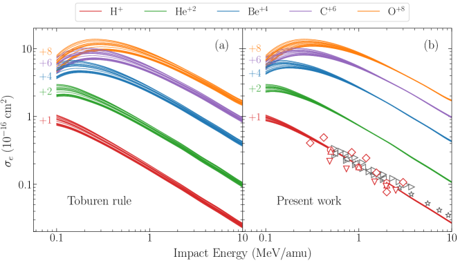
Following the Toburen scaling, we computed the scaled CDW cross sections for the molecular targets of Table 1. Our results are shown in Fig. 6a as a function of the impact energy for different projectile charges. Although the Toburen scaling holds for high energies, its performance is still not satisfactory: the universal band is quite broad, as can be noted in this figure.
III.2.2 CDW–based scaling
The departure of our theoretical results from the Toburen rule can be easily understood by inspecting Fig. 1. It can be noted that the rule , approximately constant, is not well satisfied by the CDW. For example, Fig. 1 shows that the cross sections for O are very similar to the cross sections for C, suggesting 4 active electrons in O instead of 6. In the same way, the number of active electrons for N, P, and S obtained with the CDW are also different from the of Eq. (8).
Based on the CDW results, we propose a new scaling,
| (9) |
where , and are the numbers of active electrons per atom obtained from the CDW ionization cross sections for different ions in H, C, N, O, P, and S targets, given as follows,
| (10) |
The new scaled cross sections are plotted in Fig. 6b. The experimental data for ionization of adenine iriki2011 , uracil itoh2013 , pyrimidine wolff2014 , and THF wang2016 by proton impact in Fig. 6b seems to corroborate the new scaling. We also included the electron impact ionization measurements with equivelocity conversion on pyrimidine bug2017 and THF bug2017 ; wolf2019 ; fuss2009 . It will be interesting to cross–check with future experiments, mainly for higher projectile charge states.
| Molecule | Molecule | Molecule | ||||||
|---|---|---|---|---|---|---|---|---|
| H2 | 2 | 2 | C2H7N | 19 | 20 | C4H5N3O | 37 | 42 |
| H2O | 6 | 8 | C4H8O | 28 | 30 | C5H6N2O2 | 42 | 48 |
| NH3 | 7 | 8 | C4H4N2 | 28 | 30 | C5H5N5 | 45 | 50 |
| CH4 | 8 | 8 | C6H6 | 30 | 30 | C5H5N5O | 49 | 56 |
| CH5N | 13 | 14 | C4H4N2O2 | 36 | 40 | C5H10O5P | 54.5 | 65 |

By using Eq. (10), we define new active electron numbers for molecules. In Table 2, we display the present values and ones by Toburen obtained from Eq. (8). Our values are different from the ones proposed by Toburen and used by other authors itoh2013 , mainly due to the differences in the active electron numbers of oxygen. An alternative way of testing the present scaling can be attained by plotting the ionization cross sections of molecules as a function of the from Table 2. Our findings are displayed in Fig. 7 for impact energies 0.5, 1, and 2 MeV. As can be noted, the computed CDW ionization cross sections for all the molecules show a linear dependence with the number of electrons from Table 2. We obtain similar results, even for MeV. The comparison with the experimental data available shows overall good agreement, for the smallest molecules, H2, H2O, and CH4, up to the most complex ones, like adenine. For electron impact data, the experimental data was interpolated between close neighbors. It is worth mentioning that an equivalent plot using the Toburen numbers does not exhibit the straight lines obtained with the present scaling.
While finishing the present work, we became aware of an accepted manuscript by Lüdde et al. ludde2019 on total ionization of biological molecules by proton impact, using the independent–atom–model pixel counting method ludde2016 ; ludde2018 . The authors also raised a scaling with for C, N, and O, but for P. The agreement with this independent method for proton impact reinforces our multicharged–ion findings.
III.3 Molecular structure of targets
Finally, to test the range of validity of the SSM, we performed ab initio molecular calculation of five nucleobases by employing the gamess code. The geometry optimization and single point energy calculations were performed implementing the restricted Hartree–Fock method and the 3-21G basis set.
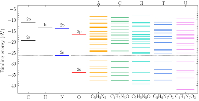
The molecular binding energies of the valence electrons for adenine, cytosine, guanine, thymine, and uracil are shown in Fig. 8. The binding energy of the highest molecular orbital (HOMO) agrees with the experimental values Hush ; Verkin ; Dougherty within 2% for all the DNA bases considered. On the left side of Fig. 8, we show the atomic Hartree–Fock energies of the constituent elements, which gives an insight into the distribution of the weakly bound electrons in the molecules. A dashed line around eV is drawn to separate the molecular band in two. We can consider the atomic energy levels above this line as the ones corresponding to the weakly bound electrons from Eq. (10). For example, the and electrons of carbon are placed above the separating line, which corresponds to the 4 electrons given by CDW–scaling. In the case of O, only the 4 electrons of the orbitals are located above the separating line, which corresponds to the number of weakly bound electron given by our new scaling. The N case is not as straightforward; the would suggest that one out of the two electrons contribute to the molecular scheme.
III.3.1 A modified stoichiometric model
The SSM considers the molecule to be assembled by isolated neutral atoms, which is definitively unrealistic. A first improvement can be suggested by assuming that the atoms are not neutral and that they have an uneven distribution of electrons within the molecule, which can be expressed as an effective charge per atom. The Mulliken charge gives a possible value for ; however, there are a wide variety of charge distributions lee2003 .
| Element | C | H | N | O | New stoichiometry |
| Adenine | +0.32 | +0.23 | –0.55 | C4.92H4.77N5.14 | |
| Cytosine | +0.28 | +0.21 | –0.56 | –0.53 | C3.93H4.79N3.14O1.13 |
| Guanine | +0.46 | +0.20 | –0.58 | –0.36 | C4.89H4.80N5.15O1.09 |
| Thymine | +0.20 | +0.19 | –0.54 | –0.52 | C4.95H5.81N2.13O2.13 |
| Uracil | +0.31 | +0.22 | –0.59 | –0.47 | C3.92H3.78N2.15O2.12 |
To take this effect into account, we can consider that the total amount of electrons on the element is equally distributed on all the atoms. Therefore, each element will have an additional charge, , which can be positive or negative. This amount will depend on the relative electronegativity respect to the other atoms rappe1991 . Following this idea, we can estimate a new number of atoms per molecule , given by
| (11) |
In the case of neutral atoms, and , as it should be. In Table 3, we display the average effective charge per atom of C, H, N, and O, for five DNA molecules, obtained from the full molecular calculation described above.
By implementing Eq. (11), it is possible to determine a new stoichiometric formula (last column of Table 3). Now, instead of having an integer number of atoms , we obtain a fractional number . New molecular cross sections can be computed considering such values. Relative errors for the ionization cross sections were computed for the DNA bases from Table 3. The differences obtained were less than 3%, which indicates that the SSM is a quite robust model to handle these type of molecules within the range error expected for this model.
IV Conclusions
In this work, we have dealt with the calculation of ionization cross sections of seventeen biological molecules containing H, C, N, O, P, and S by the impact of antiprotons, H+, He2+, Be4+, C6+, and O8+. To that end, we have employed the full CDW method and the simple stoichiometric model. The mean energy and angle of the emitted electrons, of importance in post–collisional radiation damage, has also been calculated. Our findings show a clear dependence with the ion charge . For a given target as increases, also increases, but decreases, showing a clear tendency to the forward direction. At impact energies greater than 2 MeV/amu, these values converge to the Born approximation, which embodies the simple law.
Total ionization cross sections for adenine, cytosine, thymine, guanine, uracil, DNA backbone, pyrimidine, and THF are presented and compared with the scarcely available experiments. We explored the rule of Toburen, which scales all the molecular ionization cross section normalizing with a certain number of weakly bound or valence electrons. We found that the ionization cross sections scales much better when normalizing with the number of active electrons in the collision obtained from the CDW results for atoms. This new scaling was tested with good results for the six projectiles and seventeen molecules studied here. The comparison with the experimental data reinforce our findings. Furthermore, we tested the scaling by including experimental data of ionization of H2, water, methane, and ammonia by proton impact showing good agreement at intermediate to high energies.
Finally, we performed full molecular calculations for the DNA basis. By inspecting the molecular binding energy from quantum mechanical structure calculations, we were able to understand the number of electrons proposed in our new CDW-based scaling. We attempt to improve the stoichiometric model by using the Mulliken charge to get fractional rather than integer proportions. We found no substantial correction, which indicates that the SSM works quite well.
In conclusion, the present results reinforce the reliability of the SSM to deal with complex molecules in the intermediate to high energy range. Moreover, the simple stoichiometric model and the CDW cross sections in Ref. miraglia2019 opens the possibility to describe a wide range of molecules containing H, C, N, O, P, and S, by the impact of multicharged ions.
References
- (1) O. Mohamad, B. J. Sishc, J. Saha, A. Pompos, A. Rahimi, M. D. Story, A. J. Davis, D. N. Kim, Cancers 9, 66 (2017).
- (2) M. E. Galasssi, R. D. Rivarola, M. Beuve, G. H. Olivera and P. D. Fainstein, Phys. Rev. A 62, 022701 (2000).
- (3) H. J. Lüdde, A. Achenbach, T. Kalkbrenner, H.-C. Jankowiak and T. Kirchner, Eur. Phys. J. D 70, 82 (2016).
- (4) H. J. Lüdde, M. Horbatsch and T. Kirchner, Eur. Phys. J. B 91, 99 (2018).
- (5) Fainstein P.D., Ponce V. H. and Rivarola R. D. J. Phys. B: At. Mol. Opt. Phys. 21 287 (1988).
- (6) J. E. Miraglia and M. S. Gravielle. Phys Rev A 78, 052705 (2008)
- (7) J. E. Miraglia, Phys. Rev. A 79, 022708 (2009).
- (8) Denifl S., Märk T.D., Scheier P. The Role of Secondary Electrons in Radiation Damage. In Radiation Damage in Biomolecular Systems. Biological and Medical Physics, Biomedical Engineering. Eds: García Gómez-Tejedor G., Fuss M. Springer, Dordrecht (2012)
- (9) W. E. Wilson and L. H. Toburen. Phys. Rev. A 11, 1303 (1975).
- (10) D. J. Lynch, L. H. Toburen, and W. E. Wilson. J. Chem. Phys. 64, 2616 (1976).
- (11) M. W. Schmidt, K. K. Baldridge, J. A. Boatz, S. T. Elbert, M. S. Gordon, J. H. Jensen, S. Koseki, N. Matsunaga, K. A. Nguyen, S. J. Su, T. L. Windus, M. Dupuis, J. A. Montgomery J. Comput. Chem. 14, 1347-1363 (1993).
- (12) Salvat, F., Fernández-Varea, J.M., Williamson, W. Comput. Phys. Commun. 90, 151–168 (1995)
- (13) A.M.P. Mendez, D.M. Mitnik, and J.E. Miraglia. Int. J. Quantum Chem. 24 ,116 (2016).
- (14) A.M.P. Mendez, D.M. Mitnik, and J.E. Miraglia. Adv. Quant. Chem. 76, 117–132 (2018).
- (15) C. C. Montanari, J. E. Miraglia, Nucl. Instr. Meth. Phys. Res. B 407, 236–243 (2017).
- (16) J. E. Miraglia, O8+ on H, C, N, O, P, and S atoms (to be published).
- (17) E. Surdutovich and A. V. Solov’yov, arXiv:1312.0897v, (2013)
- (18) P. de Vera, I. Abril, R. Garcia-Molina and A. V. Solov’yov, Journal of Physics: Conference Series 438, 012015 (2013).
- (19) M. E. Rudd, Y.-K. Kim,, D. H. Madison and T. J. Gay. Rev. Mod. Phys. 64, 441–490 (1992).
- (20) Y. Iriki, Y. Kikuchi, M. Imai, and A. Itoh Phys. Rev. A 84, 052719 (2011).
- (21) M. A. Rahman and E. Krishnakumar, Electron ionization of DNA bases, J. Chem. Phys. 144, 161102 (2016).
- (22) P. Mozejko and L. Sanche, Radiat Environ. Biophys 42, 201 (2003).
- (23) H. Q. Tan, Z. Mi, and A. A. Bettiol, Phys. Rev. E 97, 032403 (2018)
- (24) A. Itoh, Y. Iriki, M. Imai, C. Champion, and R. D. Rivarola, Phys. Rev. A 88, 052711 (2013).
- (25) A. N. Agnihotri, S. Kasthurirangan, S. Nandi, A. Kumar, M. E. Galassi, R. D. Rivarola, O. Fojón, C. Champion, J. Hanssen, H. Lekadir, P. F. Weck, and L. C. Tribedi. Phys. Rev. A 85, 032711 (2012).
- (26) A N Agnihotri, S Kasthurirangan, S Nandi, A Kumar, C Champion,, H Lekadir, J Hanssen, P FWeck, M E Galassi, R D Rivarola, O Fojon and L C Tribedi, J. Phys. B 46, 185201 (2013).
- (27) C Champion, M E Galassi, O Fojón, H Lekadir, J Hanssen, R D Rivarola, P F Weck, A N Agnihotri, S Nandi, and L C Tribedi. J. Phys.: Conf. Ser. 373, 012004 (2012).
- (28) W. Wolff, H. Luna, L. Sigaud, A. C. Tavares, and E. C. Montenegro J. Chem. Phys. 140, 064309 (2014).
- (29) M. U. Bug, W. Y. Baek, H. Rabus, C. Villagrasa, S. Meylan, A. B. Rosenfeld, Rad. Phys. Chem. 130, 459–479 (2017).
- (30) M. Wang, B. Rudek, D. Bennett, P. de Vera, M. Bug, T. Buhr, W. Y. Baek, G. Hilgers, H. Rabus, Phys. Rev. A 93, 052711 (2016).
- (31) W. Wolff, B. Rudek, L. A. da Silva, G. Hilgers, E. C. Montenegro, M. G. P. Homem, J. Chem. Phys. 151, 064304 (2019).
- (32) M. Fuss, A. Muñoz, J. C. Oller, F. Blanco, D. Almeida, P. Limão-Vieira, T. P. D. Do, M. J. Brunger, G. García, Phys. Rev. A 80, 052709 (2009).
- (33) R. K. Janev and L. P. Presnyakov J. Phys. B 13, 4233 (1980).
- (34) R. D. DuBois, E. C. Montenegro and G. M. Sigaud, AIP Conference Proceeding 1525, 679 (2013).
- (35) D. J. Lynch, L. H. Toburen, and W. E. Wilson, J. Chem. Phys. 64, 2616 (1976).
- (36) M.E. Rudd, Y.-K. Kim, D.H. Madison, J.W. Gallagher, Review of Modern Physics, 57, 965–994 (1985).
- (37) H. Luna, A. L. F. de Barros, J. A. Wyer, S. W. J. Scully, J. Lecointre, P. M. Y. Garcia, G. M. Sigaud, A. C. F. Santos, V. Senthil, M. B. Shah, C. J. Latimer, and E. C. Montenegro, Phys. Rev. A 75, 042711 (2007).
- (38) H J Lüdde et al. 2019 J. Phys. B: At. Mol. Opt. Phys. in press https://doi.org/10.1088/1361-6455/ab3a63
- (39) Hush, N.S.; Cheung, A.S., Chem. Phys. Lett., 34, 11 (1975).
- (40) Verkin, B.I.; Sukodub, L.F.; Yanson, I.K.,
- (41) Dougherty, D.; Younathan, E.S.; Voll, R.; Abdulnur, S.; McGlynn, S.P., J. Electron Spectrosc. Relat. Phenom., 13, 379 (1978).
- (42) Jung-Goo Lee, Ho Young Jeong, and Hosull Lee, Charges of Bull. Korean Chem. Soc. 24, 369 (2003).
- (43) A. K. Rappe, A. K.and W. A. Goddard III,. J. Phys. Chem. 95, 3358 (1991).