Learn to Segment Organs with a Few Bounding Boxes
Abstract
Semantic segmentation is an import task in the medical field to identify the exact extent and orientation of significant structures like organs and pathology. Deep neural networks can perform this task well by leveraging the information from a large well-labeled data-set. This paper aims to present a method that mitigates the necessity of an extensive well-labeled data-set. This method also addresses semi-supervision by enabling segmentation based on bounding box annotations, avoiding the need for full pixel-level annotations. The network presented consists of a single U-Net based unbranched architecture that generates a few-shot segmentation for an unseen human organ using just 4 example annotations of that specific organ. The network is trained by alternately minimizing a nearest neighbor loss for prototype learning and a weighted cross-entropy loss for segmentation learning to perform a fast 3D segmentation with a median score of 54.64%.
1 Introduction
Semantic Segmentation, reported by Hesamian et al. (2019), is used in the medical field to identify the exact shape and size of structures like organs, and pathology. Deep Learning based image segmentation using the information from large-scale fully-annotated datasets is now a robust tool for medical applications. The creation of labels for semantic segmentation is, however, a tedious task, requiring annotation of each pixel belonging to a class. According to Mondal et al. (2018), this is particularly more cumbersome in the medical domain. The annotation in the medical domain is done by highly specialized experts, who have developed their skills through years of practice. The number of man-hours spent is enormous and most of the time the output is not even optimal due to inter/intra-observer variability. In natural images, Lin et al. (2014) suggests the creation of bounding box labels is 15 times faster and cheaper compared to full pixel label annotations for the same image. This cost gap in the medical domain would, therefore, be even wider given the required expertise.
The few-shot learning (FSL), for semantic segmentation in computer vision, has been thoroughly studied by Shaban et al. (2017); Rakelly et al. (2018), and Dong and Xing (2018). All previous approaches, however, suggest a branched structure, with one branch extracting meta-information from the examples, so-called support set, and the other branch uses the meta-information to segment the required image, so-called query image. Roy et al. (2019) follow the same approach, with the help of Squeeze-and-Excite blocks (Hu et al. (2018)), to segment organs in contrast-enhanced CT scans.
In this paper, we first introduce the prototype learning into a single un-branched architecture for few-shot segmentation reducing the cost of both computation and annotation. Further, we extend our proposed method to the semi-supervised few-shot learning (SS-FSL) setup to leverage weak support annotations, i.e. bounding boxes, which is the first to be seen in few-shot approaches for medical applications.
2 Methodology
Our approach is based on prototype learning, which takes images and corresponding annotations as input and outputs a prototype. The prototype along with the feature representation of the query images are passed then to the model to predict the segmentation maps. Contrary to the branched networks seen in the Few-Shot Learning (FSL) paradigm, both the prototype learner and the segmenter are integrated into a single network. In addition, weakly annotated images, i.e. bounding boxes, are leveraged during the meta-learning phase.
Concretely, for an -shot -way segmentation problem, a model is trained on episodes consisting of a support set where each is an input instance image and is the corresponding annotation of classes, and a query set . The model then transfers the knowledge, acquired on the support set during the meta-learning, to predict the segmentation maps on the query set . In the semi-supervised FSL setup, we assume access to dataset , , where , denote all fully annotated, and weakly annotated images, respectively, and denote all annotated images of a single organ , and be all weakly annotated images . In each training episode , both support and query sets are sampled, for a particular class , i.e. organ, from both , and with different proportion, where the support of annotated samples is way larger than the support of weakly annotated ones, i.e. .
Prototype Learner: Both an image and corresponding annotation are passed as input to form a prototype , which is later passed as a meta information to the segmenter. The model parameters are optimized by minimizing the negative log likelihood,
| (1) |
where is a similarity function, i.e. cosine similarity, and is the prototype computed on the support set, . The Nearest Neighbour Loss() assists in the formation of individual clusters for all .
Segmenter: The learned prototype along with the query images are passed to the model to predict the segmentation maps, . The model parameters , and are optimized using the weighted cross-entropy loss,
| (2) |
where is additional weight to balance the the occurrence of class .
The overall loss function is minimized alternatively to enable learning from a few shot segmentation,
| (3) |
3 Experiments and Results
We evaluate our proposed model on the publicly available Visceral dataset created by Jimenez-del-Toro et al. (2016). We follow the same evaluation strategy that appeared in Roy et al. (2019)by focusing on 4 organs. Each organ class has a data set of 20 different patients, of which we use 19 for the query set and the remaining one to provide the supporting meta information. We obtain the results by following a fold wise training strategy. A fold is defined by treating one of the organs as an previously unseen testing class while utilizing the rest of the organs as for training. We train our network with the aforementioned setting both in a fully supervised fashion and a semi-supervised fashion by using weakly labeled data, i.e. bounding box labels.
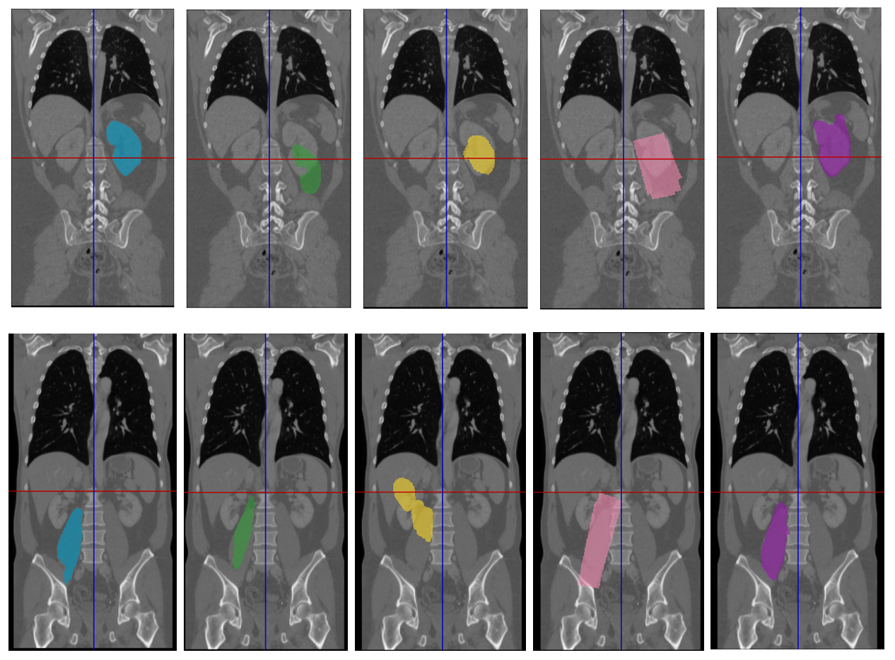
In Fig. 1, we can see that even though the support label for the fully supervised case can be considered closer to the ground truth, the result for the vague box-shaped support label yields an overall better performance in terms of results, showing an increase of the 3D dice score of around 10% on average for each organ. Another aspect is the number of used ground truth, compared to conventional full-shot techniques, we yield an annotation cost decrease of almost 500x for the fully supervised few-shot case and even up to 7500x decrease for the bonding box semi-supervised case. We were able to prove that semi-supervised few-shot learning (SS-FSL) contributes a valuable improvement to few-shot techniques, as shown in Table 1. Even though we do not reach the performance of state-of-the-art methods as full shot techniques, our approach has a great potential due to these attributes.
| Organs | |||||||
|---|---|---|---|---|---|---|---|
| Liver | Spleen | Kidney | Psoas Major | Average | Speedup | ||
| LBM:FSL | Ours | 52.78 | 53.09 | 50.52 | 35.82 | 48.06 | 2.183 |
| Roy et al. (2019) | 68.04 | 55.06 | 37.62 | 49.92 | 52.66 | 1 | |
| SS-FSL | Ours | 63.50 | 64.28 | 45.75 | 45.01 | 54.64 | 2.183 |
| UBM | INSA-Creatis | 95.11 | 91.10 | 95.00 | 81.20 | 90.61 | – |
4 Conclusion
We present a method for segmenting 3D medical scans in a low data setting. This approach can be adapted to enhance any segmentation network leaving room for improvement and further research. Our proposed SS-FSL yielded an increase of 10% performance organ-wise for most of the organs compared to full supervision. Few-Shot Learning is an interesting technique with lots of potential in the medical domain where there is a lack of good reliable large data-sets.
References
- Dong and Xing [2018] Nanqing Dong and Eric Xing. Few-shot semantic segmentation with prototype learning. In BMVC, volume 1, page 6, 2018.
- Hesamian et al. [2019] Mohammad Hesam Hesamian, Wenjing Jia, Xiangjian He, and Paul Kennedy. Deep learning techniques for medical image segmentation: Achievements and challenges. Journal of Digital Imaging, 32(4):582–596, Aug 2019. ISSN 1618-727X. doi: 10.1007/s10278-019-00227-x. URL https://doi.org/10.1007/s10278-019-00227-x.
- Hu et al. [2018] Jie Hu, Li Shen, and Gang Sun. Squeeze-and-excitation networks. In Proceedings of the IEEE conference on computer vision and pattern recognition, pages 7132–7141, 2018.
- Jimenez-del-Toro et al. [2016] O. Jimenez-del-Toro, H. Müller, M. Krenn, K. Gruenberg, A. A. Taha, M. Winterstein, I. Eggel, A. Foncubierta-Rodríguez, O. Goksel, A. Jakab, G. Kontokotsios, G. Langs, B. H. Menze, T. Salas Fernandez, R. Schaer, A. Walleyo, M. Weber, Y. Dicente Cid, T. Gass, M. Heinrich, F. Jia, F. Kahl, R. Kechichian, D. Mai, A. B. Spanier, G. Vincent, C. Wang, D. Wyeth, and A. Hanbury. Cloud-based evaluation of anatomical structure segmentation and landmark detection algorithms: Visceral anatomy benchmarks. IEEE Transactions on Medical Imaging, 35(11):2459–2475, Nov 2016. ISSN 0278-0062. doi: 10.1109/TMI.2016.2578680.
- Lin et al. [2013] Min Lin, Qiang Chen, and Shuicheng Yan. Network in network. arXiv preprint arXiv:1312.4400, 2013.
- Lin et al. [2014] Tsung-Yi Lin, Michael Maire, Serge J. Belongie, Lubomir D. Bourdev, Ross B. Girshick, James Hays, Pietro Perona, Deva Ramanan, Piotr Dollár, and C. Lawrence Zitnick. Microsoft COCO: common objects in context. CoRR, abs/1405.0312, 2014. URL http://arxiv.org/abs/1405.0312.
- Mondal et al. [2018] Arnab Kumar Mondal, Jose Dolz, and Christian Desrosiers. Few-shot 3d multi-modal medical image segmentation using generative adversarial learning. CoRR, abs/1810.12241, 2018. URL http://arxiv.org/abs/1810.12241.
- Rakelly et al. [2018] Kate Rakelly, Evan Shelhamer, Trevor Darrell, Alexei A Efros, and Sergey Levine. Meta-learning to guide segmentation. 2018.
- Ronneberger et al. [2015] Olaf Ronneberger, Philipp Fischer, and Thomas Brox. U-net: Convolutional networks for biomedical image segmentation. CoRR, abs/1505.04597, 2015. URL http://arxiv.org/abs/1505.04597.
- Roy et al. [2019] Abhijit Guha Roy, Shayan Siddiqui, Sebastian Pölsterl, Nassir Navab, and Christian Wachinger. ’squeeze & excite’ guided few-shot segmentation of volumetric images. CoRR, abs/1902.01314, 2019. URL http://arxiv.org/abs/1902.01314.
- Shaban et al. [2017] Amirreza Shaban, Shray Bansal, Zhen Liu, Irfan Essa, and Byron Boots. One-shot learning for semantic segmentation. CoRR, abs/1709.03410, 2017. URL http://arxiv.org/abs/1709.03410.
- Zhou et al. [2018] Zongwei Zhou, Md Mahfuzur Rahman Siddiquee, Nima Tajbakhsh, and Jianming Liang. Unet++: A nested u-net architecture for medical image segmentation. CoRR, abs/1807.10165, 2018. URL http://arxiv.org/abs/1807.10165.
Appendix A Appendices
A.1 Implementation
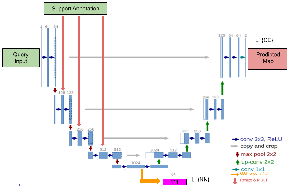
We base our model on U-Net Zhou et al. [2018], the most widely used segmentation architecture in medical imaging.
The architecture is provided with the required meta-information at every encoding stage, except at the first one. The information is supplied from the incoming data by simply doing an element-wise multiplication of the extracted features with the meta-information. The meta-information comes from the support annotation of the same organ from another patient. The support information is obtained from 1 fully labelled and 3 weakly labelled annotations.
The training is done in two phase joint training described by Dong and Xing [2018]. In the first phase, the query image and the support information leads to obtaining a 1024 dimensional bottleneck features. These features are passed through a 1x1 convolutional layer which brings down its dimensions from 1024 to 64. The 64-dimensional feature vector is then passed through a Global Average Pooling (GAP) suggested by Lin et al. [2013]. This averages out the features in a channel to obtain the prototypes. Further Dong and Xing [2018] point out that the GAP in contrast to Max or Min pooling helps prevent over-fitting to dominant or submissive features of the training organ. This obtained prototype is used to minimize the nearest neighbor loss with the learned standard prototypes of its class.
In the second phase, a contextual information is learned to segment the query. The same query and support are again passed through to optimize for the cross-entropy for the segmentation accuracy. The entire schematic for the two-phase training is given in Fig. 2.
UNet’s symmetric expanding path enables precise localization. The symmetric expansion path with the skip connection provides the decoder with boundary information from the input image to create the final segmentation mask. The idea of masking support annotations at every encoding layer may prevent the carrying of boundary related information to the decoding layer. The information may be preserved by simply not masking the support at the very first encoding block. This also serves to provide the related boundary to the very end of decoding making the data relevant to the predicted map.