An Open Source Mesh Generation Platform for Biophysical Modeling Using Realistic Cellular Geometries
Abstract
Advances in imaging methods such as electron microscopy, tomography, and other modalities are enabling high-resolution reconstructions of cellular and organelle geometries. Such advances pave the way for using these geometries for biophysical and mathematical modeling once these data can be represented as a geometric mesh, which, when carefully conditioned, enables the discretization and solution of partial differential equations. In this study, we outline the steps for a naïve user to approach GAMer 2, a mesh generation code written in C++ designed to convert structural datasets to realistic geometric meshes, while preserving the underlying shapes. We present two example cases, 1) mesh generation at the subcellular scale as informed by electron tomography, and 2) meshing a protein with structure from x-ray crystallography. We further demonstrate that the meshes generated by GAMer are suitable for use with numerical methods. Together, this collection of libraries and tools simplifies the process of constructing realistic geometric meshes from structural biology data.
[*]ctlee@ucsd.edu, prangamani@ucsd.edu
As biophysical structure determination methods improve, the rate of new structural data is increasing. New methods that allow the interpretation, analysis, and reuse of such structural information will thus take on commensurate importance. In particular, geometric meshes, such as those commonly used in graphics and mathematics, can enable a myriad of mathematical analysis. In this work, we describe GAMer 2, a mesh generation library designed for biological datasets. Using GAMer 2 and associated tools PyGAMer and BlendGAMer, biologists can robustly generate computer and algorithm friendly geometric mesh representations informed by structural biology data. We expect that GAMer 2 will be a valuable tool to bring realistic geometries to biophysical models.
Introduction
The use of Partial Differential Equations (PDEs) in mathematical modeling of cellular phenomena is becoming increasing common, particularly, for problems that include electrostatics, reaction-diffusion, fluid dynamics, and continuum mechanics. Solutions to these equations using idealized geometries have provided insight into how cell shape can affect signaling (1, 2, 3, 4), and how blood flows in vessels (5).
On the other hand, in order to gain better insight into how cellular geometry can affect the dynamics of these mechanochemical processes, using realistic geometries is necessary. Already, freely available tools such as Virtual Cell (6) and CellOrganizer (7) have paved the way for using realistic cellular geometries in simulations. With increasing availability of high-resolution images of cellular ultrastructure, including the size and shape of organelles, and the curvature of the various cellular membranes, there is a need for computational tools and algorithms that can enable us to use these data as the geometry or domain of interest and conduct simulations using numerical methods (8).
For most relevant geometries, it is impossible to obtain analytical solutions for PDEs; this necessitates the use of numerical methods to provide an approximate solution. These numerical methods are based on discretization (approximating the PDE with a discrete algebraic system) combined with solvers (typically iterative methods that converge to the solution to the algebraic system). The first step usually requires the generation of a geometric mesh over which the problem can be discretized using techniques such as finite difference, finite volume, finite element, or other methods to build the algebraic system that approximates the PDE. The numerical approximation to the PDE is then produced by solving the resulting linear or nonlinear algebraic equations using an appropriate fast solver. One of the computational challenges associated with generating meshes of biological datasets is the presence of highly irregular surfaces with curvatures at the nanometer or Ångstrom length scales. Although many tools from the graphics community exist to generate meshes for visualization, these poor quality meshes, when used to solve a PDE, can both destroy the quality of the discretization as an approximation to the PDE, and also produce algebraic systems that are badly conditioned and difficult to solve efficiently or accurately with iterative solvers.
While it is possible to design discretizations of PDE problems on surfaces using finite volume methods or other techniques, we prefer the Finite Element Method (FEM) here for a number of reasons. To begin with, the FEM first arose in the 1960s in the engineering community as a response to the poor performance of existing discretization techniques for PDEs involving shells and other complex physical shapes. In addition, methods such as the FEM that are built on basis function expansion provide a natural framework both for treating highly nonlinear problems, and for discretizing multiple PDEs that couple together to form a larger multi-physics system. Lastly, the FEM framework is quite general, and can in fact be shown to reproduce finite volume, spectral, and other discretizations through appropriate choices of basis and test functions.
One challenge preventing the routine use of experimental ultrastructural data with PDE-based mathematical modeling is the difficulty associated in generating a discretization, or commonly a mesh, which accurately represents the structures of interest. Building upon existing meshing codes, such as TetGen (9), NetGen (10), TetWild (11), MeshLab (12), Gmsh (13), CGAL (14), along with commercial codes such as ANSYS Meshing among others, we describe the development of a meshing framework, the Geometry-preserving Adaptive MeshER software version 2 (GAMer 2), which is designed specifically for biological mesh generation. This code has been completely rewritten from version 1, which was previously described by Yu et al. (15, 16), Gao et al. (17, 18), and (19). GAMer 2 features the original GAMer algorithms but with significantly improved ease of use, distributability, and maintainability. We have also developed a new Python Application Programming Interface (API): PyGAMer, and streamlined the GAMer Blender add-on: BlendGAMer. The complete explanation of the mathematics and underlying algorithms are available in (15, 16, 17, 18, 19, 20). In what follows, we provide an overview of the GAMer mesh generation capabilities for the general biophysicist.
Methods
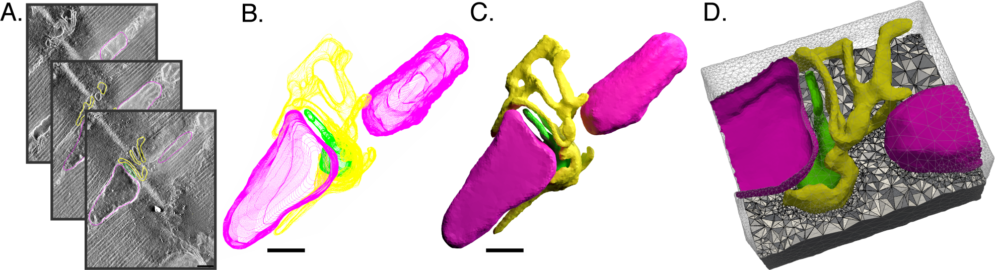
At its core, GAMer is a mesh generation and conditioning library which uses concepts from the graphics and engineering literature. It can be used to produce high quality surface and volume meshes from several types of input: 1) PDB/PQR file (Ångstrom–nanometer), 2) Volumetric dataset (Ångstrom–meters), or 3) Initial surface mesh (Ångstrom–meters). To enable FEM-based applications, GAMer also has utilities to support boundary marking. Tetrahedral meshes of a domain can be constructed in GAMer through the use of functionality provided by TetGen (21). Surface or volume meshes can be output to several common formats for use with other tools such as FEniCS (22, 23), ParaView (24), MCell (25), and FFEA (26) among others. We note that although GAMer is designed primarily with FEM-based applications in mind, conditioned meshes of realistic geometries can also be used for geometric analysis such as the estimation of curvatures, volumes, and surface areas (27). Conditioned meshes can also be used in other tools such as MCell (25) and 3D printing. Example tutorials of using GAMer 2 to generate surface and volume meshes of a protein structure (PDB ID: 2JHO) and a subcellular scene of a calcium release unit (CRU) from a murine cardiac myocyte imaged using Electron Tomography (ET) (Cell Image Library: 3603 (28)), Fig. 1, are provided in the documentation111https://gamer.readthedocs.io/en/latest/tutorial.html and described in this report. Here we will summarize the key implementation steps and refer the interested reader to (27) for technical details of the implementation.
GAMer 2 Development
One of the limitations of prior versions of GAMer is the inability to robustly represent meshes of arbitrary manifoldness and topology. This limitation prevented the safe application of GAMer to non-manifold meshes, which often produced segmentation faults or other undefined behaviors. To ameliorate this, in GAMer 2 we replaced the underlying mesh representation to use the Colored Abstract Simplicial Complex (CASC) data structure (29). CASC keeps careful track of the mesh topology and therefore makes it trivial to track the manifoldness of a given mesh object. By eliminating the need for code to handle encounters with non-manifold elements, the code base is significantly reduced and segmentation faults are eliminated. Another benefit of using CASC is that it can represent both surface meshes (2D simplices embedded in 3D) and volume meshes (3D simplices embedded in 3D), allowing users to interact with both surface and volume meshes using an identical API. This simplification contributes to the long-term maintainability of the GAMer 2 code.
In the development of GAMer 2, we have also migrated to use the cross-platform CMake build toolchain, and away from the previous GNU build system Autotools. Using CMake, GAMer 2 can now be compiled and run on major operating systems including Linux, and Mac OS–along with Windows which was previously unsupported. Detailed installation instructions are provided online222https://gamer.readthedocs.io/en/latest/install.html. We note that the Windows binary can be built using Microsoft Visual Studio and does not require the installation of Unix-like environments such as Cygwin. By supporting compilation of GAMer 2 using native and preferred tools, this improves binary compatibility with other libraries and simplifies distribution.
Collaborative Workflow
To further improve code availability and to encourage community collaboration, the GAMer code is now hosted on Github333https://github.com/ctlee/gamer. In this environment, users can file issues to report bugs or ask questions. Users are also encouraged to contribute to the code by submitting pull requests. All pull requests are subject to rigorous continuous-integration testing, using both Travis-CI (Linux and Mac OS) and Appveyor (Windows), to ensure code compatibility across a wide range of operating systems, compilers, and compiler versions prior to integration with the main deployment branches. Along with source control, GAMer 2 also implements git tagging based semantic versioning to track the software version. Compiled code can report the source version which aids in reporting and debugging. This strict versioning introduces improved ability for both users and developers to track code provenance.
Implementation of a New PyGAMer API
In addition to the complete redesign of the core library, the corresponding Python interface, now called PyGAMer, is generated using PyBind 11 (30) instead of SWIG (31). PyBind 11 was designed to expose C++ data types to Python and vice-versa while minimizing boilerplate code by capitalizing upon the capabilities of the C++ compiler. One of the benefits of this approach is the ability to bind complex template types, which are extensively used in CASC, enabling users to develop Python script to interact with various elements of the mesh and call C++ methods. Another benefit of using PyBind 11 is its support for embedding Python docstrings, which enable straightforward documentation of PyGAMer using popular Python tools such as Sphinx. As a result, documentation for GAMer 2 and PyGAMer is now automatically generated and hosted online444https://gamer.readthedocs.io.
Using scikit-build, which connects setuptools and CMake, installation of PyGAMer in any Python environment can be achieved using pip install pygamer. Versions of pip10.0 will automatically download and resolve buildtime and runtime dependencies, compile, and install the PyGAMer Python extension module. Users of older pip versions need to install any missing buildtime dependencies beforehand. Instructions on how to install these dependencies is provided in both the online documentation and demonstrated in Supplementary Movie S1.
BlendGAMer Development
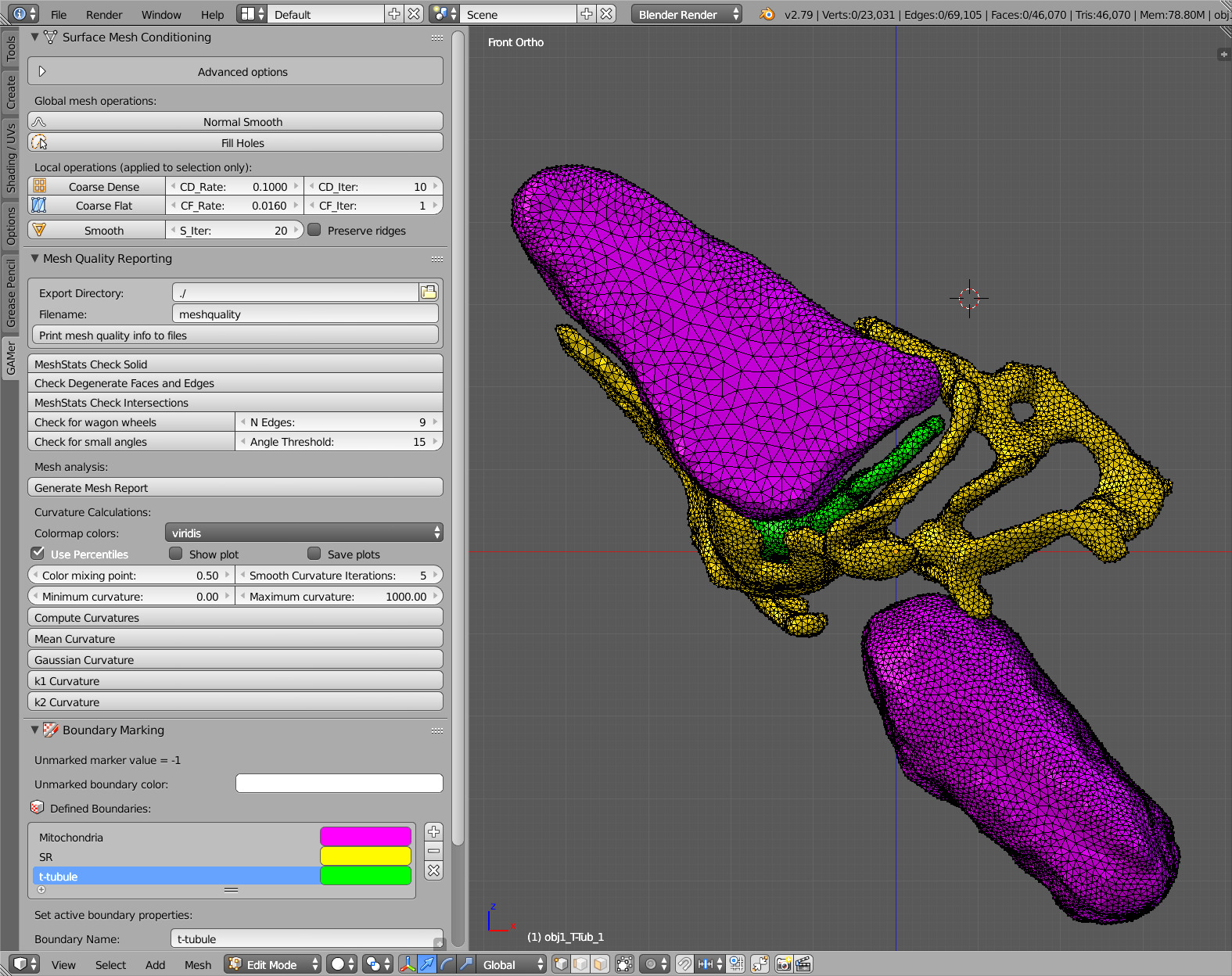
In order to support interactive modeling with graphical feedback, we have also developed a GAMer addon for Blender (32), BlendGAMer, which has also been rewritten to use PyGAMer. In addition to this update, the user interface has been redesigned to be user-friendly as shown in Fig. 2. The boundary marking capability of BlendGAMer now uses Blender attribute layers instead of lists of values. Many features now have corresponding toggles in the interface, for example, the number of neighborhood rings to consider when computing the Local Structure Tensor (LST). Several mesh conditioning operations have also been updated to operate only upon selected vertices. This provides users with the flexibility to refine local portions of the mesh as desired. There is also a new Mesh Analysis panel that contains several helpful features for analyzing the quality of a mesh and includes curvature estimation. Based upon the newly implemented semantic versioning, BlendGAMer can now track the version of metadata that is stored in a given file. Using this information, BlendGAMer can perform automatic metadata migration from version to version as improved schemes for metadata storage are created.
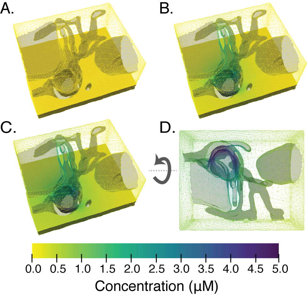
Modeling Diffusion in the CRU Geometry
To demonstrate the use of the mesh generated from GAMer 2 with the FEM, we model the diffusion of a molecule with concentration in the realistic geometry of a CRU, Fig. 3. In the volume, the dynamics of are given by
| (1) | ||||
| (2) |
where is the diffusion coefficient, is a decay constant and represents reactions that consume , is the cytosolic domain, and is time. We define the following boundary conditions:
| (3) | ||||
| (4) | ||||
| (5) |
where is the inward flux on the t-tubule membrane (). No flux boundary conditions are applied to all other boundaries. The following system is solved using FEniCS (23, 22) and visualized using ParaView (24). We note that the boundaries and are differentiated by markers applied using BlendGAMer.
Results and Discussion
We demonstrate that GAMer is capable of generating high quality surface and volume simplex meshes of geometries as informed by structural biology datasets. The incorporated Python library, PyGAMer, and Blender add-on, BlendGAMer, enable users to prototype or interact with meshes as they are conditioned. Collectively these tools facilitate mathematical modeling of biological systems using realistic geometries.
The GAMer workflow has been previously applied in several works (33, 34, 35, 36, 37, 38, 27). Several example applications are also described in the online GAMer 2 documentation. As an example, the steps to go from ET data to simulation quality mesh is shown in Fig. 1. In this example, a segmented ET dataset (Fig. 1 A) featuring a murine CRU is retrieved from the Cell Image Library, shown in Fig. 1 B. We note that this is the same starting geometry previously used by Hake et al. (36). From the model contours, we generated a preliminary mesh that was conditioned using BlendGAMer to produce Fig. 1 C. To model the cytosolic space surrounding the CRU, Blender Boolean mesh operations were used to invert the geometry followed by tetrahedralization (Fig. 1 D). The mesh is now sufficiently high quality and suitable for use with FEM-based simulations as shown in Fig. 3.
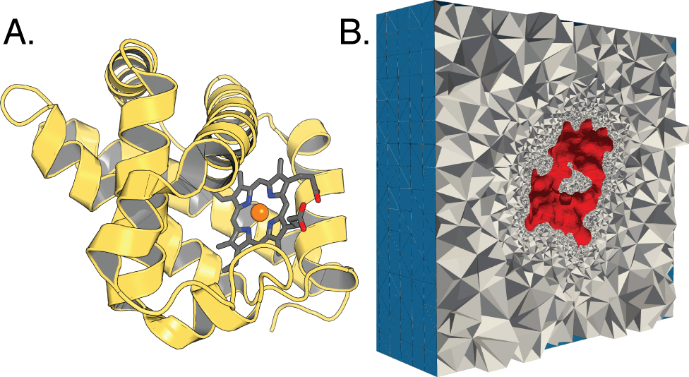
In Fig. 4, we demonstrate the mesh generation capabilities of GAMer from atomistic protein structural data such as those available from the Protein Data Bank. A volume dataset representing the approximate occupied space of all atoms is generated by applying a Gaussian kernel over the atomic positions. An isosurface of the dataset can then be meshed using the marching cubes algorithm (39). While GAMer includes a basic table of atom types to assign radii, more sophisticated atomic radius assignment tools (e.g., PDB2PQR (40)) can be used and radius information passed via the PQR file format.
Conclusion
The realism of biophysical models can be enhanced by incorporation realistic geometries from structural biology at various length scales. We have demonstrated the applicability of GAMer 2 at two different length scales and suggest that tools such as GAMer 2 brings the community closer to realizing the goal of conducting physics-based simulations in realistic cellular geometries by simplifying the mesh generation process. We believe that, moving forward, GAMer 2 can serve as an integrative platform for meshing biological mesh generation. Furthermore, our design strategies in implementing GAMer 2 encourage community collaboration, an important aspect of tool building for systems and computational biology.
Software Availability
GAMer is licensed under GNU Lesser General Public License (LGPL), version 2.1 or later. Full documentation and examples are available at the project home page, gamer.readthedocs.io, and development is hosted on GitHub at http://github.com/ctlee/gamer. The latest release v2.0.5 is archived on Zenodo (doi:10.5281/zenodo.2340294).
Author Contributions
C.T.L., J.G.L., J.B.M., and M.J.H. developed the software; C.T.L. drafted the article; C.T.L., J.G.L., J.B.M., J.A.M., R.E.A., M.J.H., and P.R. edited the article; and all authors read and approved the final article.
Acknowledgments
CTL, JBM, REA, JAM, and MJH are supported in part by the National Institutes of Health under grant number P41-GM103426. CTL, and JAM are also supported by the NIH under R01-GM31749. CTL also acknowledges support from the NIH Molecular Biophysics Training Grant T32-GM008326 and a Hartwell Foundation Postdoctoral Fellowship. MJH was supported in part by the National Science Foundation under awards DMS-CM1620366 and DMS-FRG1262982. PR was supported by the Air Force Office of Scientific Research (AFOSR) Multidisciplinary University Research Initiative (MURI) FA9550-18-1-0051 and JGL was supported by a fellowship from the UCSD Center for Transscale Structural Biology and Biophysics/Virtual Molecular Cell Consortium.
REA is a cofounder and scientific advisor of, and has equity interest in, Actavalon, Inc.
References
- (1) Rangamani, P., A. Lipshtat, E. U. Azeloglu, R. C. Calizo, M. Hu, S. Ghassemi, J. Hone, S. Scarlata, S. R. Neves, and R. Iyengar. Decoding Information in Cell Shape 154:1356–1369. http://www.sciencedirect.com/science/article/pii/S0092867413010209.
- (2) Bell, M., T. Bartol, T. Sejnowski, and P. Rangamani. Dendritic spine geometry and spine apparatus organization govern the spatiotemporal dynamics of calcium 151:1017–1034. http://jgp.rupress.org/content/151/8/1017.
- (3) Cugno, A., T. M. Bartol, T. J. Sejnowski, R. Iyengar, and P. Rangamani. Geometric principles of second messenger dynamics in dendritic spines 9:1–18. https://www.nature.com/articles/s41598-019-48028-0.
- (4) Ohadi, D., D. L. Schmitt, B. Calabrese, S. Halpain, J. Zhang, and P. Rangamani. Computational Modeling Reveals Frequency Modulation of Calcium-cAMP/PKA Pathway in Dendritic Spines http://www.sciencedirect.com/science/article/pii/S0006349519308318.
- (5) Updegrove, A., N. M. Wilson, J. Merkow, H. Lan, A. L. Marsden, and S. C. Shadden. SimVascular: An Open Source Pipeline for Cardiovascular Simulation 45:525–541.
- (6) Loew, L. M., and J. C. Schaff. The Virtual Cell: A software environment for computational cell biology.
- (7) Murphy, R. F. CellOrganizer: Image-Derived Models of Subcellular Organization and Protein Distribution. In Methods Cell Biol.
- (8) Xu, C. S., K. J. Hayworth, Z. Lu, P. Grob, A. M. Hassan, J. G. García-Cerdán, K. K. Niyogi, E. Nogales, R. J. Weinberg, and H. F. Hess. Enhanced FIB-SEM systems for large-volume 3D imaging 6. https://elifesciences.org/articles/25916.
- Si (a) Si, H. TetGen, a Delaunay-Based Quality Tetrahedral Mesh Generator 41:1–36. http://dl.acm.org/citation.cfm?id=2732672.2629697.
- (10) Schöberl, J. An advancing front 2D/3D-mesh generator based on abstract rules .
- (11) Hu, Y., Q. Zhou, X. Gao, A. Jacobson, D. Zorin, and D. Panozzo. Tetrahedral meshing in the wild 37:1–14. http://dl.acm.org/citation.cfm?doid=3197517.3201353.
- (12) Cignoni, P., M. Callieri, M. Corsini, M. Dellepiane, F. Ganovelli, and G. Ranzuglia. MeshLab: an Open-Source Mesh Processing Tool. In V. Scarano, R. De Chiara, and U. Erra, editors, Eurographics Ital. Chapter Conf. The Eurographics Association.
- (13) Geuzaine, C., and J.-F. Remacle. Gmsh: A 3-D finite element mesh generator with built-in pre- and post-processing facilities 79:1309–1331. http://doi.wiley.com/10.1002/nme.2579.
- (14) CGAL, Computational Geometry Algorithms Library. http://www.cgal.org.
- Yu et al. (a) Yu, Z., M. J. Holst, Y. Cheng, and J. McCammon. Feature-Preserving Adaptive Mesh Generation for Molecular Shape Modeling and Simulation 26:1370–1380.
- Yu et al. (b) Yu, Z., M. J. Holst, and J. Andrew McCammon. High-Fidelity Geometric Modeling for Biomedical Applications 44:715–723.
- Gao et al. (a) Gao, Z., Z. Yu, and M. Holst. Quality Tetrahedral Mesh Smoothing via Boundary-Optimized Delaunay Triangulation 29:707–721.
- Gao et al. (b) Gao, Z., Z. Yu, and M. Holst. Feature-Preserving Surface Mesh Smoothing via Suboptimal Delaunay Triangulation 75:23–38.
- (19) Chen, L., and M. Holst. Efficient Mesh Optimization Schemes Based on Optimal Delaunay Triangulations 200:967–984.
- Lee et al. (a) Lee, C. T., J. B. Moody, J. G. Laughlin, and M. J. Holst. GAMer 2.0 Software. https://github.com/ctlee/gamer.
- Si (b) Si, H. TetGen, a Delaunay-Based Quality Tetrahedral Mesh Generator 41:11:1–11:36. http://doi.acm.org/10.1145/2629697.
- (22) Logg, A., K.-A. Mardal, G. N. Wells, et al. Automated Solution of Differential Equations by the Finite Element Method. Springer.
- (23) Alnæs, M. S., J. Blechta, J. Hake, A. Johansson, B. Kehlet, A. Logg, C. Richardson, J. Ring, M. E. Rognes, and G. N. Wells. The FEniCS Project Version 1.5 3.
- (24) Ahrens, J., B. Geveci, and C. Law. ParaView: An end-user tool for large-data visualization. In Vis. Handb.
- (25) Stiles, J. R., and T. M. Bartol. Monte Carlo methods for simulating realistic synaptic microphysiology using MCell. In E. D. Schutter, editor, Computational Neuroscience: Realistic Modeling for Experimentalists, CRC Press, 87–127.
- (26) Solernou, A., B. S. Hanson, R. A. Richardson, R. Welch, D. J. Read, O. G. Harlen, and S. A. Harris. Fluctuating Finite Element Analysis (FFEA): A continuum mechanics software tool for mesoscale simulation of biomolecules 14:1–29. https://doi.org/10.1371/journal.pcbi.1005897.
- Lee et al. (b) Lee, C. T., J. G. Laughlin, N. A. de La Beaumelle, R. E. Amaro, J. A. McCammon, R. Ramamoorthi, M. J. Holst, and P. Rangamani. GAMer 2: A system for 3D mesh processing of cellular electron micrographs https://www.biorxiv.org/content/early/2019/07/23/534479.
- (28) Hoshijima, M., T. Hayashi, A. Thor, M. Terada, M. Martone, and M. Ellisman. CCDB:3603, MUS MUSCULUS, T-tubules, sarcoplasmic reticulum, myocyte. CIL. Dataset.
- Lee et al. (c) Lee, C. T., J. B. Moody, R. E. Amaro, J. A. Mccammon, and M. J. Holst. The Implementation of the Colored Abstract Simplicial Complex and Its Application to Mesh Generation 45:1–20. http://dl.acm.org/citation.cfm?doid=3349340.3321515.
- (30) Jakob, W., J. Rhinelander, and D. Moldovan. pybind11 – Seamless operability between C++11 and Python. Https://github.com/pybind/pybind11.
- (31) Beazley, D. M. SWIG: An Easy to Use Tool for Integrating Scripting Languages with C and C++. In Proceedings of the 4th Conference on USENIX Tcl/Tk Workshop, 1996 - Volume 4. USENIX Association, TCLTK’96, 15–15. http://dl.acm.org/citation.cfm?id=1267498.1267513.
- (32) Community, B. O. Blender - a 3D modelling and rendering package. Blender Foundation. http://www.blender.org.
- Yu et al. (c) Yu, Z., M. J. Holst, T. Hayashi, C. L. Bajaj, M. H. Ellisman, J. A. McCammon, and M. Hoshijima. Three-dimensional geometric modeling of membrane-bound organelles in ventricular myocytes: Bridging the gap between microscopic imaging and mathematical simulation 164:304–313. http://linkinghub.elsevier.com/retrieve/pii/S1047847708002281.
- Cheng et al. (a) Cheng, Y., Z. Yu, M. Hoshijima, M. J. Holst, A. D. McCulloch, J. A. McCammon, and A. P. Michailova. Numerical Analysis of Ca2+ Signaling in Rat Ventricular Myocytes with Realistic Transverse-Axial Tubular Geometry and Inhibited Sarcoplasmic Reticulum 6:e1000972. http://dx.plos.org/10.1371/journal.pcbi.1000972.
- Cheng et al. (b) Cheng, Y., P. Kekenes-Huskey, J. E. Hake, M. J. Holst, J. A. McCammon, and A. P. Michailova. Multi-scale continuum modeling of biological processes: from molecular electro-diffusion to sub-cellular signaling transduction 5:015002. https://iopscience.iop.org/article/10.1088/1749-4699/5/1/015002.
- (36) Hake, J., A. G. Edwards, Z. Yu, P. M. Kekenes-Huskey, A. P. Michailova, J. A. McCammon, M. J. Holst, M. Hoshijima, and A. D. McCulloch. Modelling cardiac calcium sparks in a three-dimensional reconstruction of a calcium release unit 590:4403–4422. http://doi.wiley.com/10.1113/jphysiol.2012.227926.
- (37) Kekenes-Huskey, P. M., Y. Cheng, J. E. Hake, F. B. Sachse, J. H. Bridge, M. J. Holst, J. A. McCammon, A. D. McCulloch, and A. P. Michailova. Modeling effects of L-type ca(2+) current and na(+)-ca(2+) exchanger on ca(2+) trigger flux in rabbit myocytes with realistic T-tubule geometries. 3:351. http://journal.frontiersin.org/article/10.3389/fphys.2012.00351/abstract.
- (38) Bromer, C., T. M. Bartol, J. B. Bowden, D. D. Hubbard, D. C. Hanka, P. V. Gonzalez, M. Kuwajima, J. M. Mendenhall, P. H. Parker, W. C. Abraham, T. J. Sejnowski, and K. M. Harris. Long-term potentiation expands information content of hippocampal dentate gyrus synapses 115:E2410–E2418. https://www.pnas.org/content/115/10/E2410.
- (39) Lorensen, W. E., and H. E. Cline. Marching Cubes: A High Resolution 3D Surface Construction Algorithm 21:163–169. http://doi.acm.org/10.1145/37402.37422.
- (40) Dolinsky, T. J., J. E. Nielsen, J. A. McCammon, and N. A. Baker. PDB2PQR: an automated pipeline for the setup of Poisson-Boltzmann electrostatics calculations 32:W665–W667. https://academic.oup.com/nar/article-lookup/doi/10.1093/nar/gkh381.