Pain Analysis, in Premature Infants, Using Near Infrared Spectroscopy (NIRS)
Background: The role of neonatal pain on the developing nervous system is not completely understood, but evidence suggests that sensory pathways are influenced by an infant’s pain experience. Research has shown that an infant’s previous pain experiences lead to an increased, and likely abnormal, response to subsequent painful stimuli. We are working to improve neonatal pain detection through automated devices that continuously monitor an infant. The current study outlines some of the initial steps we have taken to evaluate Near Infrared Spectroscopy (NIRS) as a technology to detect neonatal pain. Our findings may provide neonatal intensive care unit (NICU) practitioners with the data necessary to monitor and perhaps better manage an abnormal pain response.
Methods: A prospective pilot study was conducted to evaluate nociceptive evoked cortical activity in preterm infants. NIRS data were recorded for approximately 10 minutes prior to an acute painful procedure and for approximately 10 minutes after the procedure. Individual data collection events were performed at a weekly maximum frequency. Eligible infants included those admitted to the Tampa General Hospital (TGH) NICU with a birth gestational age of less than 37 weeks.
Results: A total of 15 infants were enrolled and 25 individual studies were completed. Analysis demonstrated a statistically significant difference between the median of the pre- and post-painful procedure data sets in each infant’s first NIRS collection (p value = 0.01).
Conclusions: Initial analysis shows NIRS may be useful in detecting acute pain. An acute painful procedure is typically followed by a negative deflection in NIRS readings.
I Introduction
The concept of neonatal pain has been quite fluid throughout history as perceptions and evidence have slowly morphed into what we know and believe today. The study of neonatal pain seems to have begun in the 1870s, when Dr. Flechsig proposed that it was unlikely that neonates could experience pain because their neuronal myelination was not complete (Cope, 1998). Charles Darwin agreed with this view, when he wrote in his book The Expression of the Emotions in Man and Animals, that an infant’s pain expressions were related to reflexes only (Darwin, 1872). Even as late as the 1950s some pediatric surgeries were performed without anesthesia and analgesia (Cope, 1998).
Today the prevailing belief is that not only do neonates experience pain, but their past pain experiences likely shape their futures responses to painful stimuli. This concept was first observed in infants who underwent circumcision and then were compared to age matched controls receiving immunizations at four to six months of age. Researchers found that infants who underwent circumcision demonstrated increased behavioral measures of pain compared to their age-matched controls (Taddio, 1997). Likewise, infants of diabetic mothers who required multiple heel lances in the first 24 hours of life demonstrated increased behavioral measures of pain, compared to age-matched controls, when they received venipuncture for newborn screening samples at 24 hours of life (Taddio, 2002).
Similar findings were demonstrated, in evoked neuronal activity, when researchers found that premature infants yielded significantly larger noxious evoked potentials compared to their age-matched term controls (Slater, 2010). As the concept of neonatal pain has evolved, the tools used to measure it have changed. Although behavioral measures remain the gold standard in the NICU, more objective and continuous measurement methods are becoming available. One such technology is NIRS. NIRS uses light in the 700 to 1000 nanometer range to penetrate skin, soft tissue, bone and brain to measure an infant’s deoxygenated and oxygenated hemoglobin concentrations (Ranger, 2011). These measures have shown good correlation with behavioral measures of neonatal pain (Slater, 2006).
In the current study we sought to detect neonatal pain using NIRS. This pilot study had the ultimate goal of determining if NIRS is worthy of further investigation, and possible incorporation into a device, to continuously measure neonatal pain in the NICU.
II Methods
We hypothesized that performing a painful procedure would result in a decrease in the regional oximetry of the brain as measured by NIRS. The primary outcome measure was a change in NIRS values, of the contralateral brain, following a painful procedure in the infant’s extremity.
II-A Infant Recruitment
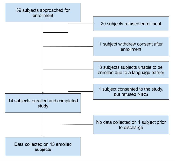
All consecutive premature infants, with a birth gestational age of less than 37 weeks, were eligible for recruitment from the TGH NICU. The birth gestational age of each infant was obtained from obstetrical records, which included antenatal ultrasounds and maternal reports of last menstrual periods. Exclusion criteria included major congenital anomalies of the central nervous system or cardiovascular system, ongoing mechanical ventilation via an endotracheal tube and infants who had received analgesic or sedative drugs in the previous twenty four hours. This study was approved by the Tampa General Hospital Institutional Review Board. Figure 1 shows the enrolment numbers for this study.
II-B Infant Data Collection
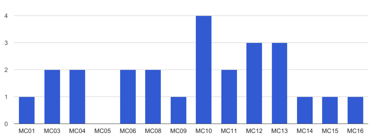
Of the fifteen infants consented for the study, data were obtained on thirteen infants. Data were not collected on two infants as consent was withdrawn on one infant and one infant was discharged prior to data collection. A total of twenty-five data collection events were performed on the fourteen infants that maintained informed consent. The number of data collection events per infant is displayed in Figure 2.
II-C Study Equipment
An INVOS 5100C NIRS meter was used to collect regional cerebral oximetry data. The meter uses near infrared light to determine the deoxyhemoglobin (HbH) and the oxy-hemoglobin (HbO2) concentrations. HbH and HbO2 are then added together to determine the total hemoglobin concentration (HbT). The meter then divides the HbO2 by the HbT to obtain a regional oximetry value (rSO2), which is displayed on the device’s screen. The meter’s sample rate was 30 seconds. A VitalSync was used to timestamp and export the NIRS data into Excel. The VitalSync was also used to log events, such as the start of pre-procedure data collection and the start of the painful procedure.
II-D Study Protocol
Once informed consent had been obtained, and no exclusion criteria were indentified, the infant’s first data collection event was performed. Painful procedures included heal sticks for point-of-care glucose checks, routine labs, routine newborn metabolic screens, and vaccinations. Additional data collection events were performed a minimum of one week apart. No painful procedures were performed for the sole purpose of obtaining NIRS data. The study team reviewed the enrolled infants’ charts to determine the date and time of a painful procedure and the bedside nurse performed it.
In an effort to have the study’s results reflect usual practice, no alterations were made to the NICU’s usual method of obtaining labs or giving vaccinations, and no alterations were made in the usual personnel performing the procedures. Prior to the placing of the NIRS probe the team verified the location of the painful procedure with the bedside nurse. The probe was then placed on the contralateral side of the forehead. Once the INVOS 5100C had established its auto-baseline, an event report was made in the VitalSync and collection of the pre-procedure data was started. Pre-procedure data were collected for approximately ten minutes, at which point, the bedside nurse would start the painful procedure and an additional event report was made on the VitalSync. Post-procedure data were then collected for an additional 10 additional minutes after the painful procedure had ended.
II-E Statistical Analysis
We investigated the change in the oximetry of the brain, as measured by NIRS, using Wilcoxson signed rank test at a significance level of 0.05. We used SPSS version23 to conduct the statistical analyses. In the analysis the ordered collections of each patient were compared to each other (the 1st, 2nd, etc. collection events were compared), as we hypothesized that any painful stimuli may impact the response to future painful stimuli.
III Results
III-A Infant Demographics
A total of thirty-nine infants were identified for enrolment. Of the thirty-nine identified infants consent was obtained for fifteen infants. Table 1 shows the patient demographics for the infants whose consent was not withdrawn. Two of the infants were found to have an intraventricular hemorrhage (IVH).
![[Uncaptioned image]](/html/1908.10240/assets/Table1.png)
III-B Data Analysis
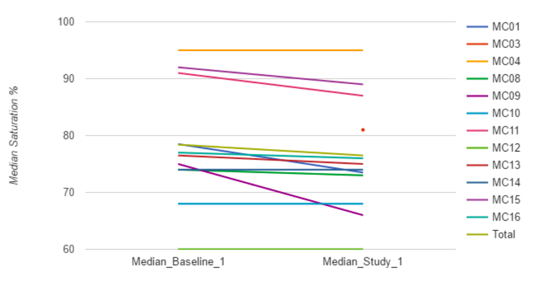
When comparing the first NIRS data collection event from each patient, we found a statistically significant negative deflection in the regional cerebral oximetry values. The median of the medians had a pre-procedure value of 78.3% and a post-procedure value of 76.5% (p value = 0.01). Figure 3 plots the median pre- and post-procedure data values for each patient’s first data collection event, as well as, the median of the medians (displayed as Total). An insufficient number of sequential data collection events hindered the determination of any meaningful conclusions about whether or not an infant’s NIRS response changes over time as the infant experiences more painful procedures.
IV Discussion
As previously described, several studies in infants have observed an increased response to painful stimuli over time. This is likely a learned response that results from a disruption in an infant’s ongoing nervous system development. We believe that continuous pain monitoring, with appropriate interventions, can alter this progression. This pilot study hypothesized that, following a painful procedure, a decrease in the contralateral regional cerebral oximetry would be seen. This hypothesis was based on literature findings that demonstrated that a painful stimulus results in an increase in the contralateral total hemoglobin concentration (Slater, 2006).
Although the NIRS device used in our study computes a regional cerebral oximetry percentage, rather than displaying total hemoglobin concentration, our results seem to agree with Slater et al. Since cortical activation, caused by a painful stimulus, likely leads to a short relative decrease in the oxygenated hemoglobin concentration despite an increase in total blood flow, the end result is a decrease in the regional oximetry percentage. In essence, the influx of oxygenated hemoglobin that should come with the increase in total hemoglobin, is not initially sufficient to overcome the increased oxygen demands of the tissues.
While our results do seem to support our primary objective, this pilot study did not yield a sufficient amount of data, due to low participant recruitment, to draw any meaningful conclusions about an infant’s cortical response to pain over time. In order to accomplish this task, we propose that future studies should allow NIRS data to be continuously collected so that many acute procedures can be captured. Our study plan limited us to weekly measures, which led to less opportunities to collect data as time went on, as infants in the NICU tend to have most of their painful procedures performed early in their stays.
The results of this pilot study are indeed promising as we believe they move us towards our ultimate goal of building a device that will continually monitor neonatal pain. We believe this information will allow NICU practitioners to better relieve neonatal pain thus breaking the abnormal developmental cycle that currently ensues.
We have previously used facial recognition software and machine learning to detect behavioural measures of neonatal pain (Zamami). While the results of our previous study did reveal a high specificity for neonatal pain, the sensitivity was inadequate. It is our hope that incorporating NIRS will eventually result in improved sensitivity and therefore improved detection of neonatal pain as we move forward.
References
-
1.
Cope D (1998) Neonatal Pain: The Evolution of an Idea. American Society of Anesthesiologists.
-
2.
Ranger M, Johnston C, Limperopoulos C, Rennick J, du Plessis A (2011) Cerebral nearinfrared spectroscopy as a measure of nociceptive evoked activity in critically ill infants. Pain Research Management 16(5):331-336.
-
3.
Slater R, Cantarella A, Gallella S, Worley A, Boyd S, Meek J, Fitzgerald M (2006) Cortical pain responses in human infants. The Journal of Neuroscience 26(14):3662- 3666.
-
4.
Slater R, Fabrizi L, Worley A, Meek J, Boyd S, Fitzgerald M (2010) Premature infants display increased noxious-evoked neuronal activity in the brain compared to healthy age-matched term-born infants. NeuroImage 583-589.
-
5.
Slater R, Worley A, Fabrizi L, Roberts S, Meek J, Boyd S, Fitzgerald M (2010) Evoked potentials generated by noxious stimulation in the human infant brain. European Journal of Pain 14:321-326.
-
6.
Slater R, Cantarella A, Franck L, Meek J, Fitzgerald M (2008) How well do clinical pain assessment tools reflect pain in infants? PLOS Medicine 5(6):0928-0930.
-
7.
Taddio A, Katz J, Ilersich A, Koren G (1997) Effect of neonatal circumcision on pain response during subsequent routine vaccination. Lancet 349:599-603.
-
8.
Taddio A, Shah V, Gilbert-MacLeod C, Katz J (2002) Conditioning and hyperalgesia in newborns exposed to repeated heel lances. JAMA 288(7):857-861.
-
9.
Zamzami, G, et al. “An approach for automated multimodal analysis of infants’ pain.” Pattern Recognition (ICPR), 2016 23rd International Conference on. IEEE, 2016.
-
10.
Darwin, C. (1872). The Expression of the Emotions in Man and Animals. (1st ed.). London, UK: John Murray.