Sensing the spin of an individual Ce adatom
Abstract
The magnetic moment of rare earth elements originates from electrons in the partially filled 4 orbitals. Accessing this moment electrically by scanning tunneling spectroscopy is hampered by shielding of outer-lying orbitals. Here we show that we can detect the magnetic moment of an individual Ce atom adsorbed on a Cu2N ultrathin film on Cu(100) by using a sensor tip that has its apex functionalized with a Kondo screened spin system. We calibrate the sensor tip by deliberately coupling it to a well characterized Fe atom. Subsequently, we use the splitting of the tip’s Kondo resonance when approaching a spectroscopically dark Ce atom to sense its magnetic moment.
Recently, the magnetism of surface-supported rare earth elements has come newly into focus, because individual atoms with electrons on ultrathin insulators have been found to show long relaxation times Donati et al. (2016), making them interesting candidates for atomic scale memory and possible qubit realization. Due to their large orbital angular momentum, which results in less extended orbitals than the orbitals of transition metals like Fe or Co, orbitals do not usually take part in chemical bonds and only hybridize weakly. This isolation is both a blessing and a curse as this promotes magnetic stability, but at the cost of easy detection and manipulation by electrical means.
Accordingly, scanning tunneling microscopy (STM) and spectroscopy measurements on Ho and Gd adatoms on Pt(111) revealed only low or no detectable interaction cross section between the tunneling electrons and the localized spin Schuh et al. (2012); Balashov et al. (2014); Steinbrecher et al. (2016). Nevertheless, the moment of Ho atoms adsorbed on a thin insulating film of MgO on Ag(100) was detected as a change in the electron spin resonance frequency of a single Fe adatom Natterer et al. (2017) and changed the spectrum of a Co atom in HoCo dimers Singha et al. (2018).
Furthermore, in compounds and thin layers of Ce, a lanthanide which hosts only one 4 electron, Kondo screening has been observed Patthey et al. (1985, 1987, 1990); Laubschat et al. (1990); Ehm et al. (2007). In Kondo systems, the magnetic moments get compensated by itinerant electrons leading to a highly correlated singlet state and a resonance at the Fermi energy below a characteristic Kondo temperature Kondo (1964, 1968); Hewson (1997); Ternes et al. (2009). Kondo features have been found on surface-supported double-decker molecules containing Dy Warner et al. (2016), however, measurements on Ce atoms on Ag(111) showed ambiguous results. While first results hinted at Kondo screening Li et al. (1998), subsequent investigations revealed that single Ce adatoms diffuse on the Ag(111) surface even at K Silly et al. (2004a, b); Negulyaev et al. (2009); Ternes et al. (2010) suggesting that the earlier measurement was taken on an immobile Ce cluster and that hydrogenated 4 atoms can show low-energy vibrational excitations mimicking a spin signal Pivetta et al. (2007). However, small Ce clusters showed a clear Kondo resonance Ternes et al. (2009).
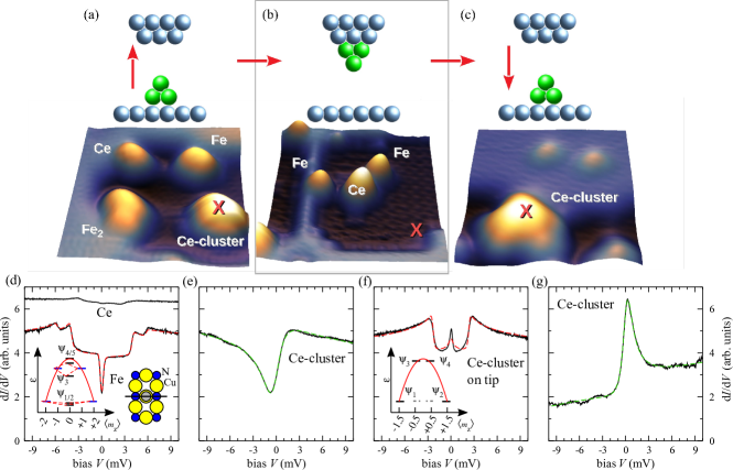
Therefore, we re-addressed the question of the magnetic moment of single Ce adatoms by co-depositing individual Fe and Ce atoms onto a monolayer of Cu2N on Cu(100) Leibsle et al. (1994) and performing experiments with a home-built STM at K. As shown in Fig. 1a, single Ce and Fe atoms as well as small clusters formed during deposition at K are imaged as stable protrusions. The single atoms adsorb on the Cu-sites of the Cu2N SOM . Measuring the differential conductance of Fe atoms revealed characteristic features originating from spin-flip excitations Heinrich et al. (2004); Hirjibehedin et al. (2007). To interpret these spectra we use a model Hamiltonian for the magnetic atom,
| (1) |
where the spin system is described by the generalized spin operator , and are the axial and transverse magnetic anisotropy parameters. The Zeeman energy is accounted for with as the gyromagnetic factor, the Bohr magneton, and the applied magnetic field.
The step-like increase in is due to excitations from the ground to energetically higher eigenstates of Equ. 1 via Kondo-like interactions between the localized magnetic moment and the tunneling electron. The tunneling electron has states and spin matrices , leading to the transition matrix elements between initial () and final () states:
| (2) |
Here, are product states, and are identity matrices in their corresponding Hilbert sub-spaces, and is a Coulomb scattering parameter which accounts not only for a background , but also leads to interference induced bias asymmetries in the spectra, when higher scattering orders are considered Ternes (2015).
We find an excellent fit to the Fe data using the previously found effective spin , easy-axis anisotropy () which favors the high magnetic moment along the N-rows of the Cu2N Hirjibehedin et al. (2007), and a transport model which includes scattering processes up to 3rd order in the matrices Ternes (2015, 2017) (Fig. 1d). For 3rd order processes, we additionally take the dimensionless coupling into account, where is the interaction strength between the many electrons of the substrate and the atom’s magnetic moment and is the density of states at Fermi energy.
Interestingly, while the of Fe atoms show strong steps that indicate spin excitations, single Ce atoms do not. Their spectra are essentially flat and featureless (Fig. 1d). This changes when measurements are taken on a small cluster of Ce atoms Ternes et al. (2009). Fig. 1e shows that we detect an asymmetric feature centered near which can be well described by a Fano line shape Fano (1961); Madhavan et al. (2001):
| (3) |
In this equation, is the normalized energy where is the position and the half-width of the resonance, is the Fano parameter, and accounts for a background . We find meV which is due to Kondo screening of the cluster’s magnetic moment with K. The indicates a relatively strong direct tunneling channel between the tip and the substrate Újsághy et al. (2000). Because the cluster is in the strong screening regime Zonda et al. (2018).
Surprisingly, when we transfer this Ce cluster to the STM tip, by applying V bias pulses at close to point contact, the spectrum changes drastically, now revealing a narrow Kondo resonance, and a spin excitation at about meV (Fig. 1f). This spectrum is typical for an effective system in which the zero-bias Kondo peak originates from scattering between the two degenerate ground states that have weights in the projections, and the steps from transitions to states that are energetically higher by (inset Fig. 1f). Similar to Fe, this spectrum can be fit with the scattering model even though the central peak is only partly represented, since Kondo screening includes multiple higher-order scattering. This spectrum is very similar to that of a single Co atom on Cu2N Otte et al. (2008); Oberg et al. (2013); Von Bergmann et al. (2015), but here the magnetic moment originates from the Ce cluster at the apex of the metallic tip, enabling us to exploit it as a mobile sensor as shown below.
Transferring the cluster back to the surface as a control experiment by moving the tip to near point contact and withdrawing while applying V leads to a similar spectrum as before pick-up (Fig. 1g). Both spectra show a Kondo signal, but the transfer of the cluster to a new location on Cu2N has changed the Kondo signal from dip to a peak. However, is changed only modestly, with changing by about , which might be due to different adsorption geometries Gao et al. (2007). In contrast to the relatively weak coupling of the cluster when attached to the tip apex, the apparently stronger coupling to bulk electrons on the surface quenches the magnetic anisotropy Oberg et al. (2013); Jacobson et al. (2015) and moves the spin system into the strong-coupling Kondo regime.
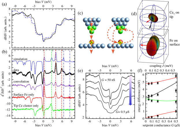
To characterize the magnetic properties of the Ce cluster at the tip apex, we probe Fe atoms using the Ce-functionalized tip, which results in a complex spectrum (Fig. 2a, b). Interestingly, the second derivative closely resembles the convolution of the individual spectra of tip and sample, similar to recent observations of coupled molecular spins in a junction Ormaza et al. (2017). However, wFhile the convolution yields the correct transition energies, the peak intensities differ significantly between data and convolution (Fig. 2b).
This difference has two origins: First, transport which excites both spin systems is complex because the scattering events can occur in different sequences, which must be summed coherently. An electron tunneling from tip (t) to sample (s) can scatter first with the spin on the tip and then with the spin on the surface, and it can also first interact with the surface spin before interacting with the tip spin (Fig. 2c). Destructive quantum interference between these different scattering channels cancel all scattering processes, except those that obey . For spins on tip and surface, this results in a different form for the interaction matrix than Equ. 2, giving SOM :
| (4) |
Here, the first two terms account for scattering in which spin-spin interaction occurs at only one of the two localized moments, while on the other only a Coulomb-like interaction take place. The last term accounts for spin-spin scattering on both the tip and sample moments.
Second, while the anisotropy axis of the Fe adatom is well known, the anisotropy axis of the Ce cluster on the tip is unknown and might point in an arbitrary direction. To determine this direction, we use a model that employs Equ. Sensing the spin of an individual Ce adatom up to 3rd order and a Hamiltonian in which the Ce cluster and the Fe adatom are both described by Equ. 1 using their individually found anisotropy and coupling parameters SOM . This model allows us to determine the relative alignment of the two spins. As illustrated in Fig. 2d we find a ° angle between the two magnetic easy axes. Furthermore, the Ce cluster’s magnetic intermediate axis, that is the -direction in Equ. 1, is tilted by only ° from the surface normal. Note, that the simplicity of the model limits its accuracy SOM .
To determine the magnetic coupling between the tip and surface spins, we proceed by changing the set-point conductance and consequently the tip-sample distance . We observe a shift in intensity and energy of the excitations (Fig. 2e). In particular the energy of the first excitation changes from meV to meV, when is varied by one order of magnitude, i. e. when is reduced by Å. This shift can be well described by assuming an antiferromagnetic Heisenberg-like exchange interaction between both spins (Fig. 2f). The linear dependence, , implies an exponential dependence on and points to an orbital overlap as origin of the interaction Muenks et al. (2017); Verlhac et al. (2019); Yang et al. (2019).
Having carefully characterized the magnetic tip, we use it to probe spectroscopically “dark” Ce adatoms. Fig. 3a shows typical spectra, which for small (tip far from the surface) are almost identical to the ones measured against the bare surface (Fig. 3a, b) except for a a slight reduction of the zero bias Kondo peak height. Prominent differences emerge at increased . We observe that the central peak becomes asymmetric and for S splits while spectra taken at similar values on the bare surface do not change significantly (Fig. 3b).
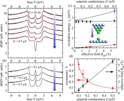
This result clearly reveals the presence of a magnetic moment in the Ce adatom that influences the spectrum of the probing tip. We can model the observed behavior by using Equ. 1 and assuming that the spectroscopically dark Ce adatom acts as an effective, set-point dependent, magnetic field oriented in a direction approximately out-of-surface having a strength of T/ (Fig. 3c, d).
While part of the spectral asymmetry can be attributed to Fano-like interference (Equ. 3), we interpret the asymmetric intensity of the split central feature to originate primarily from an imbalance between majority and minority sample states Loth et al. (2010); Von Bergmann et al. (2015). Such polarization can be induced by the spin moment of the Ce adatom Muenks et al. (2017); Verlhac et al. (2019). The decrease with suggests an antiferromagnetic interaction as the origin of the exchange field and the formation of a combined non-magnetic singlet state of both spins. This indicates a half-integer moment of the Ce adatom and assuming , the Ising-like coupling leads to a strength of meV/S. We found that using isotropic Heisenberg or dipole-dipole interactions instead of Ising interactions lead to much less adequate fits. We also have indications of a reduced tip-adatom interaction strength for Ce adsorbed close to the bare metal SOM .
To summarize, our results point to an effective spin of single Ce adatoms on the Cu2N surface. The very localized orbital is inaccessible to ordinary spectroscopy because the interaction cross section with the tunneling electron is too small. This is also the reason for the absence of Kondo screening at our accessible temperatures. Nevertheless, we sensed the magnetic moment by a “detector” spin at the apex of a functionalized STM tip. The particular coupling mechanism between the two spins is not yet fully known, but the observed exponential dependence of the Ising-like interaction points to some orbital mixing Gunnarsson and Schönhammer (1983).
As a detector we used a Ce cluster which showed a very narrow Kondo resonance at . Such a tip is ideally suited to spin detection, in particular, when is of order . Then, the tip shows a clear Kondo resonance at and its sensitivity to exchange interactions and spin polarization is only limited by the unavoidable thermal broadening of the spectra. In contrast, tips with high or very low are contra-indicative. While in the former the exchange interaction has first to overcome the Kondo energy before spectral changes can be observed Bork et al. (2011), the latter don’t show a pronounced resonance. Note, however, magnetic molecules at the tip apex can be exploited in a similar manner as sensor and may be prepared more reproducibly Verlhac et al. (2019); Czap et al. (2019).
Our results open a new route for studying elements on well-defined surfaces. For example, the method outlined here could be used to measure the interactions in artificially created or self-assembled Silly et al. (2004a); Ternes et al. (2010) atomic or molecular 1D and 2D nanostructures.
Acknowledgements.
MT acknowledges support by the Heisenberg Program (Grant No. TE 833/2-1) of the German Science Foundation and AJH from the Institute for Basic Science under grant IBS-R027-D1. We thank H.-J. Freund and A. Singha for stimulating discussions.References
- Donati et al. (2016) F. Donati, S. Rusponi, S. Stepanow, C. Wackerlin, A. Singha, L. Persichetti, R. Baltic, K. Diller, F. Patthey, E. Fernandes, et al., Science 352, 318 (2016).
- Schuh et al. (2012) T. Schuh, T. Miyamachi, S. Gerstl, M. Geilhufe, M. Hoffmann, S. Ostanin, W. Hergert, A. Ernst, and W. Wulfhekel, Nano Lett. 12, 4805 (2012).
- Balashov et al. (2014) T. Balashov, T. Miyamachi, T. Schuh, T. Märkl, C. Bresch, and W. Wulfhekel, Surf. Sci. 630, 331 (2014).
- Steinbrecher et al. (2016) M. Steinbrecher, A. Sonntag, M. Dos Santos Dias, M. Bouhassoune, S. Lounis, J. Wiebe, R. Wiesendanger, and A. A. Khajetoorians, Nature Comm. 7, 10454 (2016).
- Natterer et al. (2017) F. D. Natterer, K. Yang, W. Paul, P. Willke, T. Choi, T. Greber, A. J. Heinrich, and C. P. Lutz, Nature 543, 226 (2017).
- Singha et al. (2018) A. Singha, F. Donati, F. D. Natterer, C. Wackerlin, S. Stavrić, Z. S. Popović, Z. Sljivancanin, F. Patthey, and H. Brune, Phys. Rev. Lett. 121, 257202 (2018).
- Patthey et al. (1985) F. Patthey, B. Delley, W. D. Schneider, and Y. Baer, Phys. Rev. Lett. 55, 1518 (1985).
- Patthey et al. (1987) F. Patthey, W. D. Schneider, Y. Baer, and B. Delley, Phys. Rev. Lett. 58, 2810 (1987).
- Patthey et al. (1990) F. Patthey, J. M. Imer, W. D. Schneider, H. Beck, Y. Baer, and B. Delley, Phys. Rev. B 42, 8864 (1990).
- Laubschat et al. (1990) C. Laubschat, E. Weschke, C. Holtz, M. Domke, O. Strebel, and G. Kaindl, Phys. Rev. Lett. 65, 1639 (1990).
- Ehm et al. (2007) D. Ehm, S. Hüfner, F. Reinert, J. Kroha, P. Wolfle, O. Stockert, C. Geibel, and H. V. Lohneysen, Phys. Rev. B 76, 045117 (2007).
- Kondo (1964) J. Kondo, Prog. Theor. Phys. 32, 37 (1964).
- Kondo (1968) J. Kondo, Phys. Rev. 169, 437 (1968).
- Hewson (1997) A. C. Hewson, Kondo Problem to Heavy Fermions, The (Cambridge University Press, Cambridge, 1997).
- Ternes et al. (2009) M. Ternes, A. J. Heinrich, and W. D. Schneider, J. Phys.: Condens. Matter 21, 053001 (2009).
- Warner et al. (2016) B. Warner, F. E. Hallak, N. Atodiresei, P. Seibt, H. Prüser, V. Caciuc, M. Waters, A. J. Fisher, S. Blügel, J. Van Slageren, et al., Nature Comm. 7, 12785 (2016).
- Li et al. (1998) J. T. Li, W. D. Schneider, R. Berndt, and B. Delley, Phys. Rev. Lett. 80, 2893 (1998).
- Silly et al. (2004a) F. Silly, M. Pivetta, M. Ternes, F. Patthey, J. P. Pelz, and W. D. Schneider, Phys. Rev. Lett. 92, 016101 (2004a).
- Silly et al. (2004b) F. Silly, M. Pivetta, M. Ternes, F. Patthey, J. P. Pelz, and W. D. Schneider, New J. Phys. 6, 16 (2004b).
- Negulyaev et al. (2009) N. N. Negulyaev, V. S. Stepanyuk, L. Niebergall, P. Bruno, M. Pivetta, M. Ternes, F. Patthey, and W. D. Schneider, Phys. Rev. Lett. 102, 246102 (2009).
- Ternes et al. (2010) M. Ternes, M. Pivetta, F. Patthey, and W. D. Schneider, Prog. Surf. Sci. 85, 1 (2010).
- Pivetta et al. (2007) M. Pivetta, M. Ternes, F. Patthey, and W. D. Schneider, Phys. Rev. Lett. 99, 126104 (2007).
- Leibsle et al. (1994) F. M. Leibsle, S. S. Dhesi, S. D. Barrett, and A. W. Robinson, Surf. Sci. 317, 309 (1994).
- (24) See supplemental material [url] which includes the references [25–31].
- Appelbaum (1966) J. A. Appelbaum, Phys. Rev. Lett. 17, 91 (1966).
- Anderson (1966) P. W. Anderson, Phys. Rev. Lett. 17, 95 (1966).
- Appelbaum (1967) J. A. Appelbaum, Phys. Rev. 154, 633 (1967).
- Spinelli et al. (2015) A. Spinelli, M. Gerrits, R. Toskovic, B. Bryant, M. Ternes, and A. F. Otte, Nature Comm. 6, 10046 (2015).
- Khajetoorians et al. (2016) A. A. Khajetoorians, M. Steinbrecher, M. Ternes, M. Bouhassoune, S. Lounis, M. Dos Santos Dias, J. Wiebe, and R. Wiesendanger, Nature Comm. 7, 10620 (2016).
- Choi et al. (2017) D. J. Choi, R. Robles, S. Yan, J. A. J. Burgess, S. Rolf-Pissarczyk, J. P. Gauyacq, N. Lorente, M. Ternes, and S. Loth, Nano Lett. 17, 6203 (2017).
- Hermenau et al. (2018) J. Hermenau, M. Ternes, M. Steinbrecher, R. Wiesendanger, and J. Wiebe, Nano Lett. 18, 1978 (2018).
- Heinrich et al. (2004) A. J. Heinrich, J. A. Gupta, C. P. Lutz, and D. M. Eigler, Science 306, 466 (2004).
- Hirjibehedin et al. (2007) C. F. Hirjibehedin, C. Y. Lin, A. F. Otte, M. Ternes, C. P. Lutz, B. A. Jones, and A. J. Heinrich, Science 317, 1199 (2007).
- Ternes (2015) M. Ternes, New J. Phys. 17, 063016 (2015).
- Ternes (2017) M. Ternes, Prog. Surf. Sci. 92, 83 (2017).
- Fano (1961) U. Fano, Phys. Rev. 124, 1866 (1961).
- Madhavan et al. (2001) V. Madhavan, W. Chen, T. Jamneala, M. F. Crommie, and N. S. Wingreen, Phys. Rev. B 64, 165412 (2001).
- Újsághy et al. (2000) O. Újsághy, J. Kroha, L. Szunyogh, and A. Zawadowski, Phys. Rev. Lett. 85, 2557 (2000).
- Zonda et al. (2018) M. Zonda, O. Stetsovych, R. Korytar, M. Ternes, R. Temirov, A. Racanelli, F. S. Tautz, P. Jelinek, T. Novotny, and M. Svec, ArXiv:1811.00351v1 [cond-Mat.Mes-Hall] (2018).
- Otte et al. (2008) A. F. Otte, M. Ternes, S. Loth, K. Von Bergmann, H. Brune, C. P. Lutz, C. F. Hirjibehedin, and A. J. Heinrich, Nature Physics 4, 847 (2008).
- Oberg et al. (2013) J. C. Oberg, M. R. Calvo, F. Delgado, M. Moro-Lagares, D. Serrate, D. Jacob, J. Fernandez-Rossier, and C. F. Hirjibehedin, Nature Nanotechnology 9, 64 (2013).
- Von Bergmann et al. (2015) K. Von Bergmann, M. Ternes, S. Loth, C. P. Lutz, and A. J. Heinrich, Phys. Rev. Lett. 114, 076601 (2015).
- Gao et al. (2007) L. Gao, W. Ji, Y. B. Hu, Z. H. Cheng, Z. T. Deng, Q. Liu, N. Jiang, X. Lin, W. Guo, S. X. Du, et al., Phys. Rev. Lett. 99, 106402 (2007).
- Jacobson et al. (2015) P. Jacobson, T. Herden, M. Muenks, L. G., O. O. Brovko, V. S. Stepanyuk, M. Ternes, and K. Kern, Nature Comm. 6, 8536 (2015).
- Ormaza et al. (2017) M. Ormaza, N. Bachellier, M. N. Faraggi, B. Verlhac, P. Abufager, P. Ohresser, L. Joly, M. Romeo, F. Scheurer, M. L. Bocquet, et al., Nano Lett. 17, 1877 (2017).
- Muenks et al. (2017) M. Muenks, P. Jacobson, M. Ternes, and K. Kern, Nature Comm. 8, 14119 (2017).
- Verlhac et al. (2019) B. Verlhac, N. Bachellier, L. Garnier, M. Ormaza, P. Abufager, R. Robles, M. L. Bocquet, M. Ternes, N. Lorente, and L. Limot, Science 366, 623 (2019).
- Yang et al. (2019) K. Yang, W. Paul, F. D. Natterer, J. L. Lado, Y. Bae, P. Willke, T. Choi, A. Ferrón, J. Fernandez-Rossier, A. J. Heinrich, et al., Phys. Rev. Lett. 122, 227203 (2019).
- Loth et al. (2010) S. Loth, C. P. Lutz, and A. J. Heinrich, New J. Phys. 12, 125021 (2010).
- Gunnarsson and Schönhammer (1983) O. Gunnarsson and K. Schönhammer, Phys. Rev. Lett. 50, 604 (1983).
- Bork et al. (2011) J. Bork, Y. Zhang, L. Diekhoner, L. Borda, P. Simon, J. Kroha, P. Wahl, and K. Kern, Nature Physics 7, 901 (2011).
- Czap et al. (2019) G. Czap, P. J. Wagner, F. Xue, L. Gu, J. Li, J. Yao, R. Wu, and W. Ho, Science 364, 670 (2019).