-
July 11th, 2017
Resonant inelastic x-ray scattering study of -RuCl3: a progress report
Abstract
Ru M3-edge resonant inelastic x-ray scattering (RIXS) measurements of /̄RuCl3 with 27 meV resolution reveals a spin-orbit exciton without noticeable splitting. We extract values for the spin-orbit coupling constant ( meV) and trigonal distortion field energy ( meV) which support the nature of /̄RuCl3. We demonstrate the feasibility of M-edge RIXS for systems, which allows ultra high-resolution RIXS of systems until instrumentation for L-edge RIXS improves.
Keywords: Kitaev quantum spin liquid, resonant inelastic x-ray scattering, honeycomb lattice, spin-orbit coupling, crystal field
1 Introduction
Study of Kitaev materials has blossomed in the last decade as a promising direction to find quantum spin liquids [1]. The Kitaev honeycomb model is an exactly solvable model with quantum spin liquid (QSL) ground states [1, 2, 3, 4]. One of the key developments in this field was the proposal by Jackeli and Khaliullin that the crucial ingredient of the Kitaev model, the bond-dependent Kitaev interaction, can be realized in a magnetic insulator with a strong spin-orbit coupling (SOC), such as the iridates where Ir4+ ions take on a low-spin state due to large octahedral crystal field splitting [5]. In particular, Chaloupka, Jackeli, and Khaliullin pointed out that a honeycomb lattice formed by edge-shared IrO6 octahedra could be a realization of Kitaev’s honeycomb model [6]. The successful synthesis of a honeycomb iridate compound Na2IrO3 [7, 8] and other iridate materials based on a honeycomb lattice or its three-dimensional variants opened a new genre of Kitaev materials research [9, 10], which has become an important branch of QSL research. For a comprehensive review on Kitaev materials, see Refs. [3, 11].
One of the most promising Kitaev materials is /̄RuCl3, which has been drawing much attention ever since it was first suggested as a candidate material for Kitaev QSL by Plumb et al [12]. Subsequent observation of a broad excitation continuum by inelastic neutron scattering [13, 14] and Raman scattering [15] suggests fractionalization of spin excitations expected for a QSL. Recently, the observation of quantized thermal Hall conductivity in the field-induced state of /̄RuCl3 has caused much excitement in the field of QSLs [16].
The fact that /̄RuCl3 has emerged as a prime candidate for Kitaev QSL is somewhat surprising since its SOC is smaller than that of the iridates. However, one should realize that what is important is not necessarily the bare value of SOC, but the size of SOC in comparison to a non-cubic (i.e. trigonal or tetragonal) crystal field energy scale caused by the small distortions of RuCl6 or IrO6 octahedra. A large enough trigonal and/or tetragonal distortion could quench orbital angular momentum and spoil the Kitaev interaction between the pseudospins. Earlier structural report for /̄RuCl3 indicated that the RuCl6 octahedron is free of trigonal distortion [17], which was one of the reasons behind the original proposal by Plumb et al [12]. However, recent powder neutron diffraction [18] and single crystal x-ray diffraction [19] studies revealed that the crystal structure is quite complicated due to stacking disorder, and most importantly, there exists small trigonal distortion. Therefore, it is in fact very important to re-examine whether the description survives this trigonal crystal field effect, which is a necessary condition for realizing Kitaev physics in /̄RuCl3.
Resonant inelastic x-ray scattering (RIXS) has emerged as an invaluable technique for measuring electronic and magnetic excitations. In particular, RIXS is an exquisite tool for studying transitions between crystal-field-split orbitals. The electronic transitions between the orbitals, or transitions, are dipole-forbidden and therefore typically difficult to study with optical spectroscopy. However, RIXS is a second-order process in which two dipole-allowed transitions are combined to provide a highly sensitive probe of transitions. For example, Gretarsson et al measured the splitting of transitions in Na2IrO3 and showed that the splitting due to the trigonal crystal field (0.1 eV) is much smaller than the overall splitting between the and states, confirming the nature of its magnetism [20].
In this paper, we report our RIXS investigation of the transitions in /̄RuCl3. Similar to the Na2IrO3 case, our RIXS results show that the trigonal splitting is too small to observe with the current instrumental resolution. Our results thus suggest that the trigonal splitting is much smaller than the SOC energy scale, confirming the nature of this compound. Our result also indicates that the factor in /̄RuCl3 should be close to isotropic limit , in agreement with a recent x-ray absorption study [21].
We would like to also note that our measurements were carried out at the Ru M3 edge, not at the L3 edge which is more widely used for RIXS studies of cuprates and iridates. This study illustrates that using the M3 edge instead of the L3 edge is an option to be explored for the study of elements, which allows researchers to use the existing soft x-ray RIXS beamlines instead of tender/intermediate energy range RIXS beamlines. We will discuss the advantages and disadvantages of using the M3 edge instead of the L3 edge.
2 Experimental
Single crystals of /̄RuCl3 were prepared by vacuum sublimation of commerical RuCl3 powder (Sigma-Aldrich, Ru content 45% – 55%) in sealed quartz tubes, as in Ref. [12]. The three-zone tube furnace was cooled from 700 to 400 at 0.8/hour while maintaining a 10 difference between the tube ends. The resulting platelike crystals have their crystallographic axis perpendicular to the surface and display hexagonal facets. Unless otherwise noted, we use the hexagonal notation corresponding to the the trigonal space group from Ref. [17] in this paper ( Å, Å, ∘, ∘). The magnetic and thermodynamic properties of these single crystals have been well-characterized [22].
Single crystals were aligned and characterized using a four-circle x-ray diffractometer with a Mo-tube source. The sample we report on in this paper was relatively large, with a 55 mm2 area. Rocking curves taken at the (0,0,9) and (3,0,12) Bragg reflections showed a 1.5∘ mosaic. The crystal facets were confirmed to be high-symmetry hexagonal axes, which were used to align the sample during mounting. The sample was mounted on a copper holder using silver epoxy and cured for 3 hours at 80. X-ray diffraction of the sample on the holder confirmed that the sample was aligned with 1 0 0 and 0 0 1 in the horizontal scattering plane.
We performed RIXS at the Ru M3 edge which is at 462 eV, i.e. in the soft x-ray regime. Therefore, we used the SIX beamline [23, 24] at the NSLS-II. The VLS plane grating on the monochromator and spectrometer had line spacing of 500 mm-1 and 1250 mm-1 respectively, which coupled with a 20 m exit slit gave a combined resolution of 27 meV FWHM as measured on carbon tape. The x-ray beam size at the sample was 6 (H) 13 (V) m2. A ∘ scattering angle and -polarized incident x-rays were used to reduce the elastic line. The scattering angle gave a consant momentum transfer of Å-1. All measurements reported here were performed at 20∘ grazing incidence giving q Å-1 in-plane. In terms of reciprocal lattice units (r.l.u) we measured in-plane at , shown as a black diamond in the Brillouin zone inset of figure 3. All measurements were performed at ambient temperature. The samples were cleaved with tape before putting the holder in the vacuum load-lock.
3 Results
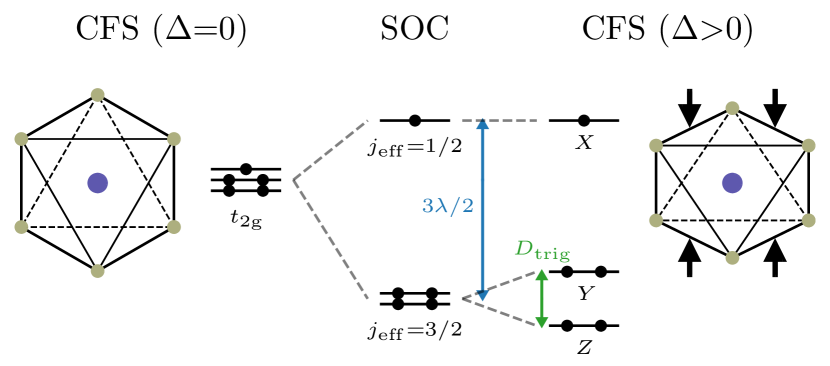
In /̄RuCl3, the Ru3+ ions are surrounded by a nearly ideal octahedral arrangement of Cl- ions (figure 1). These RuCl6 octahedra share an edge to form a honeycomb net. The octahedral cubic crystal field, , splits the Ru levels into lower and upper manifolds. It is believed that SOC further splits the manifold into upper and lower states. The electronic configurations of the Ru ions should take on a low-spin state, filling up the level completely leaving a half-filled level and leading to the so-called pseudospin. The RuCl6 octahedra however have a slight trigonal distortion which might quench the oribital angular momentum and destroy the nature of /̄RuCl3.
X-ray absorption spectroscopy (XAS) is a useful tool to shed light on the influence of SOC on electronic structure. XAS measurements by Plumb et al [12] on /̄RuCl3 at the Ru L2 () and L3 () edges observed a difference in their lineshape and found an anomalously large L3/L2 intensity ratio, the so-called branching ratio (BR). Assuming nonnegligible SOC, atomic dipole transitions must follow the selection rules. Therefore at the L2 edge, is allowed and is forbidden, while at the L3 edge, and are both allowed. This is manifested in the XAS measurements of Plumb et al, where they find the L2 absorption edge has a single peak, compared to double peaks at the L3 edge. Of these two peaks, the lower (higher) energy one is due to () states and correspondingly has a lower (higher) intensity because of its one (four) hole(s). The absence of a peak at the L2 edge is because it acquires a character due to SOC. The BR is one way of expressing this effect of SOC, where a BR is expected with SOC effects due to a reduction of dipole-allowed transitions for the L2 edge as explained above. BR is typical without SOC since the L3 edge has twice as many electrons available as the L2 edge: is a quartet and is a doublet. Plumb et al reported a BR for /̄RuCl3.
We performed XAS at the Ru M2 () and M3 () edge in total electron yield (TEY) mode, shown as a black line in figure 2(b). The first noticeable difference is that the - double peak structure is not visible at the M3 edge. The M edges correspond to shallower core holes than the L edges, i.e. lower binding energy, and therefore have shorter lifetimes. The decreased lifetime leads to broadening in the energy domain which make it appear as a single peak. Nonetheless, the peak appears asymmetric and has a slight shoulder near 460.5 eV, which with the main peak at 462.5 eV would give an estimate of the splitting between and of eV with a large error bar. In comparison, a splitting of eV was observed in the L-edge XAS data [12]. We also roughly estimated the BR by subtracting a linear background at each peak and integrating the intensity. We found BR which is consisent with the BR from L-edge XAS [12].
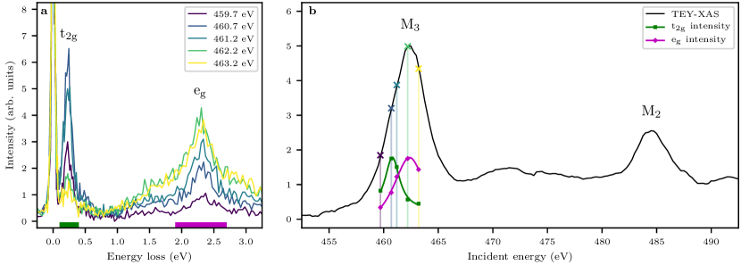
We first studied the resonant behavior of RIXS by scanning the incident energy as shown in figure 2(a). We measured RIXS spectra at five different energies, which are also shown relative to the TEY-XAS signal as vertical lines with the same color in figure 2(b). The Ru M3-edge RIXS directly probes the crystal-field split Ru levels, the so-called excitations. The two dipole transitions, followed by , allow dipole-forbidden transitions from occupied states to empty and states. The resonant enhancement is indeed strong, over an order of magnitude, and we find two different resonance regimes. The excitations to unoccupied states correspond to the spectral weight below eV while excitations to states occur above this energy. We quantitatively studied this resonant behavior by integrating the spectral weight in each region. The integration ranges are shown as solid bars at the bottom of figure 2(a): the region in green from 0.1 to 0.4 eV and the region in magenta from 1.9 to 2.7 eV. The integrated intensities are plotted in arbitrary units in figure 2(b) as green squares for and magenta diamonds for (the five points are fit with a Lorentzian function and constant background as shown by the corresponding solid lines). The and states resonate at approximately 460.8 eV and 462.4 eV respectively. The eV difference between these energies corresponds roughly to the octahedral crystal-field energy, estimated to be eV earlier.
To search for splitting due to trigonal distortions we focused on the region. Therefore, we measured a high statistics spectrum with eV, i.e. at the resonance of the states. In figure 3, we show a high-resolution Ru M3-edge RIXS spectrum on /̄RuCl3 which has many features visible in the energy loss region: three low-energy features A1, A2, and A3 (zoom in figure 4) at meV, meV, and meV; a charge gap around 1 eV; and, high-energy features , , B, , and above 1 eV. The features have been labeled with the same notation as Sandilands et al [26] with the addition of the B peak.
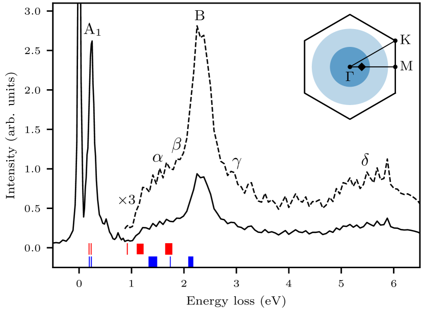
The energies of the excitations are consistent with those found with optical spectroscopy [26] and quantum chemistry calculations [27]. The quantum chemistry calculation results are indicated in figure 3. Yadav and coworkers calculated transition energies using multireference configuration-interaction (MRCI) calculations with SOC for two different structures: [17] shown in red and [19] shown in blue. Besides the B peak, the other high-energy RIXS features, , , and , are broad and difficult to compare with calculations. Nonetheless, the , , and B peaks correspond well with predicted energies for the structure, although the RIXS measurements are at a slightly higher energy. On the other hand, the structure predicts energies which are far too low compared to the RIXS data. Recent structural studies seem to agree on the fact that the room temperature structure is described by the symmetry [19, 28, 29]. Our measurements performed at 300 K clearly agrees with the structure prediction.
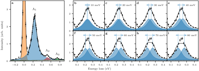
The low-energy features of the RIXS spectrum are shown in figure 4(a). The A1 peak is a excitation from to , and is therefore also known as a spin-orbit exciton [30]. Ignoring trigonal distortions, the A1 peak position corresponds to the energy difference between these levels, meV. The splitting between these levels is equal to (figure 1) from which we find a SOC constant of meV. There is no apparent energy splitting due to trigonal distortion — the A1 excitation is best fit with a single Lorentzian peak. The low energy region was fit with four peaks as shown in figure 4(a). The elastic line (orange), A2 (red), and A3 (green) peaks were fit with Gaussian functions, while the A1 peak (blue) was fit with a Lorentzian function. The fit also included a background function, shown as a dashed black line, which included a constant offset with a linear slope only in the energy loss region.
We also performed a series of fits of the A1 feature using two peaks with fixed splitting, shown in figure 4(b–i), to estimate an upper bound on the trigonal splitting (figure 1). Each panel corresponds to a fit using a fixed ranging from 10 to 80 meV. The relative energy position of the peaks is fixed by this splitting, however they are free to move in energy together during the fit. Both peaks are described by a pseudo-Voigt function with the same intensity, width, and Lorentzian/Gaussian ratio, however these values are fit for each of the individual values. We notice the fit diverging from our data at meV which we define as our upper bound.
The energy difference between the trigonally split levels is given by , however this is not equivalent to the trigonal field energy . Furthermore, the sign of both of these values depends on whether it is trigonal compression (, ) or elongation (, ). In figure 1 we show the case of trigonal compression where the energy level of is above , while in the case of trigonal elongation the opposite would be true. Our current measurements cannot differentiate between compression or elongation, therefore we have to estimate separately for each case. The splitting between the levels is given by , where [25]. From this we find for compression meV and for elongation meV
4 Discussion
We have confirmed that the trigonal splitting in /̄RuCl3 is small enough, such that the energy hierarchy needed for a ground state is satisfied: (0.065 eV) (0.154 eV) (2 eV). Our results are consistent with the MRCI calculations which predict a splitting of meV for the structure. Ru L-edge XAS reports a vanishingly small linear dichroism effect corresponding to meV [21]. It is interesting to note this report of trigonal elongation is at odds with structural studies [19, 28, 29] which show trigonal compression. Regardless of the sign of , the small magnitude found in this study and by Agrestini et al indicates the factor should be nearly isotropic.
The limit meV is likely an overestimation since our energy resolution was 27 meV and we observed no splitting of the spin-orbit exciton. The use of equal intensity peaks for our fitting in figure 4(b–i) is an approximation, the peaks should actually have different relative intensities depending on the RIXS matrix elements. Therefore, future high-resolution Ru M3-edge RIXS studies on /̄RuCl3 can use different polarizations and incident angles to determine more accurately the magnitude and perhaps even the sign of [25, 31].
Optical spectroscopy observes low-energy features at similar energies as our RIXS results: A meV, A meV, and A meV at room temperature [26, 32]. Additionally, an extremely sharp peak was observed with Raman spectroscopy at A meV ( meV) [26]. The A0 peak was initially attributed as the spin-orbit exciton, however it is at lower energy than our RIXS results and MRCI calculations [27]. The 2 meV width of the A0 Raman mode was a surprise since coupling with phonons is expected to broaden the spin-orbit exciton peak [33]. Furthermore, a recent Raman spectroscopy study on /̄RuCl3 finds that the A0 peak vanishes with increasing temperatures, which is unexpected behavior for a spin-orbit exciton [32]. This study also reports on a higher energy Raman mode at meV which agrees very well with our RIXS results. Inelastic neutron scattering has observed hints of an excitation around this energy region, albeit with poor statistics [18]. Curiously, the SOC constant we report here, meV, agrees almost exactly with the tabulated Ru3+ free ion value ( meV) [34].
Previous studies incorrectly assigned the A1–A3 peaks as transitions to SOC-split states [12, 26, 35]. The resonant behavior we observe, coupled with MRCI calculations [27], demonstrates that their energy is too low to correspond to excitations. The A1 peak observed with optical spectroscopy is the spin-orbit exciton but since it is dipole-forbidden it is a phonon-assited transition. Indeed, the optical peak is shifted 30–40 meV higher in energy, which is consistent with the energy of optical phonons measured in /̄RuCl3 [15]. The temperature dependence of all three peaks was studied by Sandilands et al [26], integrating the spectral weight from 0.1–0.87 eV, and was consistent with a phonon-assisted mechanism. However, the A1 peak is dominant in this region and Borgwardt et al [32] found that the A2 and A3 peak do not have a strong temperature dependence, i.e. A2 and A3 are not phonon-assisted transitions. This is shown clearly by comparing their energies ( meV and meV) to our RIXS energies (523 meV and 745 meV), which are nearly identical.
Borgwardt et al [32] interpreted these peaks as multiparticle excitations, i.e. double and triple spin-orbit excitons, but this seems unlikely. For example, the energy difference between multiples of a single excitation and a multiple excitation should correspond to a phonon energy, but meV which is higher than any observed phonon in /̄RuCl3. As well, we observe the same peaks with RIXS as optical spectroscopy strongly indicating a common origin. Multiparticle excitations are a nonlinear effect and thus their observation usually requires high-intensity photon beams. This could be possible in RIXS or Raman spectroscopy, however the infrared absorption experiment used a low-intensity lamp [26].
We are not completely certain of the true nature of these two peaks, however we believe that Ru -Cl hybridization plays a key role. One picture could be Cl Ru charge-transfer type excitations. For example, recent density functional theory calculations found two sharp peaks in the DOS with significant Ru-Cl hybridization at 590 meV and 730 meV above the Fermi level [21].
To our knowledge, this is the first report of M-edge RIXS on a transition metal system. Actually, there are very few reports of M-edge RIXS at all. The first M-edge RIXS experiment cleverly leveraged the low energy of the Cu M2,3 edge to achieve improved resolution on existing instrumentation [36] before the development of next-generation soft x-ray RIXS beamlines [23, 24, 37, 38, 39, 40]. Studying transition metal systems with M-edge RIXS is rare since the lifetime of the shallow core hole has a complex dependence on incident photon energy, the elastic line due to off-specular reflectivity is extremely strong and obscures low-energy features, and the inelastic cross-section is lower due to increased Auger emission [41].
These first two disadvantages are related to the eV incident energy used for M-edge RIXS of systems and not so important for systems which have higher energy M edges. In our experiment, the elastic line intensity was three times the A1 intensity, but we measured at 300 K and we found considerable quasi-elastic weight (50 meV FWHM vs. 27 meV FWHM resolution). These results are promising but further studies at higher resolutions and lower temperatures, as well as varying the scattering angle away from ∘, are important to determine if elastic intensity will be a limiting factor of M-edge RIXS in systems. The inelastic cross-section is generally lower for M vs. L edges due to the decreased fluorescence yield. For example, in Ru the fluorescence yield is and for the M3 and L3 edge respectively [42], however our results show that the resonant enhancement is still sufficient to perform experiments.
One disadvantage is that the lower energy of the M edge limits the area of the Brillouin zone which can be probed. The inset of figure 3 shows the area available with Ru M3-edge RIXS in /̄RuCl3 as dark (light) blue for ∘ (∘), while Ru L3-edge RIXS can probe a few Brillouin zones. We note that the accessible Brillouin zone in /̄RuCl3 at the M edge is still enough to search for the gapless Majorana fermions predicted by theory [43]. As well, in general the larger supercells of magnetically ordered materials will have a correspondingly smaller magnetic Brillouin zone which could possibly be probed completely at the M edge. However, the decreased SOC and lifetime of the core hole in M-edge RIXS does make it less effective at measuring magnons [44].
Nonetheless, M-edge RIXS for systems has the enormous advantage that many soft x-ray RIXS beamlines already exist or are under development which can provide high flux and sub-30 meV resolution [23, 24, 37, 38, 39, 40]. For Ru L3-edge RIXS (2840 eV) there is currently only one instrument in the world, IRIXS at P1/DESY, which is currently unavailable to general users. The latest results from IRIXS [45, 46] have a resolution approximately a factor of 5 worse than the resolution routinely available for Ru M3-edge RIXS.
5 Conclusion
We have performed Ru M3-edge resonant inelastic x-ray scattering (RIXS) on /̄RuCl3. We observe excitations in agreement with optical spectroscopy and quantum chemistry calculations. Our observation of a spin-orbit exciton allows us to extract a very accurate value for the spin-orbit coupling constant meV. The spin-orbit exciton shows no splitting due trigonal distortions and overall we find the energy hierarchy necessary for physics is satisfied in /̄RuCl3. Our results resolve some previous misconceptions about the electronic structure of /̄RuCl3 and provide a springboard for future calculations and experiments to further elucidate its nature. Measurement of systems with M-edge RIXS is a novel technique which we believe will be an important part of the x-ray spectroscopist’s toolbox since it allows ultra high-resolution RIXS measurements here and now.
References
- [1] Kitaev A 2006 Annals of Physics 321 2 – 111 ISSN 0003-4916 january Special Issue
- [2] Trebst S 2017 arXiv preprint arXiv:1701.07056
- [3] Winter S M, Tsirlin A A, Daghofer M, van den Brink J, Singh Y, Gegenwart P and Valenti R 2017 Journal of Physics - Condensed Matter 29 ISSN 0953-8984
- [4] Hermanns M, Kimchi I and Knolle J 2018 Annual Review of Condensed Matter Physics 9 17–33
- [5] Jackeli G and Khaliullin G 2009 Phys. Rev. Lett. 102(1) 017205
- [6] Chaloupka J, Jackeli G and Khaliullin G 2010 Phys. Rev. Lett. 105(2) 027204
- [7] Singh Y and Gegenwart P 2010 Phys. Rev. B 82 064412
- [8] Singh Y, Manni S, Reuther J, Berlijn T, Thomale R, Ku W, Trebst S and Gegenwart P 2012 Phys. Rev. Lett. 108(12) 127203
- [9] Modic K A, Smidt T E, Kimchi I, Breznay N P, Biffin A, Choi S, Johnson R D, Coldea R, Watkins-Curry P, McCandless G T, Chan J Y, Gandara F, Islam Z, Vishwanath A, Shekhter A, McDonald R D and Analytis J G 2014 Nature Communications 5 4203
- [10] Takayama T, Kato A, Dinnebier R, Nuss J, Kono H, Veiga L S I, Fabbris G, Haskel D and Takagi H 2015 Phys. Rev. Lett. 114(7) 077202
- [11] Takagi H, Takayama T, Jackeli G, Khaliullin G and Nagler S E 2019 Nature Reviews Physics 1 264–280
- [12] Plumb K W, Clancy J P, Sandilands L J, Shankar V V, Hu Y F, Burch K S, Kee H Y and Kim Y J 2014 Phys. Rev. B 90(4) 041112
- [13] Banerjee A, Yan J, Knolle J, Bridges C A, Stone M B, Lumsden M D, Mandrus D G, Tennant D A, Moessner R and Nagler S E 2017 Science 356 1055–1059 ISSN 0036-8075
- [14] Do S H, Park S Y, Yoshitake J, Nasu J, Motome Y, Kwon Y S, Adroja D T, Voneshen D J, Kim K, Jang T H, Park J H, Choi K Y and Ji S 2017 Nat. Phys. 13 1079–1084
- [15] Sandilands L J, Tian Y, Plumb K W, Kim Y J and Burch K S 2015 Phys. Rev. Lett. 114(14) 147201
- [16] Kasahara Y, Ohnishi T, Mizukami Y, Tanaka O, Ma S, Sugii K, Kurita N, Tanaka H, Nasu J, Motome Y, Shibauchi T and Matsuda Y 2018 Nature 559 227–231 ISSN 1476-4687
- [17] Stroganov E V and Ovchinnikov K V 1957 Ser. Fiz. i Khim. 12 152
- [18] Banerjee A, Bridges C A, Yan J Q, Aczel A A, Li L, Stone M B, Granroth G E, Lumsden M D, Yiu Y, Knolle J, Bhattacharjee S, Kovrizhin D L, Moessner R, Tennant D A, Mandrus D G and Nagler S E 2016 Nature Materials 15 733–740
- [19] Cao H B, Banerjee A, Yan J Q, Bridges C A, Lumsden M D, Mandrus D G, Tennant D A, Chakoumakos B C and Nagler S E 2016 Phys. Rev. B 93(13) 134423
- [20] Gretarsson H, Clancy J P, Liu X, Hill J P, Bozin E, Singh Y, Manni S, Gegenwart P, Kim J, Said A H, Casa D, Gog T, Upton M H, Kim H S, Yu J, Katukuri V M, Hozoi L, van den Brink J and Kim Y J 2013 Phys. Rev. Lett. 110(7) 076402
- [21] Agrestini S, Kuo C Y, Ko K T, Hu Z, Kasinathan D, Vasili H B, Herrero-Martin J, Valvidares S M, Pellegrin E, Jang L Y, Henschel A, Schmidt M, Tanaka A and Tjeng L H 2017 Phys. Rev. B 96(16) 161107
- [22] Sears J A, Songvilay M, Plumb K W, Clancy J P, Qiu Y, Zhao Y, Parshall D and Kim Y J 2015 Phys. Rev. B 91(14) 144420
- [23] Dvorak J, Jarrige I, Bisogni V, Coburn S and Leonhardt W 2016 Review of Scientific Instruments 87 115109
- [24] Jarrige I, Bisogni V, Zhu Y, Leonhardt W and Dvorak J 2018 Synchrotron Radiation News 31 7–13
- [25] Chaloupka J and Khaliullin G 2016 Phys. Rev. B 94(6) 064435
- [26] Sandilands L J, Tian Y, Reijnders A A, Kim H S, Plumb K W, Kim Y J, Kee H Y and Burch K S 2016 Phys. Rev. B 93(7) 075144
- [27] Yadav R, Bogdanov N A, Katukuri V M, Nishimoto S, Van Den Brink J and Hozoi L 2016 Scientific reports 6 37925
- [28] Johnson R D, Williams S C, Haghighirad A A, Singleton J, Zapf V, Manuel P, Mazin I I, Li Y, Jeschke H O, Valentí R and Coldea R 2015 Phys. Rev. B 92(23) 235119
- [29] Sears J A 2017 Neutron and X-ray Diffraction Studies of Structure and Magnetism in -RuCl3 Ph.D. thesis University of Toronto
- [30] Kim J, Casa D, Upton M H, Gog T, Kim Y J, Mitchell J F, van Veenendaal M, Daghofer M, van den Brink J, Khaliullin G and Kim B J 2012 Phys. Rev. Lett. 108(17) 177003
- [31] Kim J, Daghofer M, Said A H, Gog T, van den Brink J, Khaliullin G and Kim B J 2014 Nature Communications 5 4453
- [32] Borgwardt N 2019 Optics on materials with strong spin-orbit coupling: topological insulators Bi2-xSbxTe3-ySey and the j=1/2 compounds Na2IrO3 and alpha-RuCl3 Ph.D. thesis Universität zu Köln URL https://kups.ub.uni-koeln.de/9187/
- [33] Plotnikova E M, Daghofer M, van den Brink J and Wohlfeld K 2016 Phys. Rev. Lett. 116(10) 106401
- [34] Porterfield W 2013 Inorganic Chemistry (Elsevier Science) ISBN 9780323138949
- [35] Reschke S, Mayr F, Wang Z, Do S H, Choi K Y and Loidl A 2017 Phys. Rev. B 96(16) 165120
- [36] Kuiper P, Guo J H, Såthe C, Duda L C, Nordgren J, Pothuizen J J M, de Groot F M F and Sawatzky G A 1998 Phys. Rev. Lett. 80(23) 5204–5207
- [37] Lai C H, Fung H S, Wu W B, Huang H Y, Fu H W, Lin S W, Huang S W, Chiu C C, Wang D J, Huang L J, Tseng T C, Chung S C, Chen C T and Huang D J 2014 Journal of Synchrotron Radiation 21 325–332
- [38] Brookes N, Yakhou-Harris F, Kummer K, Fondacaro A, Cezar J, Betto D, Velez-Fort E, Amorese A, Ghiringhelli G, Braicovich L, Barrett R, Berruyer G, Cianciosi F, Eybert L, Marion P, van der Linden P and Zhang L 2018 Nuclear Instruments and Methods in Physics Research Section A: Accelerators, Spectrometers, Detectors and Associated Equipment 903 175 – 192 ISSN 0168-9002
- [39] Diamond light source: I21 beamline homepage https://www.diamond.ac.uk/Instruments/Magnetic-Materials/I21.html
- [40] Max iv: Veritas beamline homepage https://www.maxiv.lu.se/accelerators-beamlines/beamlines/veritas/
- [41] Wray L A, Huang S W, Jarrige I, Ikeuchi K, Ishii K, Li J, Qiu Z Q, Hussain Z and Chuang Y D 2015 Frontiers in Physics 3 32 ISSN 2296-424X
- [42] Bambynek W, Crasemann B, Fink R W, Freund H U, Mark H, Swift C D, Price R E and Rao P V 1972 Rev. Mod. Phys. 44(4) 716–813
- [43] Halász G B, Perkins N B and van den Brink J 2016 Phys. Rev. Lett. 117(12) 127203
- [44] Ament L J P, van Veenendaal M, Devereaux T P, Hill J P and van den Brink J 2011 Rev. Mod. Phys. 83(2) 705–767
- [45] Suzuki H, Gretarsson H, Ishikawa H, Ueda K, Yang Z, Liu H, Kim H, Kukusta D, Yaresko A, Minola M, Sears J A, Francoual S, Wille H C, Nuss J, Takagi H, Kim B J, Khaliullin G, Yavas H and Keimer B 2019 Nature Materials 18 563–567 ISSN 1476-4660
- [46] Gretarsson H, Suzuki H, Kim H, Ueda K, Krautloher M, Kim B J, Yavaş H, Khaliullin G and Keimer B 2019 Phys. Rev. B 100(4) 045123