MIDLMedical Imaging with Deep Learning
\jmlrpages
\jmlryear2019
\jmlrworkshopMIDL 2019 – Extended Abstract Track
\midlauthor\NameBart R. Thomson\nametag1,2 \Emailbartrthomson@gmail.com
\addr1 Department of Technical Medicine, University of Twente, Enschede, The Netherlands
\addr2 Department of Surgical Oncology, The Netherlands Cancer Institute, Amsterdam, The Netherlands
\NameJasper Nijkamp\nametag2 \Emailj.nijkamp@nki.nl
\NameOleksandra Ivashchenko\nametag2,3 \Emailo.ivashchenko@nki.nl
\addr3 Department of Radiology, Leiden University Medical Center, Leiden, The Netherlands
\NameFerdinand van der Heijden\nametag1,2 \Emailf.vanderheijden@utwente.nl
\NameJasper N. Smit\nametag2 \Emailj.smit@nki.nl
\NameNiels F.M. Kok\nametag2 \Emailn.kok@nki.nl
\NameKoert F.D. Kuhlmann\nametag2 \Emailk.kuhlmann@nki.nl
\NameTheo J.M. Ruers\nametag1,2 \Emailt.ruers@nki.nl
\NameMatteo Fusaglia\nametag2 \Emailm.fusaglia@nki.nl
Hepatic vessel segmentation using a reduced filter 3D U-Net in ultrasound imaging
Abstract
Accurate hepatic vessel segmentation on ultrasound (US) images can be an important tool in the planning and execution of surgery, however proves to be a challenging task due to noise and speckle. Our method comprises a reduced filter 3D U-Net implementation to automatically detect hepatic vasculature in 3D US volumes. A comparison is made between volumes acquired with a 3D probe and stacked 2D US images based on electromagnetic tracking. Experiments are conducted on 67 scans, where 45 are used in training, 12 in validation and 10 in testing. This network architecture yields Dice scores of 0.740 and 0.781 for 3D and stacked 2D volumes respectively, comparing promising to literature and inter-observer performance (Dice = 0.879).
keywords:
Deep learning, Segmentation, Liver, Ultrasound, U-Net1 Introduction
Surgical resections, when compared to other treatment plans, provide the best patient outcome for various types of liver malignancies [Kanas et al.(2012)Kanas, Taylor, Primrose, Langeberg, Kelsh, Mowat, Alexander, Choti, and Poston]. Due to high complexity and inter-patient variability of underlying hepatic vascular anatomy, planning and execution of safe resection is challenging in surgery. Therefore, repetitive intraoperative imaging is required to monitor surgery progress and assess the tumour-vessel relationship in 3D. Currently, US is the only imaging modality that is widely accepted and integrated into a surgical workflow. Therefore, ultrasonography is the most suitable imaging modality for intraoperative visualisation of hepatic vasculature.
Despite many advantages of intraoperative ultrasound, it is still a primary 2D imaging modality, which complicates precise localization of each 2D image in 3D for a surgeon. An interactive visualization of automatically segmented vasculature in 3D would have been of great value, yet challenging due to the complexity of US segmentation [Zhu et al.(2011)Zhu, Zhang, Shao, Cheng, Zhang, and Bai, Noble and Boukerroui(2006)]. In this work, an attempt to alleviate these challenges using 3D ultrasound imaging, in conjunction with vasculature segmentation has been proposed.
Other studies reported mean segmentation scores with a Dice of 0.5 [Wei et al.(2019)Wei, Xu, Alpers, Tianbao, Wang, Rak, and Hansen] and an intersection over union (IoU) of 0.696 [Mishra et al.(2018)Mishra, Chaudhury, Sarkar, Manohar, and Soin] when segmenting vasculature on 2D images, using a 2D U-net and a simple convolutional neural network combined with k-means clustering respectively. 3D information has been shown to improve performance in biomedical volumetric segmentation [Çiçek et al.(2016)Çiçek, Abdulkadir, Lienkamp, Brox, and Ronneberger] and can be acquired with a 3D US probe or by stacking 2D US images based on electromagnetic tracking.
In this study, a reduced filter 3D U-Net, chosen due to its popularity in medical image segmentation [Litjens et al.(2017)Litjens, Kooi, Bejnordi, Setio, Ciompi, Ghafoorian, Van Der Laak, Van Ginneken, and Sánchez], is proposed to achieve accurate vessel segmentation in true 3D (figure 1a) and stacked 2D (figure 1b & 1c) US images.
2 Materials and methods
The dataset contained 37 3D US scans and 30 2D acquired volumes, stacked based on electromagnetic tracking (Aurora Northern Digital — Ontario, Canada), data distribution is presented in table LABEL:tab:Dice. Original 3D volume sizes were and stacked 2D volumes ranged from to , depending on the zoom of the 2D slices, but were downsampled to 40% prior to training. All US imaging has been acquired intra-operatively in the NKI-AvL by 5 different observers. Each scan was delineated in 3D slicer [Kikinis et al.(2014)Kikinis, Pieper, and Vosburgh] by one out of 4 annotators, annotations have been validated with an expert radiologist. To give a sense of scale of segmentation challenges, four scans were delineated by two observers, and Dice and IoU are reported as inter-observer variation.
The 3D U-Net architecture that is used is a NiftyNet [Gibson et al.(2018)Gibson, Li, Sudre, Fidon, Shakir, Wang, Eaton-Rosen, Gray, Doel, Hu, et al.] Tensorflow implementation similar to Cicek et al. [Çiçek et al.(2016)Çiçek, Abdulkadir, Lienkamp, Brox, and Ronneberger], however with an eighth of the amount of filters in every layer, to avoid bottlenecks. Adam optimizer, with learning rate , L1 regularization with weight decay, and a batch size of 2 was used. Training was performed on four NVIDIA 1080 GTX GPUs. Twenty patches with size were used per mean value normalized volume. All volumes were padded with a volume of and were augmented by rotating (between and ), scaling (between and ) and elastically deforming (S.D. ). To reduce noise in the input images, a 3 pixel median filter was used to smooth the images. The Dice loss function was used in training of the network for 217 epochs which lasted 64 hours when there was no apparent converging of the validation loss. Segmentation accuracy was reported by means of Dice and IoU.
[5pt]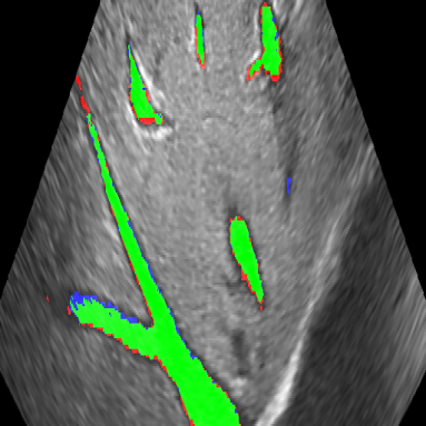 (a) Dice = 0.719
\stackunder[5pt]
(a) Dice = 0.719
\stackunder[5pt]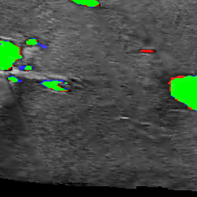 (b) Dice = 0.640
\stackunder[5pt]
(b) Dice = 0.640
\stackunder[5pt] (c) Dice = 0.861
3 Experiment and Results
The reduced filter U-Net obtained Dice scores of 0.740 () and 0.781 () for the 3D acquired and 2D stacked US images respectively compared to an inter-observer variability of 0.879 () (table LABEL:tab:Dice). IoU is reported at 0.584 (), 0.645 () and 0.785 () respectively. Figure 1 shows segmentation results for several selected cases.
tab:Dice US modality Mean Dice Mean IoU Training Validation Test Total 3D 0.740 0.584 27 7 3 37 2D 0.781 0.645 18 5 7 30 Inter-observer 0.879 0.785 4 4
4 Discussion and Conclusion
This study shows that it is possible to accurately segment hepatic vessels on US imaging with a relatively small dataset, but deviates from the inter-observer performance. Furthermore, our results seem favorable [Milko et al.(2008)Milko, Samset, and Kadir, Wei et al.(2019)Wei, Xu, Alpers, Tianbao, Wang, Rak, and Hansen], but also slightly underperform [Mishra et al.(2018)Mishra, Chaudhury, Sarkar, Manohar, and Soin] when compared to 2D segmentation literature. Overall under-segmentation of the inferior vena cava is observed, especially near the edges of the volume (figure 1b). We suspect that this is caused by incomplete vessel information (i.e. incomplete visibility of vessel cross section), strongly influencing the Dice due to its large volume. Comparing the proposed network to a network with the original amount of filters was not possible due to GPU memory limitations. The learned features between the different acquisition methods appear exchangeable as there appears no difference in segmentation performance. Whilst promising results have been demonstrated, further validation will be done by expanding the data set. In the future we will expand this methodology by discriminating between different types of vasculature (i.e. hepatic and portal vein) as well as parenchyma. Moreover, we will explore the use of these segmentations for automatic registration with a MRI model in a navigation surgery setting. These segmentations are expectantly sufficient to realize a centerline-based registration pipeline in the future.
In conclusion we demonstrate that a 3D U-Net architecture with a reduced amount of filters is able to accurately segment hepatic vasculature.
References
- [Çiçek et al.(2016)Çiçek, Abdulkadir, Lienkamp, Brox, and Ronneberger] Özgün Çiçek, Ahmed Abdulkadir, Soeren S Lienkamp, Thomas Brox, and Olaf Ronneberger. 3d u-net: learning dense volumetric segmentation from sparse annotation. In International conference on medical image computing and computer-assisted intervention, pages 424–432. Springer, 2016.
- [Gibson et al.(2018)Gibson, Li, Sudre, Fidon, Shakir, Wang, Eaton-Rosen, Gray, Doel, Hu, et al.] Eli Gibson, Wenqi Li, Carole Sudre, Lucas Fidon, Dzhoshkun I Shakir, Guotai Wang, Zach Eaton-Rosen, Robert Gray, Tom Doel, Yipeng Hu, et al. Niftynet: a deep-learning platform for medical imaging. Computer methods and programs in biomedicine, 158:113–122, 2018.
- [Kanas et al.(2012)Kanas, Taylor, Primrose, Langeberg, Kelsh, Mowat, Alexander, Choti, and Poston] Gena P Kanas, Aliki Taylor, John N Primrose, Wendy J Langeberg, Michael A Kelsh, Fionna S Mowat, Dominik D Alexander, Michael A Choti, and Graeme Poston. Survival after liver resection in metastatic colorectal cancer: review and meta-analysis of prognostic factors. Clinical epidemiology, 4:283, 2012.
- [Kikinis et al.(2014)Kikinis, Pieper, and Vosburgh] Ron Kikinis, Steve D Pieper, and Kirby G Vosburgh. 3d slicer: a platform for subject-specific image analysis, visualization, and clinical support. In Intraoperative imaging and image-guided therapy, pages 277–289. Springer, 2014.
- [Litjens et al.(2017)Litjens, Kooi, Bejnordi, Setio, Ciompi, Ghafoorian, Van Der Laak, Van Ginneken, and Sánchez] Geert Litjens, Thijs Kooi, Babak Ehteshami Bejnordi, Arnaud Arindra Adiyoso Setio, Francesco Ciompi, Mohsen Ghafoorian, Jeroen Awm Van Der Laak, Bram Van Ginneken, and Clara I Sánchez. A survey on deep learning in medical image analysis. Medical image analysis, 42:60–88, 2017.
- [Milko et al.(2008)Milko, Samset, and Kadir] Sergiy Milko, Eigil Samset, and Timor Kadir. Segmentation of the liver in ultrasound: a dynamic texture approach. International Journal of Computer Assisted Radiology and Surgery, 3(1-2):143, 2008.
- [Mishra et al.(2018)Mishra, Chaudhury, Sarkar, Manohar, and Soin] Deepak Mishra, Santanu Chaudhury, Mukul Sarkar, Sidharth Manohar, and Arvinder Singh Soin. Segmentation of vascular regions in ultrasound images: A deep learning approach. In 2018 IEEE International Symposium on Circuits and Systems (ISCAS), pages 1–5. IEEE, 2018.
- [Noble and Boukerroui(2006)] J Alison Noble and Djamal Boukerroui. Ultrasound image segmentation: a survey. IEEE Transactions on medical imaging, 25(8):987–1010, 2006.
- [Wei et al.(2019)Wei, Xu, Alpers, Tianbao, Wang, Rak, and Hansen] Wei Wei, Haishan Xu, Julian Alpers, Zhang Tianbao, Lei Wang, Marko Rak, and Christian Hansen. Fast registration for liver motion compensation in ultrasound-guided navigation, 01 2019.
- [Zhu et al.(2011)Zhu, Zhang, Shao, Cheng, Zhang, and Bai] Xinjian Zhu, Pengfei Zhang, Jinhua Shao, Yuanzhi Cheng, Yun Zhang, and Jing Bai. A snake-based method for segmentation of intravascular ultrasound images and its in vivo validation. Ultrasonics, 51(2):181–189, 2011.