MIDLMedical Imaging with Deep Learning
\jmlrpages
\jmlryear2019
\jmlrworkshopMIDL 2019 – Extended Abstract Track
\midlauthor\NameGrzegorz Chlebus\nametag1,2
\Emailgrzegorz.chlebus@mevis.fraunhofer.de
\NameNasreddin Abolmaali\midlotherjointauthor\nametag3 \EmailNasreddin.Abolmaali@klinikum-dresden.de
\NameAndrea Schenk\midlotherjointauthor\nametag1 \Emailandrea.schenk@mevis.fraunhofer.de
\NameHans Meine\nametag1,4 \Emailmeine@uni-bremen.de
\addr1 Fraunhofer Institute for Digital Medicine MEVIS, Bremen, Germany
\addr2 Diagnostic Image Analysis Group, Department of Radiology and Nuclear Medicine, Radboud
University Medical Center, Nijmegen, The Netherlands
\addr3 Department of Radiology, Städtisches Klinikum Dresden, Dresden, Germany
\addr4 University of Bremen, Medical Image Computing Group, Bremen, Germany
Relevance analysis of MRI sequences for automatic liver tumor segmentation
Abstract
Explainability of decisions made by deep neural networks is of high value as it allows for validation and improvement of models. This work proposes an approach to explain semantic segmentation networks by means of layer-wise relevance propagation. As an exemplary application, we investigate which MRI sequences are most relevant for liver tumor segmentation.
keywords:
explainability, deep learning, segmentation, MRI1 Introduction
Algorithms employing deep neural networks achieved state-of-the-art results for many computer vision tasks [Hu et al.(2018)Hu, Shen, and Sun]. Thanks to millions of parameters and nonlinear behavior deep models can learn very complex input-output dependencies allowing them to surpass human expert performance [Bejnordi et al.(2017)Bejnordi, Veta, Van Diest, Van Ginneken, Karssemeijer, Litjens, Van Der Laak, Hermsen, Manson, Balkenhol, et al.]. Understanding how such models work is of big importance because it allows to verify the reasoning of the system and possibly identify model or dataset problems [Selvaraju et al.(2017)Selvaraju, Cogswell, Das, Vedantam, Parikh, and Batra, Lapuschkin et al.(2019)Lapuschkin, Wäldchen, Binder, Montavon, Samek, and Müller]. Several approaches for classifier explainability have been proposed: guided backpropagation [Springenberg et al.(2014)Springenberg, Dosovitskiy, Brox, and Riedmiller], layer-wise relevance propagation (LRP) [Bach et al.(2015)Bach, Binder, Montavon, Klauschen, Müller, and Samek], and gradient-weighted class activation mapping [Selvaraju et al.(2017)Selvaraju, Cogswell, Das, Vedantam, Parikh, and Batra]. These methods aim at a visualization of input regions influencing the model decision and were shown to work well for image classification and reinforcement learning models [Lapuschkin et al.(2019)Lapuschkin, Wäldchen, Binder, Montavon, Samek, and Müller].
In this work, we propose a method to explain semantic segmentation models by means of LRP. We apply the proposed method to analyze the importance of input MRI sequences for the task of automatic liver tumor segmentation. Our motivation is that models requiring fewer sequences would find a broader clinical application, as not all hospitals employ the same acquisition protocols. The importance analysis would allow for an informed selection of most relevant sequences that could be used to train a segmentation model.
2 Materials and methods
2.1 Layer-wise relevance propagation
Layer-wise relevance propagation allows to obtain a pixel-wise decomposition of a model decision and is applicable for most state-of-the-art architectures for image classification and neural reinforcement learning [Bach et al.(2015)Bach, Binder, Montavon, Klauschen, Müller, and Samek]. LRP uses a notion of relevance which is equal to the model output for the output neurons and can be propagated to lower layers.
2.1.1 LRP for image classification
LRP can be employed to explain classification decisions for a given class by relevance propagation from the corresponding model output according to:
| (1) |
where refers to the layer index, to all neurons of layer , and to a relevance of neuron in layer . Typically, the relevances are propagated to the input layer () yielding a relevance map , which enables visualization of input regions influencing the model decision.
2.1.2 LRP for semantic segmentation
In order to apply LRP to semantic segmentation models, we cast the segmentation problem as a voxel-wise classification. This means that in order to explain a decision of a segmentation model for a given output region , we propose to compute the input relevance maps according to Eq. 1 for each considered output location . Then the relevance map explaining the model decision for class in the region can be calculated as:
| (2) |
We normalize by its sum to ensure that each output location equally contributes to the final relevance map .
2.2 Liver tumor segmentation model
We train and evaluate a 3D u-net [Çiçek et al.(2016)Çiçek, Abdulkadir, Lienkamp, Brox, and Ronneberger] model with a 6-channel input and 2-channel output using MRI data of 69 patients (49 training, 20 evaluation) with primary liver cancer and/or liver metastates acquired on a 3T MRI scanner (GE Healthcare, USA) at Städtisches Klinikum Dresden, Germany. Imaging data of each patient contains 6 MRI sequences: T2, non contrast enhanced T1 (plain-T1), and four dynamic contrast enhanced (DCE) T1 images acquired 20 s (T1-20s), 60 s (T1-60s), 120 s (T1-120s), and 15 min (T1-15min) after contrast agent administration (Gd-EOB-DTPA). Reference tumor segmentation was performed manually by an experienced radiologist assistant on the last DCE phase. All sequences were motion corrected using a non-rigid registration using the T1-15min image as reference [Strehlow et al.(2018)Strehlow, Spahr, Rühaak, Laue, Abolmaali, Preusser, and Schenk].
2.3 Relevance analysis of MRI sequences
We use Eq. 2 to compute a relevance map for both background () and tumor () class: . We choose regions and classified as background and tumor, respectively, to contain the same amount of voxels to prevent bias towards the more frequent background class. The importance of an input channel is corresponding to a global sum of a corresponding channel in the relevance map . As absolute values of have no meaning, we normalize such that its values sum up to 1.
3 Results and discussion
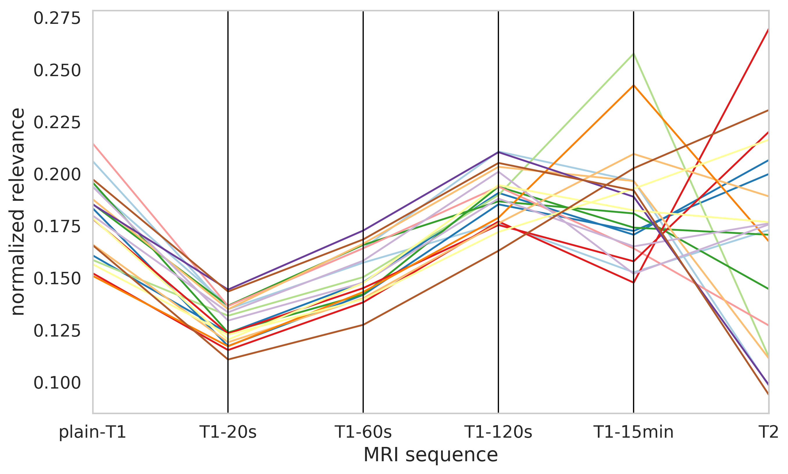
The relevance distribution across input MRI sequences for 20 test patients is shown in Fig. LABEL:fig:example. According to mean values, the most important sequence was T1-120s and the least important T1-20s. The biggest differences in attributed relevance were observed for T2 and T1-15min sequences. The relevance attribution for other sequences follows almost the same pattern for all test patients. The model learned to use information from all inputs as no sequence received a zero relevance. The T1-15min sequence, which was used to create reference segmentations, was not the most relevant for all test patients, which was contrary to our expectations.
4 Conclusion
In this work, we proposed a method to explain semantic segmentation models by means of layer-wise relevance propagation. We applied our method to analyze which MRI sequences are the most important for the task of liver tumor segmentation. Further applications of our method, for example to investigate false predictions, are future work.
fig:example
We would like to thank Maximilian Alber, Sebatian Lapuschkin, and other authors of the iNNvestigate toolbox [Alber et al.(2018)Alber, Lapuschkin, Seegerer, Hägele, Schütt, Montavon, Samek, Müller, Dähne, and Kindermans].
References
- [Alber et al.(2018)Alber, Lapuschkin, Seegerer, Hägele, Schütt, Montavon, Samek, Müller, Dähne, and Kindermans] Maximilian Alber, Sebastian Lapuschkin, Philipp Seegerer, Miriam Hägele, Kristof T Schütt, Grégoire Montavon, Wojciech Samek, Klaus-Robert Müller, Sven Dähne, and Pieter-Jan Kindermans. innvestigate neural networks! arXiv preprint arXiv:1808.04260, 2018.
- [Bach et al.(2015)Bach, Binder, Montavon, Klauschen, Müller, and Samek] Sebastian Bach, Alexander Binder, Grégoire Montavon, Frederick Klauschen, Klaus-Robert Müller, and Wojciech Samek. On pixel-wise explanations for non-linear classifier decisions by layer-wise relevance propagation. PloS one, 10(7):e0130140, 2015.
- [Bejnordi et al.(2017)Bejnordi, Veta, Van Diest, Van Ginneken, Karssemeijer, Litjens, Van Der Laak, Hermsen, Manson, Balkenhol, et al.] Babak Ehteshami Bejnordi, Mitko Veta, Paul Johannes Van Diest, Bram Van Ginneken, Nico Karssemeijer, Geert Litjens, Jeroen AWM Van Der Laak, Meyke Hermsen, Quirine F Manson, Maschenka Balkenhol, et al. Diagnostic assessment of deep learning algorithms for detection of lymph node metastases in women with breast cancer. Jama, 318(22):2199–2210, 2017.
- [Çiçek et al.(2016)Çiçek, Abdulkadir, Lienkamp, Brox, and Ronneberger] Özgün Çiçek, Ahmed Abdulkadir, Soeren S Lienkamp, Thomas Brox, and Olaf Ronneberger. 3d u-net: learning dense volumetric segmentation from sparse annotation. In International conference on medical image computing and computer-assisted intervention, pages 424–432. Springer, 2016.
- [Hu et al.(2018)Hu, Shen, and Sun] Jie Hu, Li Shen, and Gang Sun. Squeeze-and-excitation networks. In Proceedings of the IEEE conference on computer vision and pattern recognition, pages 7132–7141, 2018.
- [Lapuschkin et al.(2019)Lapuschkin, Wäldchen, Binder, Montavon, Samek, and Müller] Sebastian Lapuschkin, Stephan Wäldchen, Alexander Binder, Grégoire Montavon, Wojciech Samek, and Klaus-Robert Müller. Unmasking clever hans predictors and assessing what machines really learn. Nature communications, 10(1):1096, 2019.
- [Selvaraju et al.(2017)Selvaraju, Cogswell, Das, Vedantam, Parikh, and Batra] Ramprasaath R Selvaraju, Michael Cogswell, Abhishek Das, Ramakrishna Vedantam, Devi Parikh, and Dhruv Batra. Grad-cam: Visual explanations from deep networks via gradient-based localization. In Proceedings of the IEEE International Conference on Computer Vision, pages 618–626, 2017.
- [Springenberg et al.(2014)Springenberg, Dosovitskiy, Brox, and Riedmiller] Jost Tobias Springenberg, Alexey Dosovitskiy, Thomas Brox, and Martin Riedmiller. Striving for simplicity: The all convolutional net. arXiv preprint arXiv:1412.6806, 2014.
- [Strehlow et al.(2018)Strehlow, Spahr, Rühaak, Laue, Abolmaali, Preusser, and Schenk] Jan Strehlow, Nadine Spahr, Jan Rühaak, Hendrik Laue, Nasreddin Abolmaali, Tobias Preusser, and Andrea Schenk. Landmark-based evaluation of a deformable motion correction for dce-mri of the liver. International journal of computer assisted radiology and surgery, pages 1–10, 2018.