Spin Measurements of NV Centers Coupled to a Photonic Crystal Cavity
Abstract
Nitrogen-vacancy (NV) centers feature outstanding properties like a spin coherence time of up to one second as well as a level structure offering the possibility to initialize, coherently manipulate and optically read-out the spin degree of freedom of the ground state. However, only about three percent of their photon emission are channeled into the zero phonon line (ZPL), limiting both the rate of indistinguishable single photons and the signal-to-noise ratio (SNR) of coherent spin-photon interfaces. We here report on the enhancement of the SNR of the optical spin read-out achieved by tuning the mode of a two-dimensional photonic crystal (PhC) cavity into resonance with the NV-ZPL. PhC cavities are fabricated by focused ion beam (FIB) milling in thin reactive ion (RIE) etched ultrapure single crystal diamond membranes featuring modes with -factors of up to at mode volumes below one cubic wavelength. NV centers are produced in the cavities in a controlled fashion by a high resolution atomic force microscope (AFM) implantation technique. On cavity resonance we observe a lifetime shortening from to as well as an enhancement of the ZPL emission by almost one order of magnitude. Although on resonance the collection efficiency of ZPL photons and the spin-dependent fluorescence contrast are reduced, the SNR of the optical spin read-out is almost tripled for the cavity-coupled NV centers.
I Introduction
The nitrogen-vacancy (NV) center, a point defect in diamond consisting of a lattice vacancy and an adjacent nitrogen substitution, has attracted a lot of interest during the past years owing to its outstanding optical and spin properties.Doherty2013 The triplet ground state exhibits two sublevels attributed to two spin projections and of the NV electron spin.Doherty2011 In addition to an ultralong spin coherence time of more than one second at liquid helium temperatures,Bar-Gill2013 ; Abobeih2018 the NV center features spin-conserving optical transitions.Robledo2011 Furthermore, the electron spin may be coherently manipulated by microwave signals Jelezko2004a and purely optically initialized as well as read-out.Gruber1997 Spin initialization and read-out are enabled by a spin-selective intersystem crossing (ISC) towards the singlet system: the long lifetime in the singlet system facilitates a spin-dependent fluorescence and a preferred decay towards the ground state,Kalb2018 ; Thiering2018 allowing for fast spin initialization. Spin polarizations of about at room temperature Neumann2010 ; Robledo2011a and over at liquid helium temperature Robledo2011 may be reached, as well as spin-dependent fluorescence contrasts of up to for an optical spin read-out under non-resonant laser excitation.Dreau2011
Besides the spin-dependent fluorescence contrast, the reliability of an optical spin read-out, designated as the signal-to-noise ratio (SNR), also depends on the detected photon count rate. The SNR for an optical measurement distinguishing between the two possible spin projections and of a NV center is defined asSteiner2010
| (1) |
Here () is the expectation value for the detected photon count rate when preparing the NV center in the spin projection (). Due to the Poisson-distribution of the photon count rate we expect that the random variables and are also Poisson-distributed,Wolf2015 hence their difference is Skellam-distributed with variance .Skellam1946 As apparent in equation (1), the SNR is mainly determined by the difference . Normalized by , we obtain the already mentioned spin-dependent fluorescence contrast . With this measure we get the relation
| (2) |
As visible from equation (2), a large SNR of optical spin read-out requires both a large collected photon rate and a large contrast of spin-dependent fluorescence.Hopper2018 In view of applications, a sufficiently large SNR facilitates the use of NV centers as quantum sensors for temperature,Kucsko2013 pressure Doherty2014 as well as magnetic fields on the nanoscale Balasubramanian2008 ; Fuchs2008 ; Maze2008 ; Bernardi2017 or the verification of fundamental principles of quantum mechanics as for example demonstrated by a loophole free Bell test.Hensen2015 Furthermore, a high enough SNR in combination with the long spin coherence time enables the coupling of NV centers to nearby nuclear spins used as quantum bits Neumann2010a ; Maurer2012 ; Reiserer2016 ; Yang2016 or as building blocks of quantum repeaters.Nemoto2016 ; Rozpedek2019 In particular, for an aspired scaling of quantum systems towards quantum networks,Wehner2018 a speed-up of the entanglement generation is a central requirement and hence a preferably large SNR desirable.
The photon detection rate is usually limited by a non-perfect photon collection. Consequently, several approaches to enhance the collection efficiency have been followed by modifying the directivity of emission such as using an optimal crystal orientation,Jamali2014 fabricating solid immersion lenses around NV centers Hadden2010 ; Robledo2011 or incorporating NV centers in nanopillars,Maletinsky2012 ; Appel2016 nanowires,Babinec2010 waveguides Hausmann2012 ; Mouradian2015 or metalenses.Grote2017 Furthermore, the collection efficiency can be modified by coupling NV centers to whispering gallery Faraon2011 ; Hausmann2012 or photonic crystal (PhC) cavitiesFaraon2012 ; Hausmann2013 ; Li2015 ; Riedrich-Moeller2015 ; Schroeder2017 as well as to plasmonic structures.Choy2011 ; Bogdanov2017 In addition, by such a coupling the local density of states at the emitter’s position and hence its spontaneous emission rate may be enhanced or, correspondingly, the spontaneous emission lifetime reduced by the Purcell-factor .Purcell1946 In addition to a reduced lifetime, the coupling of NV centers to a cavity has the advantage that more than the usual of the photons are emitted into the zero phonon line (ZPL). Hence, entanglement generation by interference of ZPL photons at a beam splitter Bernien2012 ; Sipahigil2012 ; Bernien2013 ; Hensen2015 may be further sped up by coupling to a cavity featuring a mode in resonance with the NV-ZPL. However, a modification of the population dynamics, such as Purcell enhancement of emission, also influences the spin-dependent fluorescence contrast . Bogdanov et al. showed for NV ensembles in nanodiamonds, that the contrast decreases when reducing the emitter’s lifetime by coupling to plasmonic islands.Bogdanov2017 Furthermore, Babinec et al. in a theoretical study found that the spin read-out SNR achieves a maximum value for Purcell-factors on the order of .Babinec2012
In this article, we report on the SNR enhancement achieved by coupling NV centers to a two-dimensional PhC cavity. Starting from ultrapure single crystal diamond membranes bonded on a sacrificial silicon substrate and thinned by reactive ion etching (RIE), two-dimensional PhC cavities are fabricated by focused ion beam (FIB) milling at thoroughly characterized and carefully selected membrane positions.Jung2016 NV centers are subsequently incorporated into the PhC cavities by a high resolution AFM implantation technique.Pezzagna2010a A FIB-milled hole in the AFM-tip serves as an aperture, which enables the accurate implantation of nitrogen into the cavities. We activate the NV centers by extensive post processing, i. e. annealing and cleaning procedures. We here also report on the deterministic spectral tuning of a cavity mode in resonance with the NV-ZPL using thermal oxidation of diamond as well as condensation of residual gas in the cryostat. We finally experimentally measure and theoretically simulate the change in spin-dependent fluorescence contrast and SNR on resonance.
II Cavity fabrication and NV incorporation
II.1 Sample system with FIB-milled cavities
The Purcell-factor , quantifying the lifetime reduction achieved by the emitter-cavity-coupling is directly proportional to the ratio of quality factor and modal volume of a PhC cavity mode. The -factor is strongly dependent on the precision of fabrication and deviations from the design parameters. In order to produce PhC cavities with modes featuring a high -factor and a spectral position close to the NV-ZPL, diamond membranes with a precise thickness are required. However, due to polishing wedges as well as local deviations in the etching rate during RIE-thinning the thickness typically varies over the diamond film by several hundred nanometers.Jung2016 Therefore, the thinned membranes need to be characterized carefully in order to select suitable spots for the subsequent cavity production. The challenge in FIB milling of the PhC array is the fabrication of regularly hole patterns with vertical hole sidewalls. Conical shapes with typically observed inclination angles of as large as may degrade the -factor by one order of magnitude.Tanaka2003 ; Jung2016 The fabrication process and characterization methods are described in detail in our previous publication Jung2016 .
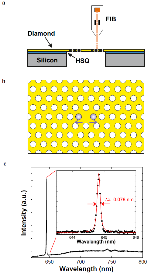
The sample system is depicted in Fig. 1a. As starting material we use thick, chemical vapor deposition (CVD) grown, (001)-oriented, ultrapure, single crystal diamond membranes (electronic grade quality, ElementSix) with a nitrogen concentration below . At first, the diamond film is etched by RIE in an -plasma to remove of the surface material as its quality is degraded due to the polishing process.Volpe2009 After acid-cleaning, the remaining thick diamond film is, mediated by a spin-coated thick layer of hydrogen silsesquioxane (HSQ XR-1541-002, DowCorning), bonded to a silicon substrate containing pre-fabricated windows. Curing the sample system at renders the bond persistent during the post-processing and cleaning procedure required after FIB milling as well as for removing damages in the diamond crystal lattice after nitrogen implantation (see section II.2). Furthermore, the stable bond allows measurements at liquid helium temperatures. The final membrane thickness of a few hundred nanometers is finally reached by further RIE thinning from topside.
The membranes are subsequently characterized by laser-scanning microscopy, cross-section measurements, and quantitative dispersive X-ray spectroscopy. The combination of these methods allows us to map the thickness of the diamond films as well as the surface structure with high resolution, enabling the selection of defect-free spots featuring a suitable thickness for the fabrication of PhC cavities.
M0-cavities generated by a shift of two adjacent holes (Fig. 1b) are fabricated by FIB milling. The hole radii of and the lattice constant of of the photonic crystal array are chosen such, that the cavity modes match the NV-ZPL position at . In addition, the M0-cavity is optimized by slight changes in position and/or radii of holes close by the point-defect.Riedrich-Moeller2010 The simulated -factor of the cavity mode is at a mode volume of .
Optimizations in the FIB milling process as for instance the use of overmilling and drift control programs, realizing a chamber pressure below and a temperature stabilization of the ion-column as well as the deposition of a metal protection layer prior to FIB milling reduce the conical hole shape to below , optimize the hole positions within the pattern and lead to sharp and well-defined hole edges. After an extended post-processing, consisting of annealing steps in vacuum at and acid-cleaning, the fabricated cavities are analyzed in a home build confocal setup. The PhC cavities feature modes with -factors up to , as depicted in Fig. 1c. Hence, the obtained -factors reach the same order of -factors of two-dimensional PhC cavities with small modal volumes (around one cubic wavelength) and a spectral mode position close to the NV-ZPL as currently observed from RIE fabrication methods.Faraon2012 ; Li2015a ; Schroeder2017
II.2 NV incorporation by AFM implantation
This section summarizes how we generate NV centers located at the field maximum of the PhC cavity.
As we used ultrapure diamond as starting material, at first nitrogen has to be implanted. In the past few years several high resolution implantation techniques were developed, most of them masking the sample surface by spin-coated photoresist films,Toyli2010 ; Spinicelli2011 Mica layers,Pezzagna2011 transferred silicon hard masks Bayn2015 as well as structured AFM-tips,Pezzagna2010a each featuring small holes as apertures. Also a maskless FIB-implanter was realized.Lesik2013 The last two techniques have the outstanding advantage, that the implantation spot can be positioned relative to previously produced nanostructures with an accuracy at the nanoscale. Whereas the lateral resolution of a nitrogen FIB implantation is about at a typical acceleration voltage of , nitrogen may be implanted through an AFM-tip with a lateral resolution of up to at a typical implantation energy of .Pezzagna2011a As precise positioning is crucial for efficient cavity coupling, we here use the AFM-tip technique.
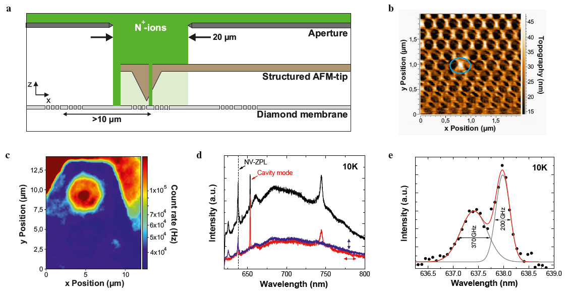
The home built AFM-setup combines a conventional low-energy ion source for generation and acceleration of nitrogen ions with an AFM-unit. A small hole is FIB-milled close to the apex of the pyramidal AFM-tip which is positioned over the implantation spot (Fig. 2a). To align the AFM-tip relative to the sample, AFM-scans are performed prior to every implantation (Fig. 2b). The lateral implantation resolution with regard to the diameter of the AFM-aperture of , the alignment accuracy of the tip to the sample of about , and the ion straggle in diamond of at the chosen implantation energy of yields a total accuracy of . Note that small straggling effects of ions at the edges of the aperture are not included in the analysis above and are under present investigation. The expected implantation depth of the nitrogen ions in -oriented diamond samples is on average Lehtinen2016 and the expected yield about .Pezzagna2010 The implantation dose aiming at one NV center per cavity is calculated to . As the generation of NV centers is a statistical process, the final implantation doses were varied between half and triple of the calculated value.
After implantation, an extensive post-processing is required in order to restore a good diamond crystal quality and to activate NV centers. At first, the samples are therefore annealed in vacuum () at for . Subsequently, oxidation of the samples at for in air atmosphere and acid cleaning ( in a tri-acid mixture of perchloric, sulfuric and nitric acid) removes graphitic residuals from the surface and provides an oxygen termination. Hence, the possibility to obtain negatively charged NV centers within the PhC cavity is enhanced.Groot-Berning2014
In Fig. 2c a typical spatially resolved photoluminescence (PL) image after post-processing is shown. Generated NV centers are localized in the PhC cavities as well as outside the area masked by the AFM-cantilever. This observation is in agreement with the results of additionally performed optically detected magnetic resonance (ODMR) measurements, which reveal a dip at frequencies around , characteristic for NV centers,Gruber1997 inside the cavities and no hints of NV centers within the surrounding PhC arrays. The cavities were further examined with PL spectroscopy at low temperatures. In Fig. 2d and 2e PL spectra of the M0-cavity with the lowest applied implantation dose are shown. Besides the cavity mode spectral lines around are visible featuring Gaussian shapes with half widths (FWHM) of and , respectively. We performed photon correlation () measurements to estimate the number of generated NV centers. As the results did not show a dip but Poissonian photon statistics we have to assume that a few NV centers were created inside the cavity volume. The deviation from the expected implantation yield might be explained by a large number of defects in the material as a consequence of the PhC fabrication process, providing a high density of vacancies for NV center creation. In addition, the emission lines might be shifted by local strain and strongly broadened by spectral diffusion due to nearby charges.Fu2010 ; Robledo2010
III Resonance Tuning
In the past few years several techniques have been developed for the deterministic tuning of cavity modes into resonance with the ZPL of color centers.
A well-established but irreversible method is the oxidation of diamond in an air or oxygen atmosphere.Riedrich-Moeller2012 ; Hausmann2013 At temperatures above the diamond surface starts to oxidize, the thickness of the diamond membrane decreases with an accompanying enlargement of the hole diameters. Larger holes as well as thinner diamond films lead to a spectral blue shift of the cavity modes. Finite Difference Time Domain (FDTD) simulations predict a blue shift of for our M0-cavity if diamond are removed from the surface. On the other hand, cavity modes have been successfully redshifted by condensing inert gases like xenon or nitrogen on the sample’s surface,Lee2014 ; Hausmann2013 ; Mosor2005 thereby increasing the sample thickness and reducing the hole radii. In contrast to the oxidation technique, a tuning by gas adsorption is a reversible process where heating the sample enables the residual-free removal of the condensed layers.Mosor2005
In the following we focus on the M0-cavity implanted with the lowest implantation dose of , featuring a cavity mode with a -factor of (spectra in Fig. 2d and 2e). At first, the oxidation technique described above, using a temperature of , is applied to tune the cavity mode to shorter wavelengths. After tuning and acid-cleaning the resulting mode position is . In a second step, the residual gas present in the sample chamber of the cryostat (Attodry 2100, Attocube) is adsorbed to the sample, allowing a redshift of the cavity mode under continuous optical control: we observed that during PL measurements at room-temperature and a pressure of cavity modes show a red shift under continuous laser excitation at . Furthermore, only the modes of a cavity directly irradiated with laser light are affected and the observed shift rates scale with the power. All these observations may be explained as follows: as the vacuum pump is attached at the topside of the cryostat, pumping leads to an efficient removal of lightweight molecules, whereas heavy gas molecules, e. g. hydrocarbons, partly remain in the sample chamber. Based on the observations we assume a light-assisted adsorption of residual gas molecules onto the sample’s surface.Mosor2005 ; Preclikova2010 Under illumination with of laser light at we observe a shift rate of the cavity modes of approximately . The -factor remains almost unchanged with after tuning. Importantly, after further pumping and cooling to liquid helium temperatures no further mode shifts are observable under laser illumination. We point out that the described tuning by gas adsorption is a reversible process. Heating the sample to in air atmosphere resets the sample to the initial state before the gas adsorption.
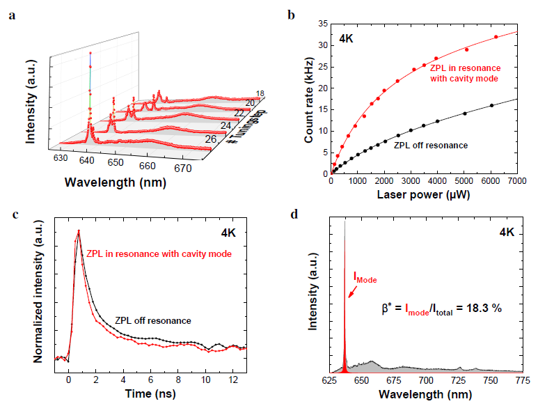
The PL spectra depicted in Fig. 3a show a strong enhancement of the ZPL intensity by tuning the cavity mode on resonance. We further observe an enhancement of the saturation count rate filtered in a wide window by a factor of from to (Fig. 3b). Furthermore, the emitter’s lifetime decreases from at a spectral mode position of to in resonance with the NV-ZPL. The recorded lifetime traces (Fig. 3c) feature a double exponential decay. The second time constant on the order of may be attributed to fast decaying fluorescence in the diamond membrane (background). Moreover, PL spectra allow for the estimation of the fraction of emission into the cavity mode as well as the resulting Purcell-factor. The deduced spontaneous emission coupling factor on resonance is (Fig. 3d). As the incorporated NV centers feature an off-resonant Debye-Waller factor of , we conclude that the emission fraction into the ZPL is strongly enhanced due to the cavity coupling.
The -factor finally enables us to calculate the total Purcell-factor to be . We modeled the total Purcell-factor on resonance by using a phonon assisted cavity coupling model presented in Albrecht2013 . With the -factor and modal volume of the cavity mode as well as the measured linewidths of the ZPL and the sideband transitions of the NV centers a Purcell-factor of is predicted. The experimentally determined value of is reduced due to a non-perfect lateral positioning of the implanted NV centers relative to the mode field, a shallow implantation depth as well as a non-perfect dipole orientation in the used (001)-oriented diamond samples. The model further correctly predicts the experimentally observed lifetime reduction.
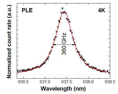
To analyze the NV-ZPL in detail despite the existing resonance with the cavity mode, a photoluminescence excitation (PLE) spectrum was recorded for wavelengths between and . In the spectrum depicted in Fig. 4 it is noticeable, that only one of the previously detected two emission lines (see Fig. 2e) remains. In the PLE spectrum this line at has a half width of , in good agreement with the value of deduced from the PL spectra. The absence of the second line is most likely due to the removal of shallow implanted NV centers during the extended tuning steps by oxidation.
IV SNR enhancement
To estimate the SNR enhancement due to the emitter-cavity-coupling we at first have to consider the change in the photon count rate . For this, we have to take into account the shortening of the emitter’s lifetime as well as the change in collection efficiency. Furthermore, for the SNR the change in spin-dependent fluorescence contrast has to be considered. To this end, a reliable methodology is required to compare the contrast before and after tuning the cavity mode in resonance with the NV-ZPL.
IV.1 Light extraction from a PhC cavity
We simulate the emission of the cavity-coupled NV centers using a finite element software (FDTD solutions, Lumerical) by modeling the emission of an electric dipole point source positioned in the mode field maximum. The simulated photonic nanostructure features the same number of holes as the produced M0-cavity where the holes have a conical shape with an inclination angle of (see section II). We calculate the emission fractions in the two half-spaces above and below the PhC as well as the collection efficiency for a microscope objective () positioned vertically above the diamond membrane. Each simulation is performed for a dipole oriented vertically to the diamond film as well as for the in-plane dipoles and . These dipoles are oriented such that the cavity mode is fed by the dipole whereas the dipole has orthogonal polarization. The orthogonality of the dipoles also allows us to calculate the emission for arbitrary dipole orientations as discussed below.
As an example we show in Fig. 5a a simulation for the dipole. We find that the collection efficiency drops to at the resonance wavelength of the cavity mode, whereas for other wavelengths within the photonic band gap collection efficiencies around are achieved.
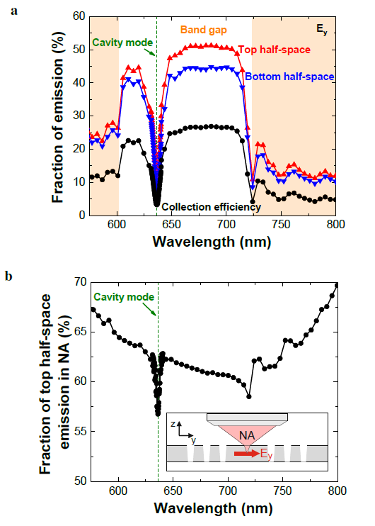
This minimum arises from a modified directional characteristic of the emitted light. Two factors play a role here: first, the fraction of photons emitted in the top half-space decreases at the mode’s resonance wavelength from values over to values below (Fig. 5a). Second, a smaller part of the photons emitted in the top half-space (below instead of over ) can be collected within the NA at the resonance wavelength of the mode (Fig. 5b).
The NV emission dipoles are oriented in the -plane of the diamond lattice. For the small NV ensemble in our experiment we average over all possible dipole orientations within the -plane. As the used diamond material features a -orientation, the -axis exhibits an angle of to the -plane (membrane plane). The projection of the averaged NV dipoles onto the or -axis amounts to and onto the -axis. To calculate the resulting collection efficiency for our NV ensemble, also the Purcell-factors for the three dipole orientations have to be considered. The emission directivity of photons emitted into the cavity mode is only determined by the mode emission directivity. Further, the emission directivity of off-resonant photons is defined by the photonic nanostructure. Considering this, we first calculated the spontaneous emission rates of the projections of the averaged NV dipoles onto the , and -axis considering the corresponding Purcell-factors as follows:
| (3) |
with the free-space spontaneous emission rate , for and for .
Subsequently, the fraction of emission produced by the averaged NV dipole projected to the , or -axis may be calculated by normalizing the spontaneous emission rates to the sum over all these rates. To finally calculate the collection efficiency at the given dipole orientation, the simulated collection efficiencies for a dipole oriented in , or -direction are weighted with the respective fraction of emission and the resulting values summed up.
For the optical read-out of the NV electron spin we can distinguish between two fundamental cases. In the first case only ZPL photons are considered. This is for instance the case, if indistinguishable photons are required. On the other hand, for projective spin read-out generally all the emitted photons are used, e. g. if NV centers are used as magnetic field sensors on the nanoscale.Balasubramanian2008 ; Fuchs2008 ; Maze2008 ; Bernardi2017 The simulations predict a collection efficiency of for the off-resonant mode position at , which is slightly reduced to in resonance with the NV-ZPL. Hence if for the spin read-out only ZPL photons are used, the collection efficiency drops by a factor of . If in the second case all emitted photons are used for the spin read-out, the collection efficiency drops only by a factor of . The reduction in the second case is smaller, as only a fraction of the emitted photons is affected by the dominant decrease of collection efficiency at the resonance wavelength of the cavity mode.
IV.2 Fluorescence contrast measurements
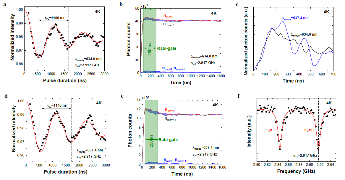
We apply a protocol composed of three succeeding measurements for the reliable and reproducible determination of the time-resolved, spin-dependent fluorescence contrast of the cavity-coupled NV centers. For the considered off-resonant spectral mode position at at first a ODMR-measurement under continuous laser and microwave excitation is performed. An external magnetic field of is applied to split the two ground state sublevels and . Hence, in the ODMR-spectrum in Fig. 6f, two dips are visible belonging to the resonance frequencies of the ground state transitions from to and respectively. In the following we only focus on the transition to the sublevel featuring a resonance frequency of . For all presented measurements the optical excitation of the NV centers coupled to the M0-cavity is carried out in the PL saturation regime.
To determine the population inversion time a Rabi measurement on the transition to is performed (Fig. 6a). From the observed damped Rabi oscillations a -time of and a spin coherence time of is deduced. The value of is in accordance with typical spin coherence times observed for shallow NV centers in nanophotonic structures based on single crystal diamond.Appel2015 This indicates that our PhC fabrication process does not adversely influence the NV spin coherence properties.
In Fig. 6b we present the time-resolved, spin-dependent fluorescence. Here, after spin initialization a resonant microwave -pulse is applied, resulting in a preparation of the NV centers in the state. During the spin read-out a wide photon detection gate is temporally shifted. For each gate position the measurement is repeated for a NV preparation in the state and the difference of the two fluorescence curves, the time-resolved fluorescence contrast, calculated. As expected, the fluorescence curves at first rise for both spin states whereas the count rate for the state (bright state) is generally higher as for the state (dark state) with a maximum contrast of . With progressing laser excitation time both fluorescence curves converge towards a joint value, indicating an equilibrium, spin-mixed state. Whereas the fluorescence contrast measured on single NV centers under zero magnetic field can reach values of Rondin2014 to Dreau2011 , for NV ensembles in a magnetic field contrasts of only a few percent are expected.Chipaux2015 Possible reasons are lattice and surface defects introduced by FIB milling, impairing charge state stability and spin coherence of the shallow implanted emitters.Ofori-Okai2012 ; Chipaux2015 Furthermore, a strain gradient may locally alter the ground state splitting. Hence, different emitters of the considered small NV ensemble may feature slightly different resonance frequencies.
Subsequently, the cavity mode is tuned into resonance with the NV-ZPL (see section III). As for the tuning process the sample has to be dismounted from the cryostat, a repositioning of the microwave antenna, an air suspended gold wire loop mounted on a positioner, is required after remounting the sample. To establish comparable experimental conditions we position the antenna in such a way that a -time of is reached (Fig. 6d), in good agreement with the -time of (Fig. 6a) for the NV-ZPL off resonance with the cavity mode. We follow the same protocol as in the off-resonant case to measure Rabi oscillations (Fig. 6d) as well as fluorescence curves (Fig. 6e) on resonance. When comparing the resulting time-resolved fluorescence contrast on- and off-resonance (Fig. 6c), we find a small reduction of contrast of on resonance (integrated over the entire time interval in Fig. 6c).
IV.3 Rate equation model
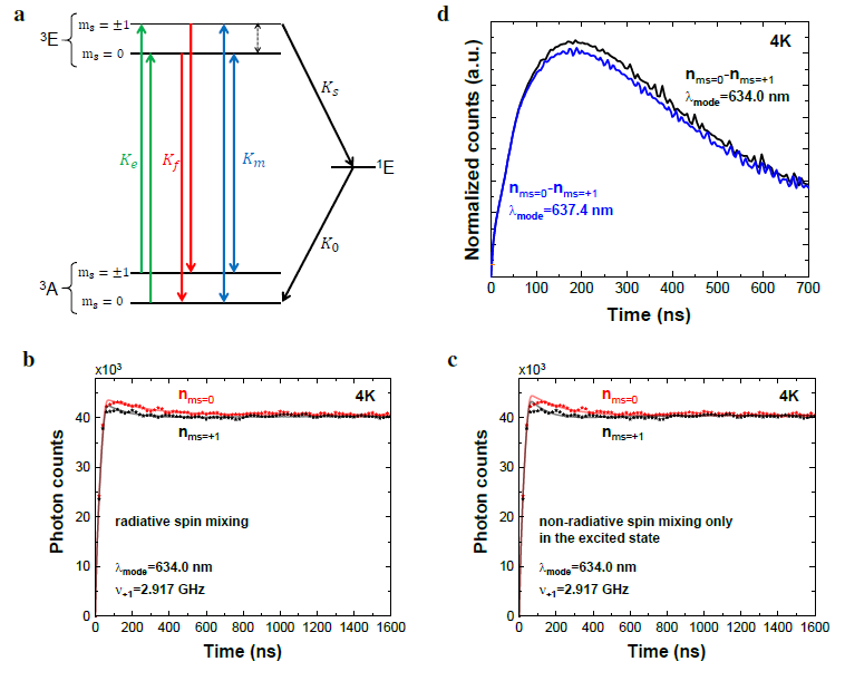
To analyze the measured spin-dependent fluorescence contrast we set up a rate equation model following Wolf et al. .Wolf2015 In this model a NV center is adopted as a five level system (Fig. 7a), consisting of the two ground states and , respectively, the corresponding excited states and a long-living singlet state. For the modeling we assume that the excitation rates out of the two ground states are identical for the non-resonant laser excitation.Manson2006 Furthermore it is assumed that the emission rates from the excited to the ground states of the triplet system are spin independent.Goldman2015 In addition, the transition rate from the excited state to the singlet state is neglected as this rate is four orders of magnitude smaller than the corresponding transition rate for the state.Young2009 ; Goldman2015 ; Kalb2018 As a further assumption the transition rate from the singlet state to the ground state is neglected, because the rate to the ground state is about six times larger.Thiering2018 ; Kalb2018 By reason of these assumptions a spin-mixing, leading to a statistical mixture of and states due to the spin-selective intersystem crossing is already included in the model. Such a spin-mixing was both theoretically predicted Wolf2015 ; Bogdanov2017 ; Thiering2018 and experimentally confirmed.Kalb2018
If, however, the only spin-mixing process was due to ISC, the modeled population would end up in the state for sufficient long-lasting laser excitation and the modeled fluorescence would thus be maximized. Instead, the spin-dependent fluorescence curves in our experiment (Fig. 6b and 6e) as well as for other NV centers (see e. g. Steiner2010 ; Robledo2011a ) show a convergence of the fluorescence and hence the populations towards a steady state. Therefore a further spin-mixing has to be included in the model. Wolf et al. propose as additional spin-mixing mechanisms on the one hand a radiative spin-mixing between the excited and ground states of the triplet system and on the other hand a purely non-radiative spin-mixing between the excited states.Wolf2015 As can be seen in Fig. 7b and 7c, respectively, the measured data may be fitted well under the assumption of a radiative spin-mixing and, in particular, better than under the assumption of a non-radiative spin-mixing in the excited state. However, Kalb et al. demonstrated that a radiative spin-mixing occurs only with a probability of well below and hence should not be the dominant spin-mixing mechanism in our experiments.Kalb2018 Instead, the reason for the additionally observed spin-mixing mechanism is that in our experiments we apply an external magnetic field to split the ground states of the NV centers (see section IV.2). As the diamond sample features a -surface and the magnetic field an orientation vertical to the diamond’s surface, the magnetic field exhibits an angle of here to the magnetic dipole axis of the NV centers. As now both ground and excited states consist of superpositions of bare, zero-field spin states, optical transitions with spin flips become allowed resulting in an effective radiative spin mixing.Tetienne2012 Furthermore, the rates of ISC transitions are modified. Therefore, in summary, the data are fitted appropriately under the assumption of a spin-mixing due to ISC and by radiative transitions in the triplet system.
In the following we assume for simplicity that the spin-mixing induced by the off-axis magnetic field is the same for excitation and emission.
With this, the following rate equations are set up, determining the internal dynamics of a NV center:
| (4) | |||||
and are the populations of the ground states and , and the populations of the corresponding excited states and the population of the singlet state. and are the spin-preserving excitation and emission rates, the transition rate from the excited -state to the singlet state, the transition rate from the singlet state to the ground state and the rate of the radiative spin-mixing induced by the off-axis magnetic field. As the measurements are performed in the PL saturation regime, the relation holds in the following.
According to the measured off-resonant lifetime value of , a decay rate of is set in the model. With the parameters and fixed, the rate equations are solved and the simulated time-resolved fluorescence is fitted to the measured curves by varying the free parameters , and (Fig. 7b). From this fit a spin-mixing rate of , a transition rate to the singlet state of and a transition rate to the ground state of are determined.
These rates allow us to theoretically predict the time-resolved, spin-dependent fluorescence contrast on resonance. Fig. 7d shows the fluorescence contrast simulated by the rate equation model for the cavity mode on and off resonance with the NV-ZPL. The theoretically predicted fluorescence contrast on resonance is slightly lower than for the off-resonant case. This is expected, as the emission rate scales with the Purcell-factor and the probability for spin-mixing is the larger the more fluorescence cycles occur per time unit. Depending on the position as well as the size of the temporal read-out window, a reduction of the fluorescence contrast by tuning the mode into resonance with the NV-ZPL of up to is theoretically predicted. For a realistic read-out photon gate of , as for instance typically used for the acquisition of Rabi measurements, the fluorescence contrast would be reduced by . Such a reduction is conformable with the experimental results (see section IV.2).
IV.4 Implications for the SNR
We now discuss the modification of the spin measurement SNR induced by the modified photon collection and modified spin-dependent fluorescence contrast due to the cavity coupling. With equation (1) as well as the appropriate assumption of small contrasts, we arrive at:
| (5) |
where variables with (without) a star indicate the on- (off-) resonant case. Based on the presented measurements as well as the theoretical modeling we assume in the following a reduction of the spin-dependent fluorescence contrast by , i. e. . Due to the lifetime reduction the number of emitted photons is increased by a factor of , when the mode is tuned into resonance with the NV-ZPL. If we, in a first scenario, consider only ZPL photons for spin read-out, the collection efficiency is reduced by a factor of (see section IV.1). Furthermore, the fraction of photons emitted into the ZPL is modified from off resonance to on resonance (see Fig. 3d), resulting in an enhancement factor of . The total estimated enhancement of the collected photons by applying a narrow-band photon detection around the ZPL is hence and the SNR, also taking the contrast reduction of into account, thus enhanced by a factor of . As a result, the SNR is almost tripled by tuning the mode into resonance with the NV-ZPL. If we, in a second scenario, consider the collection of all photons for the spin read-out, we cannot benefit from the higher emission fraction into the ZPL any more, but the number of emitted photons is still enhanced by a factor of due to the lifetime shortening on resonance. The collection efficiency is here reduced by a factor of (see section IV.1). Altogether the number of detected photons by applying a broad-band photon detection is enhanced by a factor of , resulting in a very small SNR enhancement of about . In summary, the cavity coupling of NV centers as demonstrated here leads to a spin read-out SNR enhancement of up to a factor of . This factor is within the same order of magnitude as the SNR enhancement achieved by other methods for improving the photon collection efficiency.Jamali2014 ; Robledo2011 ; Steiner2010
V Conclusions
In summary, we reported on the SNR enhancement of the optical spin read-out achieved by tuning the mode of a two-dimensional PhC cavity into resonance with the NV-ZPL. To achieve this, ultrapure -oriented CVD-grown diamond films were used as starting material for the RIE fabrication of thin, air-suspended membranes. An extended characterization allowed us to select defect-free spots featuring a suitable thickness for the subsequent fabrication of PhC cavities by FIB milling. The analyzed cavity modes showed -factors of up to at mode volumes of less than one cubic wavelength. The application of a high-resolution implantation technique using a pierced AFM-tip allowed the subsequent generation of NV centers in the cavities. The combination of two spectral tuning methods, an oxidation technique for the blue shift and a gas adsorption technique for the red shift, facilitated the reliable and precise tuning of a cavity mode. For the considered cavity-coupled NV centers the SNR was almost tripled. A theoretical model, taking into account the measured shortening of the emitter’s lifetime, the measured and theoretically predicted change in fluorescence contrast as well as the simulated modification of the collection efficiency reproduces the experimental findings very well.
Whereas the reported SNR enhancement is on par with simpler methods for photon collection enhancement,Jamali2014 ; Steiner2010 ; Robledo2011 it could be still increased by a further optimized cavity coupling: a -oriented diamond sample with an optimal dipole orientation may lead to a fourfold enhancement of the total Purcell-factor. For an optimal implantation depth in the center of the diamond film, instead of the shallow implantation, the Purcell-factor may be further increased by a factor of . In total, for an optimal cavity coupling our theoretical model predicts a SNR enhancement by a factor of more than . A possible method to reach this is a change of the fabrication order such that at first NV centers are implanted into the diamond film and subsequently a PhC cavity is fabricated around. This would allow a maskless implantation with higher energies resulting in deeper implanted NV centers. Subsequently, NV centers could be pre-characterized and suitable single emitters chosen, featuring an ideal dipole orientation. Alternatively, also improved nanoimplantation techniques such as a maskless FIB-implantation of ions with high lateral resolution are in reach.Schroeder2017a Eventually, the here generated NV centers feature an optical linewidth of several hundred GHz, whereas narrow line widths below are accessible,Ruf2019 at least in m-thin RIE-etched diamond membranes. Smaller linewidths would further increase the total Purcell-factor. Therefore, in conclusion, higher SNR enhancements are within reach with the method presented here.
Acknowledgements.
We thank B. Lägel and S. Wolff (Nano Structuring Center, University of Kaiserslautern) for helpful discussions on nano-fabrication and use of their facilities. We further thank Alexander Huck and Simeon Bogdanov for helpful discussions on fluorescence contrast measurements and modeling. This research has been partially funded by the European Quantum Technology Flagship Horizon 2020 (H2020-EU1.2.3/2014-2020) under Grant No. 820394 (ASTERIQS). EU funding for the project AME-Lab (European Regional Development Fund C/4-EFRE 13/2009/Br) for the FIB/SEM is acknowledged. E. Neu acknowledges funding via the NanoMatFutur program of the German Ministry of Education and Research (BMBF) under Grant No. FKZ13N13547 as well as a PostDoc Fellowship by the Daimler and Benz Foundation.References
- (1) M. W. Doherty, N. B. Manson, P. Delaney, F. Jelezko, J. Wrachtrup and L. C. L. Hollenberg. The nitrogen-vacancy colour centre in diamond. Physics Reports - Review Section of Physics Letters, 528, 1–45, 2013.
- (2) M. W. Doherty, N. B. Manson, P. Delaney and L. C. L. Hollenberg. The negatively charged nitrogen-vacancy centre in diamond: the electronic solution. New Journal of Physics, 13, 025019, 2011.
- (3) N. Bar-Gill, L. Pham, A. Jarmola, D. Budker and R. Walsworth. Solid-state electronic spin coherence time approaching one second. Nature Communications, 4, 1743, 2013.
- (4) M. H. Abobeih, J. Cramer, M. A. Bakker, N. Kalb, M. Markham, D. J. Twitchen and T. H. Taminiau. One-second coherence for a single electron spin coupled to a multi-qubit nuclear-spin environment. Nature Communications, 9, 2552, 2018.
- (5) L. Robledo, L. Childress, H. Bernien, B. Hensen, P. Alkemade and R. Hanson. High-fidelity projective read-out of a solid-state spin quantum register. Nature, 477, 574–578, 2011.
- (6) F. Jelezko, T. Gaebel, I. Popa, A. Gruber and J. Wrachtrup. Observation of Coherent Oscillations in a Single Electron Spin. Physical Review Letters, 92, 076401, 2004.
- (7) A. Gruber, A. Dräbenstedt, C. Tietz, L. Fleury, J. Wrachtrup and C. v. Borczyskowski. Scanning Confocal Optical Microscopy and Magnetic Resonance on Single Defect Centers. Science, 276, 2012–2014, 1997.
- (8) N. Kalb, P. C. Humphreys, J. J. Slim and R. Hanson. Dephasing mechanisms of diamond-based nuclear-spin memories for quantum networks. Physical Review A, 97, 062330, 2018.
- (9) G. Thiering and A. Gali. Theory of the optical spin-polarization loop of the nitrogen-vacancy center in diamond. Physical Review B, 98, 085207, 2018.
- (10) P. Neumann, R. Kolesov, B. Naydenov, J. Beck, F. Rempp, M. Steiner, V. Jacques, G. Balasubramanian, M. L. Markham, D. J. Twitchen, S. Pezzagna, J. Meijer, J. Twamley, F. Jelezko and J. Wrachtrup. Quantum register based on coupled electron spins in a room-temperature solid. Nature Physics, 6, 249–253, 2010.
- (11) L. Robledo, H. Bernien, T. van der Sar and R. Hanson. Spin dynamics in the optical cycle of single nitrogen-vacancy centres in diamond. New Journal of Physics, 13, 025013, 2011.
- (12) A. Dréau, M. Lesik, L. Rondin, P. Spinicelli, O. Arcizet, J.-F. Roch and V. Jacques. Avoiding power broadening in optically detected magnetic resonance of single NV defects for enhanced dc magnetic field sensitivity. Physical Review B, 84, 195204, 2011.
- (13) M. Steiner, P. Neumann, J. Beck, F. Jelezko and J. Wrachtrup. Universal enhancement of the optical readout fidelity of single electron spins at nitrogen-vacancy centers in diamond. Physical Review B, 81, 035205, 2010.
- (14) S. A. Wolf, I. Rosenberg, R. Rapaport and N. Bar-Gill. Purcell-enhanced optical spin readout of nitrogen-vacancy centers in diamond. Physical Review B, 92, 235410, 2015.
- (15) J. G. Skellam. The frequency distribution of the difference between two Poisson variates belonging to different populations. Journal of the Royal Statistical Society Series A, 109, 296, 1946.
- (16) D. A. Hopper, R. R. Grote, S. M. Parks and L. C. Bassett. Amplified Sensitivity of Nitrogen-Vacancy Spins in Nanodiamonds Using All-Optical Charge Readout. ACS Nano, 12, 4678–4686, 2018.
- (17) G. Kucsko, P. C. Maurer, N. Y. Yao, M. Kubo, H. J. Noh, P. K. Lo, H. Park and M. D. Lukin. Nanometre-scale thermometry in a living cell. Nature, 500, 54–58, 2013.
- (18) M. W. Doherty, V. V. Struzhkin, D. A. Simpson, L. P. McGuinness, Y. Meng, A. Stacey, T. J. Karle, R. J. Hemley, N. B. Manson, L. C. L. Hollenberg and S. Prawer. Electronic Properties and Metrology Applications of the Diamond Center under Pressure. Physical Review Letters, 112, 047601, 2014.
- (19) G. Balasubramanian, I. Y. Chan, R. Kolesov, M. Al-Hmoud, J. Tisler, C. Shin, C. Kim, A. Wojcik, P. R. Hemmer, A. Krüger, T. Hanke, A. Leitenstorfer, R. Bratschitsch, F. Jelezko and J. Wrachtrup. Nanoscale imaging magnetometry with diamond spins under ambient conditions. Nature, 455, 648–651, 2008.
- (20) G. D. Fuchs, V. V. Dobrovitski, R. Hanson, A. Batra, C. D. Weis, T. Schenkel and D. D. Awschalom. Excited-State Spectroscopy Using Single Spin Manipulation in Diamond. Physical Review Letters, 101, 117601, 2008.
- (21) J. Maze, P. Stanwix, J. Hodges, S. Hong, J. Taylor, P. Cappellaro, L. Jiang, M. Dutt, E. Togan, A. Zibrov, A. Yacoby, R. Walsworth and M. Lukin. Nanoscale magnetic sensing with an individual electronic spin in diamond. Nature, 455, 644–647, 2008.
- (22) E. Bernardi, R. Nelz, S. Sonusen and E. Neu. Nanoscale Sensing Using Point Defects in Single-Crystal Diamond: Recent Progress on Nitrogen Vacancy Center-Based Sensors. Crystals, 7, 124, 2017.
- (23) B. Hensen, H. Bernien, A. E. Dréau, A. Reiserer, N. Kalb, M. S. Blok, J. Ruitenberg, R. F. L. Vermeulen, R. N. Schouten, C. Abellán, W. Amaya, V. Pruneri, M. W. Mitchell, M. Markham, D. J. Twitchen, D. Elkouss, S. Wehner, T. H. Taminiau and R. Hanson. Loophole-free Bell inequality violation using electron spins separated by 1.3 kilometres. Nature, 526, 682–686, 2015.
- (24) P. Neumann, J. Beck, M. Steiner, F. Rempp, H. Fedder, P. R. Hemmer, J. Wrachtrup and F. Jelezko. Single-Shot Readout of a Single Nuclear Spin. Science, 329, 542–544, 2010.
- (25) P. C. Maurer, G. Kucsko, C. Latta, L. Jiang, N. Y. Yao, S. D. Bennett, F. Pastawski, D. Hunger, N. Chisholm, M. Markham, D. J. Twitchen, J. I. Cirac and M. D. Lukin. Room-Temperature Quantum Bit Memory Exceeding One Second. Science, 336, 1283–1286, 2012.
- (26) A. Reiserer, N. Kalb, M. S. Blok, K. J. M. van Bemmelen, T. H. Taminiau, R. Hanson, D. J. Twitchen and M. Markham. Robust Quantum-Network Memory Using Decoherence-Protected Subspaces of Nuclear Spins. Physical Review X, 6, 021040, 2016.
- (27) S. Yang, Y. Wang, D. D. B. Rao, T. H. Tran, A. S. Momenzadeh, M. Markham, D. J. Twitchen, P. Wang, W. Yang, R. Stöhr, P. Neumann, H. Kosaka and J. Wrachtrup. High-fidelity transfer and storage of photon states in a single nuclear spin. Nature Photonics, 10, 507–511, 2016.
- (28) K. Nemoto, M. Trupke, S. J. Devitt, B. Scharfenberger, K. Buczak, J. Schmiedmayer and W. J. Munro. Photonic Quantum Networks formed from NV centers. Scientific Reports, 6, 26284, 2016.
- (29) F. Rozpedek, R. Yehia, K. Goodenough, M. Ruf, P. C. Humphreys, R. Hanson, S. Wehner and D. Elkouss. Near-term quantum-repeater experiments with nitrogen-vacancy centers: Overcoming the limitations of direct transmission. Physical Review A, 99, 052330, 2019.
- (30) S. Wehner, D. Elkouss and R. Hanson. Quantum internet: A vision for the road ahead. Science, 362, eaam9288, 2018.
- (31) M. Jamali, I. Gerhardt, M. Rezai, K. Frenner, H. Fedder and J. Wrachtrup. Microscopic diamond solid-immersion-lenses fabricated around single defect centers by focused ion beam milling. Review of Scientific Instruments, 85, 123703, 2014.
- (32) J. P. Hadden, J. P. Harrison, A. C. Stanley-Clarke, L. Marseglia, Y.-L. D. Ho, B. R. Patton, J. L. O’Brien and J. G. Rarity. Strongly enhanced photon collection from diamond defect centers under microfabricated integrated solid immersion lenses. Applied Physics Letters, 97, 241901, 2010.
- (33) P. Maletinsky, S. Hong, M. S. Grinolds, B. Hausmann, M. D. Lukin, R. L. Walsworth, M. Loncar and A. Yacoby. A robust scanning diamond sensor for nanoscale imaging with single nitrogen-vacancy centres. Nature Nanotechnology, 7, 320–324, 2012.
- (34) P. Appel, E. Neu, M. Ganzhorn, A. Barfuss, M. Batzer, M. Gratz, A. Tschöpe and P. Maletinsky. Fabrication of all diamond scanning probes for nanoscale magnetometry. Review of Scientific Instruments, 87, 063703, 2016.
- (35) T. M. Babinec, B. J. M. Hausmann, M. Khan, Y. Zhang, J. R. Maze, P. R. Hemmer and M. Loncar. A diamond nanowire single-photon source. Nature Nanotechnology, 5, 195–199, 2010.
- (36) B. J. M. Hausmann, B. Shields, Q. Quan, P. Maletinsky, M. McCutcheon, J. T. Choy, T. M. Babinec, A. Kubanek, A. Yacoby, M. D. Lukin and M. Loncar. Integrated Diamond Networks for Quantum Nanophotonics. Nano Letters, 12, 1578–1582, 2012.
- (37) S. L. Mouradian, T. Schröder, C. B. Poitras, L. Li, J. Goldstein, E. H. Chen, M. Walsh, J. Cardenas, M. L. Markham, D. J. Twitchen, M. Lipson and D. Englund. Scalable Integration of Long-Lived Quantum Memories into a Photonic Circuit. Physical Review X, 5, 031009, 2015.
- (38) R. R. Grote, T. Y. Huang, S. A. Mann, D. A. Hopper, A. L. Exahos, G. G. Lopez, E. C. Garnett and L. C. Bassett. Imaging a Nitrogen-Vacancy Center with a Diamond Immersion Metalens. arxiv, 1711.00901, 2017.
- (39) A. Faraon, P. E. Barclay, C. Santori, K.-M. C. Fu and R. G. Beausoleil. Resonant enhancement of the zero-phonon emission from a colour centre in a diamond cavity. Nature Photonics, 5, 301–305, 2011.
- (40) A. Faraon, C. Santori, Z. Huang, V. M. Acosta and R. G. Beausoleil. Coupling of Nitrogen-Vacancy Centers to Photonic Crystal Cavities in Monocrystalline Diamond. Physical Review Letters, 109, 033604, 2012.
- (41) B. J. M. Hausmann, B. J. Shields, Q. Quan, Y. Chu, N. P. de Leon, R. Evans, M. J. Burek, A. S. Zibrov, M. Markham, D. J. Twitchen, H. Park, M. D. Lukin and M. Loncar. Coupling of NV Centers to Photonic Crystal Nanobeams in Diamond. Nano Letters, 13, 5791–5796, 2013.
- (42) L. Li, T. Schröder, E. H. Chen, M. Walsh, I. Bayn, J. Goldstein, O. Gaathon, M. E. Trusheim, M. Lu, J. Mower, M. Cotlet, M. L. Markham, D. J. Twitchen and D. Englund. Coherent spin control of a nanocavity-enhanced qubit in diamond. Nature Communications, 6, 6173, 2015.
- (43) J. Riedrich-Möller, S. Pezzagna, J. Meijer, C. Pauly, F. Mücklich, M. Markham, A. M. Edmonds and C. Becher. Nanoimplantation and Purcell enhancement of single nitrogen-vacancy centers in photonic crystal cavities in diamond. Applied Physics Letters, 106, 221103, 2015.
- (44) T. Schröder, M. Walsh, J. Zheng, S. Mouradian, L. Li, G. Malladi, H. Bakhru, M. Lu, A. Stein, M. Heuck and D. Englund. Scalable fabrication of coupled NV center - photonic crystal cavity systems by self-aligned N ion implantation. Optics Materials Express, 7, 1514–1524, 2017.
- (45) J. T. Choy, B. J. M. Hausmann, T. M. Babinec, I. Bulu, M. Khan, P. Maletinsky, A. Yacoby and M. Lončar. Enhanced single-photon emission from a diamond–silver aperture. Nature Photonics, 5, 738–743, 2011.
- (46) S. Bogdanov, M. Y. Shalaginov, A. Akimov, A. S. Lagutchev, P. Kapitanova, J. Liu, D. Woods, M. Ferrera, P. Belov, J. Irudayaraj, A. Boltasseva and V. M. Shalaev. Electron spin contrast of Purcell-enhanced nitrogen-vacancy ensembles in nanodiamonds. Physical Review B, 96, 035146, 2017.
- (47) E. M. Purcell. Spontaneous emission probabilities at radio frequencies. Physical Review, 69, 681, 1946.
- (48) H. Bernien, L. Childress, L. Robledo, M. Markham, D. Twitchen and R. Hanson. Two-Photon Quantum Interference from Separate Nitrogen Vacancy Centers in Diamond. Physical Review Letters, 108, 043604, 2012.
- (49) A. Sipahigil, M. L. Goldman, E. Togan, Y. Chu, M. Markham, D. J. Twitchen, A. S. Zibrov, A. Kubanek and M. D. Lukin. Quantum Interference of Single Photons from Remote Nitrogen-Vacancy Centers in Diamond. Physical Review Letters, 108, 143601, 2012.
- (50) H. Bernien, B. Hensen, W. Pfaff, G. Koolstra, M. S. Blok, L. Robledo, T. H. Taminiau, M. Markham, D. J. Twitchen, L. Childress and R. Hanson. Heralded entanglement between solid-state qubits separated by three metres. Nature, 497, 86–90, 2013.
- (51) T. M. Babinec, H. Fedder, J. T. Choy, I. Bulu, M. W. Doherty, P. R. Hemmer, J. Wrachtrup and M. Loncar. Design of Diamond Photonic Devices for Spintronics. In Conference on Lasers and Electro-Optics 2012, page JW1I.6. Optical Society of America, 2012.
- (52) T. Jung, L. Kreiner, C. Pauly, F. Mücklich, A. M. Edmonds, M. Markham and C. Becher. Reproducible fabrication and characterization of diamond membranes for photonic crystal cavities. physica status solidi (a), 213, 3254–3264, 2016.
- (53) S. Pezzagna, D. Wildanger, P. Mazarov, A. D. Wieck, Y. Sarov, I. Rangeow, B. Naydenov, F. Jelezko, S. W. Hell and J. Meijer. Nanoscale Engineering and Optical Addressing of Single Spins in Diamond. Small, 6, 2117–2121, 2010.
- (54) Y. Tanaka, T. Asano, Y. Akahane, B. S. Song and S. Noda. Theoretical investigation of a two-dimensional photonic crystal slab with truncated cone air holes. Applied Physics Letters, 82, 1661–1663, 2003.
- (55) P.-N. Volpe, P. Muret, F. Omnes, J. Achard, F. Silva, O. Brinza and A. Gicquel. Defect analysis and excitons diffusion in undoped homoepitaxial diamond films after polishing and oxygen plasma etching. Diamond and Related Materials, 18, 1205 – 1210, 2009.
- (56) J. Riedrich-Möller, E. Neu and C. Becher. Design of microcavities in diamond-based photonic crystals by Fourier- and real-space analysis of cavity fields. Photonics and Nanostructures-Fundamentals and Applications, 8, 150–162, 2010.
- (57) L. Li, I. Bayn, M. Lu, C.-Y. Nam, T. Schroeder, A. Stein, N. C. Harris and D. Englund. Nanofabrication on unconventional substrates using transferred hard masks. Scientific Reports, 5, 7802, 2015.
- (58) D. M. Toyli, C. D. Weis, G. D. Fuchs, T. Schenkel and D. D. Awschalom. Chip-Scale Nanofabrication of Single Spins and Spin Arrays in Diamond. Nano Letters, 10, 3168–3172, 2010.
- (59) P. Spinicelli, A. Dréau, L. Rondin, F. Silva, J. Achard, S. Xavier, S. Bansropun, T. Debuisschert, S. Pezzagna, J. Meijer, V. Jacques and J.-F. Roch. Engineered arrays of nitrogen-vacancy color centers in diamond based on implantation of CN molecules through nanoapertures. New Journal of Physics, 13, 025014, 2011.
- (60) S. Pezzagna, D. Rogalla, H.-W. Becker, I. Jakobi, F. Dolde, B. Naydenov, J. Wrachtrup, F. Jelezko, C. Trautmann and J. Meijer. Creation of colour centres in diamond by collimated ion-implantation through nano-channels in mica. physica status solidi (a), 208, 2017–2022, 2011.
- (61) I. Bayn, E. H. Chen, M. E. Trusheim, L. Li, T. Schröder, O. Gaathon, M. Lu, A. Stein, M. Liu, K. Kisslinger, H. Clevenson and D. Englund. Generation of Ensembles of Individually Resolvable Nitrogen Vacancies Using Nanometer-Scale Apertures in Ultrahigh-Aspect Ratio Planar Implantation Masks. Nano Letters, 15, 1751–1758, 2015.
- (62) M. Lesik, P. Spinicelli, S. Pezzagna, P. Happel, V. Jacques, O. Salord, B. Rasser, A. Delobbe, P. Sudraud, A. Tallaire, J. Meijer and J.-F. Roch. Maskless and targeted creation of arrays of colour centres in diamond using focused ion beam technology. physica status solidi (a), 210, 2055–2059, 2013.
- (63) S. Pezzagna, D. Rogalla, D. Wildanger, J. Meijer and A. Zaitsev. Creation and nature of optical centres in diamond for single-photon emission - overview and critical remarks. New Journal of Physics, 13, 035024, 2011.
- (64) O. Lehtinen, B. Naydenov, P. Börner, K. Melentjevic, C. Müller, L. P. McGuinness, S. Pezzagna, J. Meijer, U. Kaiser and F. Jelezko. Molecular dynamics simulations of shallow nitrogen and silicon implantation into diamond. Physical Review B, 93, 035202, 2016.
- (65) S. Pezzagna, B. Naydenov, F. Jelezko, J. Wrachtrup and J. Meijer. Creation efficiency of nitrogen-vacancy centres in diamond. New Journal of Physics, 12, 065017, 2010.
- (66) K. Groot-Berning, N. Raatz, I. Dobrinets, M. Lesik, P. Spinicelli, A. Tallaire, J. Achard, V. Jacques, J.-F. Roch, A. M. Zaitsev, J. Meijer and S. Pezzagna. Passive charge state control of nitrogen-vacancy centres in diamond using phosphorous and boron doping. physica status solidi (a), 211, 2268–2273, 2014.
- (67) K.-M. C. Fu, C. Santori, P. E. Barclay and R. G. Beausoleil. Conversion of neutral nitrogen-vacancy centers to negatively charged nitrogen-vacancy centers through selective oxidation. Applied Physics Letters, 96, 121907, 2010.
- (68) L. Robledo, H. Bernien, I. van Weperen and R. Hanson. Control and Coherence of the Optical Transition of Single Nitrogen Vacancy Centers in Diamond. Physical Review Letters, 105, 177403, 2010.
- (69) J. Riedrich-Möller, L. Kipfstuhl, C. Hepp, E. Neu, C. Pauly, F. Mücklich, A. Baur, M. Wandt, S. Wolff, M. Fischer, S. Gsell, M. Schreck and C. Becher. One- and two-dimensional photonic crystal microcavities in single crystal diamond. Nature Nanotechnology, 7, 69–74, 2012.
- (70) J. C. Lee, D. O. Bracher, S. Cui, K. Ohno, C. A. McLellan, X. Zhang, P. Andrich, B. Aleman, K. J. Russell, A. P. Magyar, I. Aharonovich, A. B. Jayich, D. Awschalom and E. L. Hu. Deterministic coupling of delta-doped nitrogen vacancy centers to a nanobeam photonic crystal cavity. Applied Physics Letters, 105, 261101, 2014.
- (71) S. Mosor, J. Hendrickson, B. C. Richards, J. Sweet, G. Khitrova, H. M. Gibbs, T. Yoshie, A. Scherer, O. B. Shchekin and D. G. Deppe. Scanning a photonic crystal slab nanocavity by condensation of xenon. Applied Physics Letters, 87, 141105, 2005.
- (72) J. Preclíková, F. Trojánek, B. Dzurňák, P. Malý, A. Kromka and B. Rezek. Light-assisted adsorption processes in nanocrystalline diamond membranes studied by femtosecond laser spectroscopy. Diamond and Related Materials, 19, 918 – 922, 2010.
- (73) R. Albrecht, A. Bommer, C. Deutsch, J. Reichel and C. Becher. Coupling of a Single Nitrogen-Vacancy Center in Diamond to a Fiber-Based Microcavity. Physical Review Letters, 110, 243602, 2013.
- (74) P. Appel, M. Ganzhorn, E. Neu and P. Maletinsky. Nanoscale microwave imaging with a single electron spin in diamond. New Journal of Physics, 17, 112001, 2015.
- (75) L. Rondin, J.-P. Tetienne, T. Hingant, J.-F. Roch, P. Maletinsky and V. Jacques. Magnetometry with nitrogen-vacancy defects in diamond. Reports on Progress in Physics, 77, 056503, 2014.
- (76) M. Chipaux, A. Tallaire, J. Achard, S. Pezzagna, J. Meijer, V. Jacques, J.-F. Roch and T. Debuisschert. Magnetic imaging with an ensemble of nitrogen-vacancy centers in diamond. The European Physical Journal D, 69, 166, 2015.
- (77) B. K. Ofori-Okai, S. Pezzagna, K. Chang, M. Loretz, R. Schirhagl, Y. Tao, B. A. Moores, K. Groot-Berning, J. Meijer and C. L. Degen. Spin properties of very shallow nitrogen vacancy defects in diamond. Physical Review B, 86, 081406, 2012.
- (78) N. B. Manson, J. P. Harrison and M. J. Sellars. Nitrogen-vacancy center in diamond: Model of the electronic structure and associated dynamics. Physical Review B, 74, 104303, 2006.
- (79) M. L. Goldman, A. Sipahigil, M. W. Doherty, N. Y. Yao, S. D. Bennett, M. Markham, D. J. Twitchen, N. B. Manson, A. Kubanek and M. D. Lukin. Phonon-Induced Population Dynamics and Intersystem Crossing in Nitrogen-Vacancy Centers. Physical Review Letters, 114, 145502, 2015.
- (80) A. Young, C. Y. Hu, L. Marseglia, J. P. Harrison, J. L. O’Brien and J. G. Rarity. Cavity enhanced spin measurement of the ground state spin of an NV center in diamond. New Journal of Physics, 11, 013007, 2009.
- (81) J.-P. Tetienne, L. Rondin, P. Spinicelli, M. Chipaux, T. Debuisschert, J.-F. Roch and V. Jacques. Magnetic-field-dependent photodynamics of single NV defects in diamond: an application to qualitative all-optical magnetic imaging. New Journal of Physics, 14, 103033, 2012.
- (82) T. Schröder, M. E. Trusheim, M. Walsh, L. Li, J. Zheng, M. Schukraft, A. Sipahigil, R. E. Evans, D. D. Sukachev, C. T. Nguyen, J. L. Pacheco, R. M. Camacho, E. S. Bielejec, M. D. Lukin and D. Englund. Scalable focused ion beam creation of nearly lifetime-limited single quantum emitters in diamond nanostructures. Nature Communications, 8, 15376, 2017.
- (83) M. Ruf, M. IJspeert, S. van Dam, N. de Jong, H. van den Berg, G. Evers and R. Hanson. Optically Coherent Nitrogen-Vacancy Centers in Micrometer-Thin Etched Diamond Membranes. Nano Letters, 19, 3987–3992, 2019.